


| Case Report | ||
Open Vet J. 2021; 11(4): 645-650 Open Veterinary Journal, (2021), Vol. 11(4): 645–650 Case Report Laterally applied single bone plate option for fixation of complete diaphyseal fracture of a third metatarsal bone in a circus work ponyIsabel R. Dias1,2,3*, Luís M. Maia4, Miguel Quaresma2,4, Mário Cotovio1,2 and Filipe C. Silva1,21Department of Veterinary Sciences, Agricultural and Veterinary Sciences School (ECAV), University of Trás-os-Montes and Alto Douro (UTAD), Vila Real, Portugal 2CECAV – Animal and Veterinary Research Centre, ECAV, UTAD, Vila Real, Portugal 3CITAB – Centre for the Research and Technology of Agro-Environmental and Biological Sciences, UTAD, Vila Real, Portugal 4Veterinary Teaching Hospital, ECAV, UTAD, Vila Real, Portugal *Corresponding Author: Isabel R. Dias. Agricultural and Veterinary Sciences School (ECAV), University of Trás-os-Montes and Alto Douro (UTAD), Vila Real, Portugal. Email: idias [at] utad.pt Submitted: 16/08/2021 Accepted: 26/10/2021 Published: 14/11/2021 © 2021 Open Veterinary Journal
AbstractBackground: Complete fractures of the third metacarpal/metatarsal bones (Mc/t3) are not common, but can occur in various situations and all types of horses, contributing for approximately one third of all long bone fractures in the horse, mostly related to external trauma or to high energy injuries. To stabilize Mc/t3 fractures in the horse, conservative management with walking casts and/or open reduction and internal fixation techniques are referred in the scientific literature, these last one generally by double platting application to bone surface in 90 degrees. In the present case will be described a Mt3 complete diaphyseal fracture stabilization on an adult pony from a circus by applying only one bone plate. This work pony could return to its previous activity fully recovered 3 months after fracture stabilization. Case Description: A 7-year-old, male, Shetland pony (103 kg), used for performance work at a circus was admitted to the Veterinary Teaching Hospital of University of Tras-os-Montes and Alto Douro, presenting on lateral recumbency after being struck by a truck. On physical examination, a penetrating wound of small dimension was detected at the medial aspect of the right hindlimb. After radiographic examination, a complete diaphyseal fracture with slight obliquity of the right Mt3 was confirmed. The owner opted for surgical repair thus fracture stabilization was achieved by the placement of just a single 8-hole 4.5 mm broad dynamic compression plate applied to the lateral face of Mt3 in compression function with 7 cortical screws (4.5 mm). Post-operatively a full limb cast was performed and maintained for 4 weeks, while the animal was kept under movement limitation at the hospital facilities for this period of time although the animal stayed until his full recovery 3 months’ post-surgery. Conclusion: A complete recovery was achieved and the animal returned to the previous level of exercise without any significant postoperative complications or degree of lameness. In the authors knowledge, this is the first case report where stabilization of a complete Mt3 diaphyseal fracture was attain using a single compression plate contoured to the lateral face of the bone, instead of double plating, on an adult work pony from a circus subjected to a very demanding and intense physical activity. Keywords: Dynamic compression plate, Internal fixation, Complete diaphyseal third metatarsal fracture, Pony. IntroductionComplete fractures of the third metacarpal/metatarsal bones (Mc/t3) are not common, but can occur in various situations and all types of horses (Bischofberger et al., 2009). This type of fractures contributes for approximately one third of all long bone fractures in the horse, mostly related to external trauma or to high energy injuries (Bischofberger et al., 2009). Conservative management with walking casts is referred in the scientific literature to repair Mc/t3 fractures in horses (Nemeth and Back, 1991; Lloyd et al., 2008) and small equids (Nemeth and Back, 1991; Ladefoged et al., 2017). However, in recent decades, open reduction and internal fixation (ORIF), involving anatomical reconstruction and fixation of the bone fragments, generally applying directly to the bone surface two standard or limited contact dynamic compression plates (DCPs) or locking compression plates (LCPs), placed at 90 degrees, have been recommended as the most adequate method for complete Mc/t3 fractures treatment (McClure et al., 1998; Beinlich and Bramlage, 2002; Bischofberger et al., 2009). In the presence of propagating condylar fractures of the Mc/t3 bones also cortical screws placed in lag fashion (Zekas et al., 1999b; Moulin et al., 2018) and/or DCP/LCP fixation have been recommended (Goodrich et al., 2014). The advantages of this option are due to the fact that ORIF decreases the time required for the fracture healing process and minimizing post-operative disability of Mc/t3 fractures (Bischofberger et al., 2009). Also, external skeletal fixation techniques are referred to treat Mc/t3 fractures, alone or in association with internal fixation, in highly comminuted open fractures and fractures with severe lesions of the surrounding soft tissues (Lescun et al., 2007; Bischofberger et al., 2009). The main complication in the time of presentation of these fractures is the fact that the weak soft tissue envelope that surrounds the Mc/t3, composed only of skin and tendons, facilitates the occurrence of open fractures due to the original trauma or to the tip of the bony fractures that penetrate the skin (Orsini and Nunamaker, 1988; McClure et al., 1998; Schneider and Sampson, 2020). On the other hand, the most frequently observed post-operative complications are osteomyelitis, sequestrum formation, bone lysis, delayed or non-union processes, and implant loosening with consequent fracture fixation failure (Bramlage, 1983). The overload failure of the contralateral limb, with angular limb deformities in foals and laminitis in adult horses, is reported (Bramlage, 1983; McClure et al., 1998). Although the management of Mc/t3 fractures in horses have been reported in the scientific literature, the present case becomes original because is the first description of a Mt3 complete diaphyseal fracture in a mature pony from a circus, with a very demanding and intense work activity. A clinical case of a complete fracture of the Mc3 diaphysis in a filly of 180 kg, stabilized through a single 4.5 mm broad DCP plate applied to the cranial surface of the Mc3, with a successful outcome, is described in the scientific literature (Prabhakar et al., 2013). In the same way, the purpose of this case report was to describe the treatment option and outcome of fracture stabilization with a laterally applied Mt3 single 4.5 mm broad DCP plate and 4.5 mm cortical screws. Due to the weight of the pony, it was proposed to use only one plate, instead of two, in order to facilitate surgical wound closure, minimize the risk of infection and to reduce the intervention costs. Case DetailsA 7-year-old male Shetland pony, weighing 103 kg, whose performed on a circus, entered the Veterinary Teaching Hospital from the University of Trás-os-Montes e Alto Douro, Portugal, following a trauma caused by a truck accident. At presentation, the animal was on lateral recumbency and agitated, but revealed normal color of the mucous membranes, capillary refill time < 2 and a rectal temperature of 38.4°C. However, the animal was with mild tachycardia (70 beats per minute) and increased respiratory rate (36 breaths per minute). Hemogram revealed a mild anemia (erythrocytes count 6.31 M/μl, range: 6.2–10.2 M/μl; hematocrit 27.0%, range: 31.0%–50.0%; hemoglobin 10.1 g/dl, range: 11.4–17.3 g/dl), with the remain parameters within the reference range for the species. The biochemistry analysis revealed total plasma protein (5.4 g/dl, range: 4.6–6.9 g/dl), blood urea nitrogen (3.5 mmol/l, range: 2.9–9.6 mmol/l), creatinine (60.2 μmol/l, range: 53.1–159.2 μmol/l), aspartate aminotransferase (250 U/l, range: 205–555 U/l), and gamma glutamyltransferase (12 U/l, range: 12–45 U/l) within the reference range. The alterations found can be attributed to the acute hemorrhage and mild dehydration. A deep puncture wound on the right hind limb at the medial aspect of the canon was detected. Moderated soft tissue swelling and blood were detected. Exploration of the wound revealed a movable bone fragment protruding through the wound and after performing radiographs this was confirmed to be part of the third metatarsus, thus this fracture could be classified has type II – open fracture with large skin laceration but little actual tissue loss (Nixon, 1996). Radiographic examination confirmed that the presented fracture was a simple complete diaphyseal fracture with slight obliquity of the right Mt3 (Fig. 1a and b). After discussion about prognosis with the owner, surgical repair was opted. The anaesthetic protocol was performed through premedication with detomidine (Domidine™, Dechra, United Kingdom; 0.01 mg/kg IV), acepromazine (Calmivet™, Vetoquinol, Portugal; 0.04 mg/kg IV) and butorphanol (Butomidor™, Richter Pharma, Austria; 0.04 mg/kg IV) and, after 10 minutes, anesthesia was induced with ketamine (Ketamidor™, Richter Pharma, Austria; 2.0 mg/kg IV) and diazepam (Diazepam, Labesfal, Portugal; 0.1 mg/kg IV). After placement of the endotracheal tube, the anesthesia was maintained through isoflurane (IsoVet®, BBraun, Germany; 1.3%) in oxygen (rebreathing circular circuit system) for 3 hours (Tafonius Wind God—Hallowell EMC model, Vetronic Services Ltd, Devon, UK). The animal was placed in left lateral recumbency with the lateral aspect of the right hindlimb uppermost for a dorsolateral approach to Mt3 for plate application. A curvilinear skin incision was performed from the tarsometatarsal joint to the tarsophalangeal joint and the long digital extensor tendon and overlying fascia were retracted (Schneider and Sampson, 2020). The application of a single 8-hole 4.5 mm broad DCP in compression function, fixed with seven cortical screws (4.5 mm), was carried out to stabilize the fracture (with four screws in the proximal fragment and three in the distal one) in the Mt3 lateral bone surface (Fig. 1c). During the surgical procedure the surgeon opted to do not place a lag compressive screw through the fracture line since the obliquity of the same was very slight. The incision was routinely closed in two layers, and an adhesive elastic pressure bandage was applied. Post-operative antimicrobial treatments with penicillin associated to dihydrostreptomycin (Pendistrept™, Syva, Spain; 25,000 UI/kg and 31.25 mg/kg IM, respectively) and enrofloxacin (Baytril 10%, Bayer, Germany; 5.5 mg/kg IV) were administered for 15 days. Post-operative analgesia was mainly provided by phenylbutazone (Phenylarthrite Inj., Vetóquinol, Portugal; 4.4 mg/kg IV). Additional external coaptation with full limb cast composed internally by orthopedic padding and externally with veterinary casting tape (Vetcast Plus®), was maintained postoperatively during 1 month for additional movement limitation (Fig. 2). This cast was cut with an oscillating saw and taken off to revaluation of the surgical wound 15 days after surgery and a new cast was then applied again as described. Postoperative radiographic evaluation (dorsopalmar and lateromedial views) was performed in a biweekly period during the first post-surgery month and thereafter radiographs were taken monthly (Fig. 1d and e). The animal stayed for a 3 months period at a hospital stall with 1.5 meters width by 2.5 meters length. At this moment, due to a slight lameness and swelling of the metatarsophalangeal joint and since bone repair was proven by radiographic monitoring (Fig. 1f), it was decided to remove the internal implants, having the pony fully recovered after this procedure (Fig. 3).
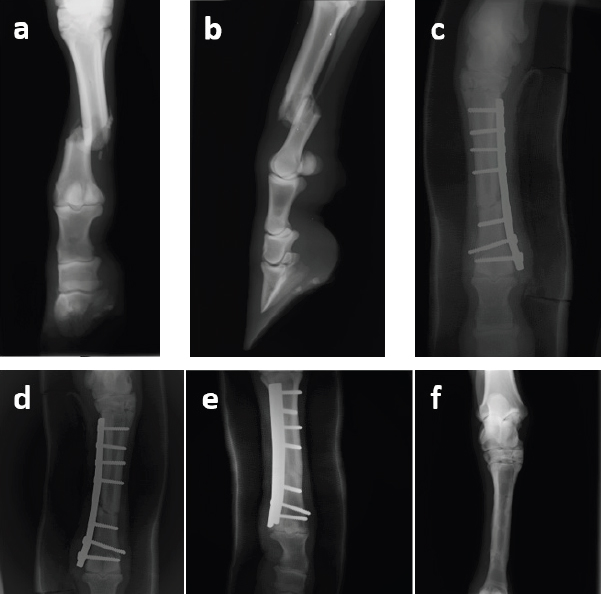
Fig. 1. Radiographic images of the third right metatarsus: (a) and (b) in dorsopalmar and lateromedial views in the preoperative period, and dorsopalmar views in the (c) immediate postoperative period, (d) fourth postoperative week, (e) second postoperative month and (f) third month after surgery and after the removal of the implants. Ethical approvalThere were no ethical concerns with the collection of data or with the management of the case. All data used in this manuscript were collected in a retrospective manner. The clients and owners of the animal approved the management of the case and this report. DiscussionHere, the authors report a successful surgical management and clinical outcome of a complete diaphyseal Mt3 fracture treatment in a pony from a circus, subject to a very demanding working activity, with a laterally applied single 4.5 mm broad DCP and respective 4.5 mm cortical screws.
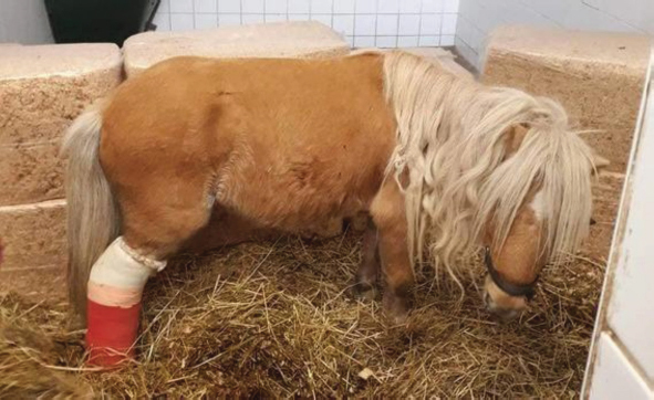
Fig. 2. Image of the pony a few days after surgery with the immobilization on the right hindlimb reinforced by the application of a full limb cast.
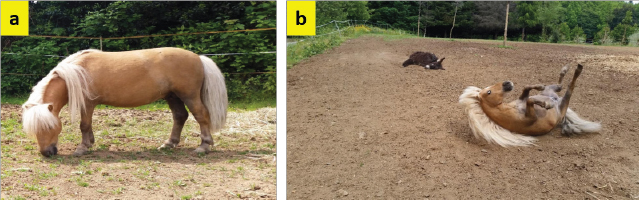
Fig. 3. Photos of the pony (a) and (b), without the cast, and fully recovered 3 months after the surgery. The etiology of Mc/t3 fractures is generally associated to intense and repeated stress efforts or trauma, namely during horse race courses and hurdle races, often with catastrophic fractures occurring specially at the lateral condyle level (Zekas et al., 1999a; Parkin et al., 2005, 2006). Also, frequently the occurrence of these type of fractures is associated to pre-existent pathology (Parkin et al., 2006). In foals generally occur simple fractures, mostly related to external trauma (Schneider and Sampson, 2020), whereas in mature horse severe comminution could occur due to high energy injuries (Bischofberger et al., 2009). The selection of animals for Mc/t3 fractures treatment is highly conditioned by the type of fracture present and the weight of the animal, especially in mature sport horses. Instable fracture fixation, mature age, bodyweight over 320 kg, infection, and increased age are strongly associated with the occurrence of postoperative complications and poor outcome in treatment of complete Mc/t3 fractures (Bischofberger et al., 2009). Thus, in highly comminuted high-energy fractures in racehorses or other athletic horses, or in open type III fractures—open, contaminated fracture with severe tissue loss (Nixon, 1996), the treatment is not indicated due to their poor prognosis (Bischofberger et al., 2009). When there is an indication for the treatment, namely in simple transverse or oblique fractures, then the scientific literature refers that rigid fixation using plates and screws is the most appropriate method for the treatment of closed or open, complete diaphyseal Mc/t3 fractures in mature horses and foals (Bischofberger et al., 2009). However, namely in small equids, the possibility of conservative treatment for this type of fracture is referred to using only immobilization of the limb with cast, especially in incomplete/non-displaced fractures (Nemeth and Back, 1991; Ladefoged et al., 2017). The clinical outcome of the present case report confirms the above mentioned, since the ORIF option with plate and screws choice was fully successful for the fracture healing in this case. However, the internal fixation was associated to a full limb cast and movement limitation during the first four postoperatively weeks to increase the likelihood of successful limb recovery. Although Bischofberger et al. (2009) recommend the use of one or two 3.5 mm broad plates or 4.5 mm narrow plates in foals or ponies, since the thickness and width of the plates should be kept as small as possible to allow the closure of the surgical wound on them, in the present clinical case we opted for the placement of a single one 4.5 mm broad plate due to the bodyweight of the pony (slightly over 100 kg) and the 4.5 mm screws stronger. However, the option of 4.5 mm DCP was less convenient in the particular aspect of the greater spacing between the holes in relation to a 3.5 mm DCP. This made it difficult to place three screws distally to the fracture focus, causing the plate distal end to get too close to the fetlock joint. The option of applying the contoured plate to the lateral, rather than cranial, surface of the Mt3 also allowed avoiding the long digital extensor tendon, minimizing possible complications involving this tendon in the postoperative period. Other reasons for the success of this case were the emergency management of soft tissue swelling with the stabilization of the fractured hindlimb in anatomically normal position, to prevent increased damage to bone and soft tissues at arrival to the hospital facilities and until the surgical fracture fixation. Also, pre- and postoperative medical management, namely through the implementation of a broad-spectrum antibiotic protocol, essential for the prevention of infection. In this clinical case, the only postoperative complications observed were a slight lysis around the first screws inserted in the distal fragment around the 20th postoperative day. Also, an articular impingement with a slight lameness and swelling of the metatarsophalangeal joint, probably caused by the plate distal end has become too close to the fetlock joint, as mentioned above. This was the main reason why the plate was removed as soon as possible, with the articular impingement fully resolved after the implant removal. Three months after fracture fixation the animal fully recovered, with clinical and radiological evidence of fracture healing, and could return to its previous activity. The originality of this clinical case results from the fact that for small equids of this weight, even with an extremely demanding physical activity such as in the present case, the option of a single plate can be used, instead of double plating, reducing the cost and risk of infection. AcknowledgmentsThis work was supported by the Portuguese Foundation for Science and Technology under the projects UIDB/AGR/04033/2020 and UIDB/CVT/00772/2020. The authors thank Dr. Maria F. Alexandre and Dr. Pedro P. Garcia for their support in the management of this clinical case. Conflict of interestThe authors declare no potential conflict of interest with the respect to this case report study, authorship, and/or publication of this article. Authors’ contributionsI. R. Dias contributed to the clinical case resolution, data collection, and writing of the first draft of the manuscript. L. M. Maia and F. C. Silva contributed to the clinical case resolution, data collection, and critical revision of the manuscript. M. Quaresma and M. Cotovio aided with the data collection, analysis and interpretation of the data, and critical revision of the manuscript. All the authors read and approved the final version of the manuscript. ReferencesBeinlich, C.P. and Bramlage, L.R. 2002. Results of plate fixation of third metacarpal and metatarsal diaphyseal fractures. Proc. Lexington, KY: AAEP, vol. 48, pp: 247–248.vol. 48, pp: 247–248. Bischofberger, A.S., Fürst, A., Auer, J. and Lischer, C. 2009. Surgical management of complete diaphyseal third metacarpal and metatarsal bone fractures: clinical outcome in 10 mature horses and 11 foals. Equine. Vet. J. 41(5), 465–473. Bramlage, L.R. 1983. Long bone fractures. Vet. Clin. North Am. Large. Anim. Pract. 5(2), 285–310. Goodrich, L.R., Nixon, A.J., Conway, J.D., Morley, P.S., Bladon, B.M. and Hogan, P.M. 2014. Dynamic compression plate (DCP) fixation of propagating medial condylar fractures of the third metacarpal/metatarsal bone in 30 racehorses: retrospective analysis (1990-2005). Equine. Vet. J. 46(6), 695–700. Ladefoged, S., Grulke, S., Busoni, V., Serteyn, D., Salciccia, A. and Verwilghen, D. 2017. Modified Thomas splint-cast combination for the management of limb fractures in small equids. Vet. Surg. 46(3), 381–388. Lescun, T.B., McClure, S.R., Ward, M.P., Downs, C., Wilson, D.A., Adams, S.B., Hawkins, J.F. and Reinerton, E.L. 2007. Evaluation of transfixation casting for treatment of third metacarpal, third metatarsal, and phalangeal fractures in horses: 37 cases (1994-2004). J. Am. Vet. Med. Assoc. 230, 1340–1349. Lloyd, D., Johanson, C. and Phillips, T. 2008. Treatment of medial condylar fractures of the third metatarsus in three horses with fibreglass casts under standing neuroleptanalgesia. Vet. Rec. 162(18), 586–589. McClure, S.R., Watkins, J.P., Glickman, N.W., Hawkins, J.F. and Glickman, L.T. 1998. Complete fractures of the third metacarpal or metatarsal bone in horses: 25 cases (1980-1996). J. Am. Vet. Med. Assoc. 213(6), 847–850. Moulin, N., François, I., Coté, N., Alford, C., Cleary, O. and Desjardins, M.R. 2018. Surgical repair of propagating condylar fractures of the third metacarpal/metatarsal bones with cortical screws placed in lag fashion in 46 racehorses (2007-2015). Equine. Vet. J. 50(5), 629–635. Nemeth, F. and Back, W. 1991. The use of the walking cast to repair fractures in horses and ponies. Equine. Vet. J. 23(1), 32–36. Nixon, A.J. 1996. General considerations in selecting cases for fracture repair. In Equine fracture repair. Ed., Nixon, A.J., Philadelphia, PA: W.B. Saunders, pp: 30–35. Orsini, J.A. and Nunamaker, D.N. 1988. Management of a severely cumminuted fracture of the third metacarpal bone on a horse. J. Am. Vet. Med. Assoc. 193(6), 683–686. Parkin, T.D.H., Clegg, P.D., French, N.P., Proudman, C.J., Riggs, C.M., Singer, E.R., Webbon, P.M. and Morgan, K.L. 2006. Catastrophic fracture of the lateral condyle of the third metacarpus/metatarsus in UK racehorses – fracture descriptions and pre-existing pathology. Vet. J. 171(1), 157–165. Parkin, T.D.H., Clegg, P.D., French, N.P., Proudman, C.J., Riggs, C.M., Singer, E.R., Webbon, P.M. and Morgan, K.L. 2005. Risk factors for fatal lateral condylar fracture of the third metacarpus/metatarsus in UK racing. Equine. Vet. J. 37(3), 192–199. Prabhakar, V., Raghunath, M., Singh, T., Saini, N.S., Mohindroo, J. and Mahajan, S.K. 2013. Use of bone plate for treatment of an open third metacarpal fracture in a foal. J. Equine. Vet. Sci. 33(8), 640–644. Schneider, R.K. and Sampson, S.N. 2020. Fractures of the third metacarpus and metatarsus. In Equine fracture repair, Ed., Nixon, A.J., 2nd ed. Hoboken, NJ: Wiley Blackwell, pp: 436–451. Zekas, L.J., Bramlage, L.R., Embertson, R.M. and Hance, S.R. 1999a. Characterization of the type and location of fractures of the third metacarpal metatarsal condyles in 135 horses in central Kentucky (1986-1994). Equine. Vet. J. 31(4), 304–308. Zekas, L.J., Bramlage, L.R., Embertson, R.M. and Hance, S.R. 1999b. Results of treatment of 145 fractures of the third metacarpal metatarsal condyles in 135 horses (1986-1994). Equine. Vet. J. 31(4), 309–313. | ||
| How to Cite this Article |
| Pubmed Style IRD, Maia LM, Quaresma M, Cotovio M, Silva FC. Laterally applied single bone plate option for fixation of complete diaphyseal fracture of a third metatarsal bone in a circus work pony. Open Vet J. 2021; 11(4): 645-650. doi:10.5455/OVJ.2021.v11.i4.14 Web Style IRD, Maia LM, Quaresma M, Cotovio M, Silva FC. Laterally applied single bone plate option for fixation of complete diaphyseal fracture of a third metatarsal bone in a circus work pony. https://www.openveterinaryjournal.com/?mno=100004 [Access: July 27, 2024]. doi:10.5455/OVJ.2021.v11.i4.14 AMA (American Medical Association) Style IRD, Maia LM, Quaresma M, Cotovio M, Silva FC. Laterally applied single bone plate option for fixation of complete diaphyseal fracture of a third metatarsal bone in a circus work pony. Open Vet J. 2021; 11(4): 645-650. doi:10.5455/OVJ.2021.v11.i4.14 Vancouver/ICMJE Style IRD, Maia LM, Quaresma M, Cotovio M, Silva FC. Laterally applied single bone plate option for fixation of complete diaphyseal fracture of a third metatarsal bone in a circus work pony. Open Vet J. (2021), [cited July 27, 2024]; 11(4): 645-650. doi:10.5455/OVJ.2021.v11.i4.14 Harvard Style , I. R. D., Maia, . L. M., Quaresma, . M., Cotovio, . M. & Silva, . F. C. (2021) Laterally applied single bone plate option for fixation of complete diaphyseal fracture of a third metatarsal bone in a circus work pony. Open Vet J, 11 (4), 645-650. doi:10.5455/OVJ.2021.v11.i4.14 Turabian Style , Isabel R. Dias, Lus M. Maia, Miguel Quaresma, Mario Cotovio, and Filipe C. Silva. 2021. Laterally applied single bone plate option for fixation of complete diaphyseal fracture of a third metatarsal bone in a circus work pony. Open Veterinary Journal, 11 (4), 645-650. doi:10.5455/OVJ.2021.v11.i4.14 Chicago Style , Isabel R. Dias, Lus M. Maia, Miguel Quaresma, Mario Cotovio, and Filipe C. Silva. "Laterally applied single bone plate option for fixation of complete diaphyseal fracture of a third metatarsal bone in a circus work pony." Open Veterinary Journal 11 (2021), 645-650. doi:10.5455/OVJ.2021.v11.i4.14 MLA (The Modern Language Association) Style , Isabel R. Dias, Lus M. Maia, Miguel Quaresma, Mario Cotovio, and Filipe C. Silva. "Laterally applied single bone plate option for fixation of complete diaphyseal fracture of a third metatarsal bone in a circus work pony." Open Veterinary Journal 11.4 (2021), 645-650. Print. doi:10.5455/OVJ.2021.v11.i4.14 APA (American Psychological Association) Style , I. R. D., Maia, . L. M., Quaresma, . M., Cotovio, . M. & Silva, . F. C. (2021) Laterally applied single bone plate option for fixation of complete diaphyseal fracture of a third metatarsal bone in a circus work pony. Open Veterinary Journal, 11 (4), 645-650. doi:10.5455/OVJ.2021.v11.i4.14 |

