


| Short Communication | ||
Open Vet J. 2021; 11(4): 695-699 Open Veterinary Journal, (2021), Vol. 11(4): 695–699 Short Communication Schirmer tear test and strip meniscometry in healthy catsLīga Kovaļčuka*, Līga Šarpio and Aija MālnieceFaculty of Veterinary Medicine, Clinical Institute, Latvia University of Life Sciences and Technologies, Jelgava, Latvia *Corresponding Author: Līga Kovaļčuka. Faculty of Veterinary Medicine, Clinical Institute, Latvia University of Life Sciences and Technologies, Jelgava, Latvia. Email: kovalcuka [at] gmail.com Submitted: 07/08/2021 Accepted: 15/11/2021 Published: 09/12/2021 © 2021 Open Veterinary Journal
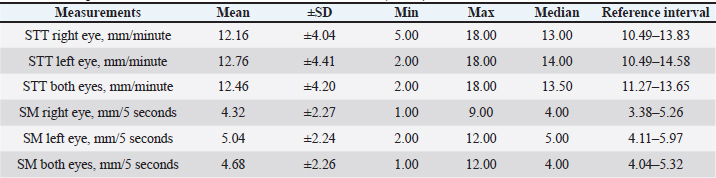
AbstractBackground: The surface of the eye is covered by the preocular tear film, which is critical for maintaining a normal, healthy, visual, and comfortable vision. The Schirmer tear test (STT) and, more recently, strip meniscometry (SM) are used to evaluate tear production. Aim: To establish the normal values for STT and SM in healthy cats and to discover the correlation between these tests. Methods: A total of 25 mixed breed cats, aging from 8 months to 13 years of both genders (10 females and 15 males) were included in the study. All the cats were assigned to the study as being both clinically and ophthalmologically healthy. For the SM test, the tip of the strip was used to evaluate the meniscus without touching the eyelid or the cornea for 5 seconds. After a full tear washout period of 10 minutes, the STT was performed using a standard STT strip. Results: In the right eyes, the mean ± standard deviation (SD) of SM was 4.32 ± 2.27 mm/5 seconds, and in the left eyes it was 5.04 ± 2.24 mm/5 seconds (for both eyes combined: 4.68 ± 2.26 mm/5 seconds), with a median of 4 in both eyes; the reference values ranged from 4.04 to 5.32 mm/5 seconds. No significant differences were recorded in the SM between the right and left eyes of the cats when using the SM (p > 0.05). When the STT was used, the mean ± SD for the cats’ right eyes was 12.16 ± 4.04 mm/minute, and for the left eyes, it was 12.76 ± 4.1 mm/minute (for both eyes combined: 12.46 ± 4.20 mm/minute), with a median of 13.50 for both eyes. Reference values were calculated and ranged from 11.27 to 13.65 mm/minute. No significant differences were recorded between the STT for the right and left eyes (p > 0.05). Conclusion: Both tests can, therefore, be used to assess tear production in cats. For more precise results, SM should be evaluated according to the cat’s eye position—whether it is a brachiocephalic cat or a normaloid cat—and according to the age. In all cases, STT and SM should be evaluated according to the animal’s clinical status and the results of other diagnostic tools. Keywords: Cat, STT-1, Meniscometry, Tears. IntroductionThe eye’s surface is covered by the precorneal tear film, which is critical for maintaining a normal, healthy, visual, and comfortable eye (Dilly, 1994; Ohashi et al., 2006). The aqueous portion, the middle layer of the tear film, plays an essential role in providing the necessary ocular surface moisture, a normal nutrient supply, and the oxygenation needed to maintain a smooth and transparent cornea (Grahn and Storey, 2004). If the tear film is lost, the eye loses its protective ability, which may lead to ocular infection, corneal abrasion, and erosion, or even a corneal ulcer and keratitis, which can lead to keratoconjunctivitis sicca. The Schirmer tear test (STT) provides a vital measurement in all animals as part of an initial ophthalmic examination. For years, the use of the STT with cats was controversial, with low STT values being interpreted as indicative of cat stress (Lim et al., 2009). In a clinical setting, the STT is the primary quantitative tear test. More recently, the strip meniscometry (SM) test, which was initially used in human medicine, has also been used in animals (Dogru et al., 2006; Miyasaka et al., 2019). However, limited data are available on the normal values for SM and the correlation between STT and SM although some positive correlations have been found in both humans and dogs (Dogru et al., 2006; Kazama et al., 2014; Miyasaka et al., 2019). The STT is the conventional test, and it requires the placement of a paper strip in the ventral conjunctival fornix for one minute. In contrast, SM is performed by placing the tip of a strip in the meniscus for just 5 seconds. This study aimed to establish the normal values for STT and SM in healthy cats and to discover the correlation between the results of these tests. Materials and MethodsThe study was performed with full respect for ethical criteria and the welfare of the cats involved. All the animals examined were privately owned and were outpatients at the Latvia University of Life Sciences and Technologies (LLU) veterinary clinic. Twenty five mixed breeds cats, aged from 8 months to 13 years of both genders (10 females and 15 males) were included. To ensure uniformity in the results, the ophthalmological examination of all animals was conducted by the same person, the veterinarian/ophthalmologist of the Clinical Institute of the Faculty of Veterinary Medicine at the LLU. The ophthalmic examination included direct ophthalmoscopy (Keeler Practitioner, Windsor, UK), monocular ophthalmoscopy with the PanOptic ophthalmoscope (Welch Alynn, Romford, UK), slit-lamp biomicroscopy (Kowa SL15, Nagoya, Aichi, Japan), and rebound tonometry (TonoVet®, Tiolat Ltd., Finland). All cats were assessed as being both clinically and ophthalmologically healthy.
Fig. 1. SM in a cat. For the SM (SMTube, Fukushima-ken, Japan), the tip of the measurement strip was placed precisely at the edge of the lower tear meniscus without touching the eyelid or the cornea (Fig. 1) for 5 seconds, and the result was immediately determined (Dogru et al., 2006). After 10 minutes of a complete tear washout period (Sebbag et al., 2019), STT was performed using standardized sterile strips (Eickemeyer, Tuttlingen, Germany). The strip tip was inserted over the lower lateral eyelid margin into the conjunctival fornix for 60 seconds. After removing the test strip, the length of the wet part of the strip was immediately measured in millimetres. Statistical analysisStatistical analysis of the data was performed using the statistical software programs Statistical Package for the Social Sciences and Microsoft Excel. The arithmetic mean values (X) as mean ± standard deviation (SD) and the reference values for STT and SM were calculated for each eye separately and for both eyes combined. Normality was tested using the Shapiro–Wilk test. The Mann-Whitney U test compared the STT and SM mean values obtained from the right and left eyes. The Pearson Correlation was used to determine the correlation between STT and SM. p values of less than 0.05 were considered to be statistically significant. Ethical approvalThe present study did not require specific ethical approval, and all examinations were performed during the routine clinical examination before the cats were neutered or spayed. In all cases, informed consent was obtained from the pet owners for the study. Results and DiscussionSTT is a standard method commonly used during an ophthalmic examination. However, some difficulties may occur with animals showing aggression or being uncooperative. There also may be medical issues, such as eyelid trauma, deep corneal ulcers, or laceration, for which STT is not applicable. In these cases, other tear film assessment methods should be considered. SM was initially used in human ophthalmology and has only relatively recently been introduced into veterinary ophthalmology. Some data has been obtained from dogs and, recently, from cats and rabbits in veterinary studies. Similar to what Rajaei et al. (2018) describe in their study, we also observed that some cats showed fear-induced restlessness when the test was done, but this did not affect the SM results. In our study, the SM average for the right eye was 4.32 ± 2.27 mm/5 seconds; in the left eye, it was 5.04 ± 2.24 mm/5 seconds (in both eyes combined: 4.68 ± 2.26 mm/5 seconds), with a median of 4 in both eyes. Reference values ranged from 4.04 to 5.32 mm/5 seconds (Table 1). These results were much lower than the 10.50 ± 0.7 mm/5 seconds reported previously by Rajaei et al. (2018). The difference between our and Rajaei results can be more likely due to the human factor or different cat breed and age. When the SM test was applied, no significant differences were recorded between the right and left eyes (p > 0.05). During the meniscometry, the cats generally did not show any pronounced discomfort or awareness of the test being applied, as confirmed by other studies (Ibrahim et al., 2011). However, in some cases, this test was more difficult for the researcher to administer than the STT due to the precise application of the strip to the meniscus of the eye and, as mentioned, the slight fear-induced restlessness shown by some cats. Therefore, the cooperation of the cat and, moreover, human factors can influence these results. To analyse the correlation between SM and STT, the classical tear test was also performed. The average STT for the right eyes was 12.16 ± 4.04 mm/minute, and for the left eyes, it was 12.76 ± 4.41 mm/minute (for both eyes: 12.46 ± 4.20 mm/minute), with a median of 13.50 for both eyes (Table 1). Reference values were calculated, and they ranged from 11.27 to 13.65 mm/minute. No significant differences were recorded in the STT for the right and left eyes of the cats (p > 0.05). Our results were slightly lower than in our previous observations (15.8 ± 6.1 mm/minute in the right eye and 17.3 ± 5.6 mm/minute in the left eye) (Kovalcuka and Nikolajenko, 2020). In other studies, STT-1 has ranged between 11.00 ± 1.41 and 20.80 ± 2.25 mm/minute (Aftab et al., 2018; Rajaei et al., 2018; Sebbag et al., 2020). This variability is high, but none of the researchers have shown less than 10 mm/minute. In the course of our study, no significant signs of ocular pain were detected in any of the animals examined at any time. Still, some of the cats showed mild discomfort and impatience, which clinically could cause reflex tearing both during and after the examination. Table 1. Descriptive statistics for STT and SM measurements in cats (n=25).
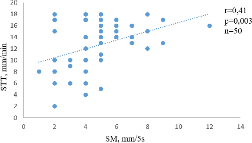
Fig. 2. Correlation between STT and SM.
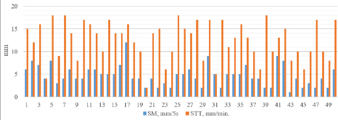
Fig. 3. Individual results of STT and SM in 50 eyes.c On average, STT is higher in dogs, ranging from 20.4 ± 2.89 to 23.56 ± 3.98 mm/minute (Hartly et al., 2006; Visser et al., 2017), than in cats, where it ranges from 13.7 ± 4.6 to 15.7 ± 3.7 mm/minute (Sebbag et al., 2020). This difference possibly correlates with SM results. Rajaei et al. (2018) reported results for dogs where STT averaged 15.10 mm/minute and SM averaged 9.66 mm/5 seconds. However, in cats they reported STT as 11 mm/minute and SM as 10.5 mm/5 seconds. These results for cats do not correlate with those obtained for dogs. As for the cats, the results from STT and SM were similar. In our research, we found a significant medium positive correlation between the STT and SM results for cats (r=0.411; p=0.003) (Fig. 2). The STT and SM results for the individual cats are shown in Figure 3. The SM should be evaluated according to the cat’s eye position—brachiocephalic cats or normaloid cats—and age for more precise results. However, the authors of this study advise clinicians to perform the STT-1 as part of their ophthalmic examination in cats, particularly in cats with an ocular surface disease. More data is needed to assess the values for SM at different stages of keratoconjunctivitis and understand at what level SM values increase during painful conditions. In every case, both the STT and SM should be evaluated according to the animal’s clinical status and the results of other diagnostic tests. Conflict of interestThe authors declare that there is no conflict of interest. Authors contributionsLK conceptualized the aim of the study, designed, planned, and supervised the analysis and corrected the manuscript. LŠ and LK conceived the work and performed all animal examinations and tests. AM analysed the data, prepared the graphs, figures and tables, and drafted the manuscript. All authors read, reviewed, discussed, and agreed to their individual contributions prior to and during submission of this article. AcknowledgmentsThis work was supported by the framework of LLU, specifically: “The effects of drug use habits on the eye microbiota antimicrobial resistance in dogs” (LLU P18). ReferencesAftab, G., Rajaei, S.M. and Faghihi, H. 2018. Comparison of the Schirmer tear test I values after placement in ventral and dorsal conjunctival fornices in healthy cats. J. Feline Med. Surg. 20(12), 1169–1172. Dilly, P.N. 1994. Structure and function of the tear film. Adv. Exp. Med. Biol. 350, 239–247. Dogru, M., Ishida, K., Matsumoto, Y., Goto, E., Ishioka, M., Kojima, T., Goto, T., Saeki, M. and Tsubota, K. 2006. Strip meniscometry: a new and simple method of tear meniscus evaluation. Invest. Ophthalmol. Vis. Sci. 5, 1895–1901. Grahn, B.H. and Storey, E. S. 2004. Lacrimostimulants and lacrimomimetics. Vet. Clin. North Am. Small Anim. Pract. 3, 739–753. Hartly, C., Williams, D. and Adams, V.J. 2006. Effect of age, gender, weight, and time of day on tear production in normal dogs. Vet. Ophthalmol. 9(1), 53–57. Ibrahim, O.M., Dogru, M., Ward, S.K., Matsumoto, Y., Wakamatsu, T.H., Ishida, K., Tsuyama, A., Kojima, T., Shimazaki, J. and Tsubota, K. 2011. The efficacy, sensitivity, and specificity of strip meniscometry in conjunction with tear function tests in the assessment of tear meniscus. Invest. Ophthalmol. Vis. Sci. 5, 2194–2198. Kazama, Y., Wakaiki, S., Washita, H.I. and Saito, A. 2014. A novel method of lacrimal function testing: strip meniscometry. In First International Conference on Ocular Surface Diseases in Dogs and Cat, 2014 March 6–8, Niseko, Japan, 2014. Kovalcuka, L. and Nikolajenko, M. 2020. Changes in intraocular pressure, horizontal pupil diameter, and tear production during the use of topical 1% cyclopentolate in cats and rabbits. Open Vet. J. 10(1), 59–67. Lim, C.C., Reilly, C.M., Thomasy, S.M., Kass, P.H. and Maggs, D.J. 2009. Effects of feline herpesvirus type 1 on tear film break-up time: Schirmer tear test results, and conjunctival goblet cell density in experimentally infected cats. Am. J. Vet. Res. 70, 394–403. Miyasaka, K., Kazama, Y., Iwashita, H., Wakaiki, S. and Saito, A. 2019. A novel strip meniscometry method for measuring aqueous tear volume in dogs: clinical correlations with the Schirmer tear and phenol red thread tests. Vet. Ophthalmol. 22(6), 864–871. Ohashi, Y., Dogru, M. and Tsubota, K. 2006. Laboratory findings in tear fluid analysis. Clin. Chim. Acta. 369, 17–28. Rajaei, S.M., Mood, M.A., Asadi, F., Rajabian, M.R. and Aghajanpour, L. 2018. Strip meniscometry in dogs, cats, and rabbits. Vet. Ophthalmol. 21(2), 210–213. Sebbag, L., Allbaugh, R.A., Wehrman, R.F., Uhl, L.K., Ben-Shlomo, G., Chen, T. and Mochel, J.P. 2019. Fluorophotometric assessment of tear volume and turnover rate in healthy dogs and cats. J. Ocul. Pharmacol. Ther. 35(9), 497–502. Sebbag, L., Uhl, L.K., Schneider, B., Hayes, B., Olds, J. and Mochel, J.P. 2020. Investigation of Schirmer tear test-1 for measurement of tear production in cats in various environmental settings and with different test durations. J. Am. Vet. Med. Assoc. 256(6), 681–686. Visser, H.E., Toffemire, K.L., Love-Myers, K.R., Allbaugh, R.A., Ellinwood, N.M., Dees, D.D., Ben-Shlomo, G. and Whitley, R.D. 2017. Schirmer tear test I in dogs: Results comparing placement in the ventral vs. dorsal conjunctival fornix. Vet. Ophthalmol. 20(6), 522–525. | ||

| How to Cite this Article |
| Pubmed Style Kovalcuka L, LS, Malniece A. Schirmer tear test and strip meniscometry in healthy cats.. Open Vet J. 2021; 11(4): 695-699. doi:10.5455/OVJ.2021.v11.i4.21 Web Style Kovalcuka L, LS, Malniece A. Schirmer tear test and strip meniscometry in healthy cats.. https://www.openveterinaryjournal.com/?mno=105114 [Access: July 27, 2024]. doi:10.5455/OVJ.2021.v11.i4.21 AMA (American Medical Association) Style Kovalcuka L, LS, Malniece A. Schirmer tear test and strip meniscometry in healthy cats.. Open Vet J. 2021; 11(4): 695-699. doi:10.5455/OVJ.2021.v11.i4.21 Vancouver/ICMJE Style Kovalcuka L, LS, Malniece A. Schirmer tear test and strip meniscometry in healthy cats.. Open Vet J. (2021), [cited July 27, 2024]; 11(4): 695-699. doi:10.5455/OVJ.2021.v11.i4.21 Harvard Style Kovalcuka, L., , . L. S. & Malniece, . A. (2021) Schirmer tear test and strip meniscometry in healthy cats.. Open Vet J, 11 (4), 695-699. doi:10.5455/OVJ.2021.v11.i4.21 Turabian Style Kovalcuka, Liga, Liga Sarpio, and Aija Malniece. 2021. Schirmer tear test and strip meniscometry in healthy cats.. Open Veterinary Journal, 11 (4), 695-699. doi:10.5455/OVJ.2021.v11.i4.21 Chicago Style Kovalcuka, Liga, Liga Sarpio, and Aija Malniece. "Schirmer tear test and strip meniscometry in healthy cats.." Open Veterinary Journal 11 (2021), 695-699. doi:10.5455/OVJ.2021.v11.i4.21 MLA (The Modern Language Association) Style Kovalcuka, Liga, Liga Sarpio, and Aija Malniece. "Schirmer tear test and strip meniscometry in healthy cats.." Open Veterinary Journal 11.4 (2021), 695-699. Print. doi:10.5455/OVJ.2021.v11.i4.21 APA (American Psychological Association) Style Kovalcuka, L., , . L. S. & Malniece, . A. (2021) Schirmer tear test and strip meniscometry in healthy cats.. Open Veterinary Journal, 11 (4), 695-699. doi:10.5455/OVJ.2021.v11.i4.21 |

