


| Case Report | ||
Open Vet J. 2021; 11(1): 96-99 doi: 10.4314/ovj.v11i1.14 Open Veterinary Journal, (2021), Vol. 11(1): 96–99 Case Report http://dx.doi.org/10.4314/ovj.v11i1.14 Rabies in bovine: First case report of rabies in Al Jabal Al Akhdar, LibyaMonier Sharif1*, Aiman Arhaiem1, Osama Giadan1, Abdulkarim Adam1, Fawzia Abdalla1, Abdunaser Dayhum2 and Mohammed Bengoumi31University of Omar Al-Mukhtar, Albeida, Libya 2Department of Preventive Medicine, Faculty of Veterinary Medicine, University of Tripoli, Tripoli, Libya 3FAO Subregional Office for North Africa, Tunis, Tunisia *Corresponding Author: Monier Sharif. Department of Basic Veterinary Medical Sciences, University of Omar Al-Mukhtar, Albeida, Libya. Email: monier.sharif [at] omu.edu.ly Submitted: 21/05/2020 Accepted: 09/01/2021 Published: 03/02/2021 © 2021 Open Veterinary Journal
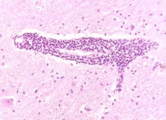
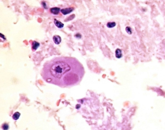
AbstractBackground: Rabies is still one of the most neglected diseases in developing countries. It is endemic to North Africa, although rabies incidence in North Africa is certainly underestimated. Case Description: On 18 December 2018 in the region of Al-Jabal Al-Akhdar, an 8-month-old calf died after a period of characteristic clinical symptoms of rabies. This is the first case of rabies in bovine which is confirmed through histopathological examination in the Laboratory of Veterinary Pathology, Omar Al-Mukhtar University. Microscopic examination clearly revealed encephalitis with the pathognomonic Negri bodies in the cerebellar neurons. Conclusion: Since the characteristic lesions in the histopathological examination are sufficient to confirm the diagnosis and report infected cases, we recommend that the next version of the OIE Terrestrial Manual should add and clarify that the results of the use of histopathological techniques in the diagnosis of rabies are significant. Keywords: Rabies, Bovine, Negri bodies, Histopathology, Libya. IntroductionRabies poses a threat to more than 3.3 billion people worldwide (Giesen et al., 2015) and is estimated to cause about 60,000 deaths a year. However, according to the World Health Organization (WHO), rabies is still one of the most neglected diseases in developing countries (Giesen et al., 2015). Rabies is endemic to North Africa, although rabies incidence in North Africa is certainly underestimated (Gautret et al., 2011; Bengoumi et al., 2018). Rabies is caused by neurotropic viruses of the genus Lyssavirus in the family Rhabdoviridae and is transmissible to all warm-blooded mammals. Eleven distinct species can be distinguished within the genus according to the World Organization for Animal Health (OIE) Terrestrial Manual [World Organization for Animal Health (OIE), 2012]. The viral reservoirs include wild and inland animals (Alknawy et al., 2018). The Lyssavirus genus consists of 14 recognized species that are all capable of causing rabies, a fatal encephalitic disease. The prototype species of this genus is Rabies lyssavirus and the rest of the species are known as the Rabies-related lyssaviruses (Afonso et al., 2016). Case DetailsOn 18 December 2018, the Veterinary Teaching Hospital of Omar Al-Mukhtar University, Al-Bayda, Libya, received a complaint from an owner of an 8-month-old calf suffering from neurological issues and a change in the normal behavior as the owner isolated the calf from five meat-producing cows and preventively treated them with ivermectin, albendazole, and multivitamins. The calf had received intravenous fluids and antibiotics and had been injected intramuscularly with anti-inflammatory drugs and multivitamins for a week. The calf’s condition did not improve, so it was transferred to the Teaching Veterinary Hospital. Clinically, the calf produced profuse butter-like salivation, with congestion of the conjunctiva in both eyes. Also, the calf bellowed loudly and showed neurological symptoms such as head extension, gait asymmetry, especially in the hind limbs, loss of appetite, and excessive excitement of anything around. The calf was also overly aggressive. Finally, the calf showed paralysis in the forelimb and death occurred the next day. According to the owner, there were similar cases in neighboring farms where animals experienced the same symptoms, and rabies was highly suspected. Therefore, we accepted to confirm the disease by a histopathological examination. Nervous tissue samples, including tissue from the cerebellum and cerebrum, were collected and fixed for a week in 10% neutral buffered formalin solution. These tissues were routinely processed through ascending grades of alcohol, cleared in xylene, and embedded in paraffin wax. The paraffin sections were cut into four to 5-μ thickness and stained by routine hematoxylin and eosin stain (H&E). Finally, the slides were examined under a light microscope. On a macroscopic examination, no apparent pathological changes were observed. On histopathological examination, the brain tissues showed mild to moderate perivascular, lymphocytic encephalitis (Fig. 1) with clear intracytoplasmic, sharply outlined, dense eosinophilic, pathognomonic inclusion bodies (Negri bodies) (Fig. 2), which are compatible with rabies virus infection. These inclusions were numerous and most readily seen in the Purkinje cells of the cerebellum. DiscussionThe confirmed case of rabies in animals (Cattle) in the Jabal Al-Akhdar of Libya means that there is always a risk of transmission to humans, as well as livestock, due to various factors. Rabies virus is always deadly when the symptoms appear and it has been recorded to have the highest mortality rate among all other known infectious diseases (Huang et al., 2015). Most rabies cases are transmitted from the bite of infected animals (Giesen et al., 2015). In North Africa, cattle represent the second most highly affected species by rabies after dogs (Bengoumi et al., 2018). Therefore, investigations of the disease are necessary as dogs are the main reservoir of rabies, to prevent further cases of high consequence of the disease [World Health Organization (WHO) et al., 2019]. Furthermore, it is listed as a notifiable disease to the World Organization for Animal Health OIE [World Organization for Animal Health (OIE), 2019] and the tripartite Food and Agriculture Organization of the United Nations (FAO)/the World Organization for Animal Health (OIE)/World Health Organization (WHO) developed a global strategic plan to control human deaths from dog-mediated rabies by 2030, which was launched in June 2018 [World Health Organization (WHO) et al., 2019]. This prompted us to perform a rapid diagnosis to minimize the risk of the disease for both humans and animals.
Fig. 1. Brain tissue showing moderate lymphocytic perivascular cuffing. H&E 400×.
Fig. 2. Section of the cerebellum of rabid cattle showing a clear cytoplasmic inclusion of the Negri body in Purkinje cells. H&E 400×. This description of the current rabies situation in Al Jabal Al Akhdar, Libya, was only based on this reported case and thus it does not necessarily represent the actual epidemiological feature in the area, given the underreported results from the non-exhaustive surveillance. So, the epidemiological situation might be very serious in animals and very dangerous for human health and might need an emergency declaration. As no clinical sign or gross post-mortem lesion can be considered as pathognomonic in domestic or wild animals, the diagnosis of rabies relies on laboratory testing. Serological testing is rarely useful for ante-mortem diagnosis because of late seroconversion and the high mortality rate of host species [World Organization for Animal Health (OIE), 2012]. According to the World Organization for Animal Health (OIE), the laboratory demonstration of rabies viral antigen, nucleic acid, or viable virus will be necessary for a positive diagnosis and that can be conducted using immunochemical identification of virus antigen or by detection of rabies virus after inoculation. The Fluorescent Antibody Test (FAT) is the gold standard diagnostic test that detects rabies virus antigen in brain samples [World Organization for Animal Health (OIE), 2014] with a sensitivity of 100% (Sharma et al., 2014). In addition to that, the reverse transcription-polymerase chain reaction has more than 97% sensitivity even in decomposed samples (Prabhu et al., 2018). However, it is not currently recommended for routine post-mortem diagnosis of rabies, if brain tissue is available then the FAT test should be used [World Organization for Animal Health (OIE), 2014]. The presence of Negri bodies and eosinophilic inclusions in the cytoplasm of infected neurons is a unique and pathognomonic finding in many cases of rabies virus infection [World Health Organization (WHO), 2017]. The sensitivity of the histopathological demonstration of Negri bodies was found to be 83.33%, while the sensitivity of the immunohistochemistry (IHC) method in rabies diagnosis was found to be 100%, exhibiting good agreement to the FAT. IHC provides a promising alternative approach to the gold standard when FAT is not feasible. Otherwise, it should always be the preferred method of choice over histopathology technique (Sharma et al., 2014). Receiving fresh samples for authentic diagnosis in our laboratory is impractical; moreover, working with fresh brain samples renders increased chances of infection. Therefore, formalin-fixed tissue samples offer better promise as a practically feasible approach for diagnostic investigation in our laboratory at present, maybe with a lower sensitivity but with specificity up to 100%, which means that there are no false-positive results upon using histopathological techniques by an experienced specialist and the diagnosis of this case is certain and undebatable. Due to all the above factors and the need of rapid notification in such diseases in addition to many veterinarians who are not pathologists following everything mentioned in the World Organization for Animal Health (OIE) reports and manuals, we recommend that the next version of the World Organization for Animal Health (OIE) Terrestrial Manual add and clarify the results of the use of histopathological techniques in the diagnosis of rabies which is significant if the pathognomonic inclusion bodies (Negri bodies) are detected, although it is not the preferred laboratory method for laboratory diagnosis if the authentic diagnostic methods are available. We also recommend that our laboratories be included in the Zero by 30 plan of the rehabilitation program to assist in applying the recommended techniques for the diagnosis of rabies in the future. AcknowledgmentsWe would like to express our deepest appreciation to the staff of the Veterinary Hospital at Omar Al-Mukhtar University for field visits and collection of the data. Conflict of interestThe authors declare that they have no conflict of interest. ReferencesAfonso, C.L., Amarasinghe, G.K., Bányai, K., Bào, Y., Basler, C.F., Bavari, S., Bejerman, N., Blasdell, K.R., Briand, F.X., Briese, T., Bukreyev, A., Calisher, C.H., Chandran, K., Chéng, J., Clawson, A.N., Collins, P.L., Dietzgen, R.G., Dolnik, O., Domier, L.L., Dürrwald, R., Dye, J.M., Easton, A.J., Ebihara, H., Farkas, S.L., Freitas-Astúa, J., Formenty, P., Fouchier, R.A., Fù, Y., Ghedin, E., Goodin, M.M., Hewson, R., Horie, M., Hyndman, T.H., Jiāng, D., Kitajima, E.W., Kobinger, G.P., Kondo, H., Kurath, G., Lamb, R.A., Lenardon, S., Leroy, E.M., Li, C.X., Lin, X.D., Liú, L., Longdon, B., Marton, S., Maisner, A., Mühlberger, E., Netesov, S.V., Nowotny, N., Patterson, J.L., Payne, S.L., Paweska, J.T., Randall, R.E., Rima, B.K., Rota, P., Rubbenstroth, D., Schwemmle, M., Shi, M., Smither, S.J., Stenglein, M.D., Stone, D.M., Takada, A., Terregino, C., Tesh, R.B., Tian, J.H., Tomonaga, K., Tordo, N., Towner, J.S., Vasilakis, N., Verbeek, M., Volchkov, V.E., Wahl-Jensen, V., Walsh, J.A., Walker, P.J., Wang, D., Wang, L.F., Wetzel, T., Whitfield, A.E., Xiè, J.T., Yuen, K.Y., Zhang, Y.Z. and Kuhn, J.H. 2006. Taxonomy of the order Mononegavirales: update 2016. Arch Virol. 161(8), 2351–2360. Alknawy, M., Mohammed, I., Ulla, S.N. and Al Aboud, A. 2018. First confirmed case of human rabies in Saudi Arabia. IDCases. 12, 29–31. Bengoumi, M., Mansouri, R., Ghram, B. and Mérot, J. 2018. Rabies in North Africa and the Middle East: current situation, strategies and outlook. Rev. Sci. Tech. 37(2), 497–510. Gautret, P., Ribadeau-Dumas, F., Parola, P., Brouqui, P. and Bourhy, H. 2011. Risk for rabies importation from North Africa. Emerg. Infect. Dis. 17(12), 2187–2193. Giesen, A., Gniel, D. and Malerczyk, C. 2015. 30 years of rabies vaccination with Rabipur: a summary of clinical data and global experience. Expert Rev. Vaccines 14(3), 351–367. Huang, Y., Chen, Z., Huang, J., Fu, Z. and He, B. 2015. Parainfluenza virus 5 expressing the G protein of rabies virus protects mice after rabies virus infection. J. Virol. 89(6), 3427–3429. Prabhu, K.N., Isloor, S., Veeresh, B.H., Rathnamma, D., Sharada, R., Das, L.J., Satyanarayana, M.L., Hegde, N.R. and Rahman, S.A. 2018. Application and comparative evaluation of fluorescent antibody, immunohistochemistry and reverse transcription polymerase chain reaction tests for the detection of rabies virus antigen or nucleic acid in brain samples of animals suspected of rabies in India. Vet. Sci. 5(1), 24; doi: 10.3390/vetsci5010024. Sharma, P., Singh, C., Sood, N., Sandhu, B., Gupta, K. and Brar, A. 2014. Diagnosis of rabies from brain: comparison of histochemical and histopathological approaches. Indian J. Vet. Pathol. 38(4), 269–272. World Health Organization (WHO). 2017. The immunological basis for immunization series: module 17: rabies. Available via https://apps.who.int/iris/bitstream/handle/10665/259511/9789241513371-eng.pdf;jsessionid=B52ECF598E45567E65F6FCFEA30F2F1C?sequence=1. (Accessed 5 January 2020). World Health Organization (WHO), Food and Agriculture Organization of the United Nations (FAO) and World Organization for Animal Health (OIE). (2019). United against rabies collaboration. First annual progress report: global strategic plan to end human deaths from dog-mediated rabies by 2030. Available via https://apps.who.int/iris/bitstream/handle/10665/328053/WHO-CDS-NTD-NZD-2019.04-eng.pdf?ua=1. (Accessed 10 March 2020). World Organization for Animal Health (OIE). 2012. Manual of diagnostic tests and vaccines for terrestrial animals (mammals, birds and bees), 7th ed. volume 2. Paris, France: World Organization for Animal Health (OIE). Retrieved from: https://www.oie.int/doc/ged/D12008.PDF. (Accessed 27 March 2020). World Organization for Animal Health (OIE). 2014. Technical disease card. Rabies, aetiology epidemiology diagnosis prevention and control references. Available via https://www.oie.int/fileadmin/Home/eng/Animal_Health_in_the_World/docs/pdf/Disease_cards/RABIES_FINAL.pdf. (Accessed 17 January 2020]. World Organization for Animal Health (OIE). 2019. Old classification of diseases notifiable to the OIE List B. Available via https://www.oie.int/en/animal-health-in-the-world/the-world-animal-health-information-system/old-classification-of-diseases-notifiable-to-the-oie-list-b/ (Accessed 16 February 2020). | ||
| How to Cite this Article |
| Pubmed Style Sharif MAM, Arhaiem AM, Giadan OKS, Adam AFH, Abdalla FFM, Dayhum ASR, Bengoumi M, . Rabies in Bovine: First Case Report of Rabies in Al Jabal Al Akhdar Libya. Open Vet J. 2021; 11(1): 96-99. doi:10.4314/ovj.v11i1.14 Web Style Sharif MAM, Arhaiem AM, Giadan OKS, Adam AFH, Abdalla FFM, Dayhum ASR, Bengoumi M, . Rabies in Bovine: First Case Report of Rabies in Al Jabal Al Akhdar Libya. https://www.openveterinaryjournal.com/?mno=108676 [Access: November 08, 2024]. doi:10.4314/ovj.v11i1.14 AMA (American Medical Association) Style Sharif MAM, Arhaiem AM, Giadan OKS, Adam AFH, Abdalla FFM, Dayhum ASR, Bengoumi M, . Rabies in Bovine:
First Case Report of Rabies in Al Jabal Al Akhdar Libya. Open Vet J. 2021; 11(1): 96-99. doi:10.4314/ovj.v11i1.14 Vancouver/ICMJE Style Sharif MAM, Arhaiem AM, Giadan OKS, Adam AFH, Abdalla FFM, Dayhum ASR, Bengoumi M, . Rabies in Bovine:
First Case Report of Rabies in Al Jabal Al Akhdar Libya. Open Vet J. (2021), [cited November 08, 2024]; 11(1): 96-99. doi:10.4314/ovj.v11i1.14 Harvard Style Sharif, M. A. M., Arhaiem, A. M., Giadan, O. K. S., Adam, A. F. H., Abdalla, F. F. M., Dayhum, A. S. R., Bengoumi, M. & (2021) Rabies in Bovine:
First Case Report of Rabies in Al Jabal Al Akhdar Libya. Open Vet J, 11 (1), 96-99. doi:10.4314/ovj.v11i1.14 Turabian Style Sharif, Monier A. M., Aiman M. Arhaiem, Osama K. S. Giadan, Abdulkarim F. H. Adam, Fawzia F. M. Abdalla, Abdunaser S. R. Dayhum, Mohammed Bengoumi, and . 2021. Rabies in Bovine:
First Case Report of Rabies in Al Jabal Al Akhdar Libya. Open Veterinary Journal, 11 (1), 96-99. doi:10.4314/ovj.v11i1.14 Chicago Style Sharif, Monier A. M., Aiman M. Arhaiem, Osama K. S. Giadan, Abdulkarim F. H. Adam, Fawzia F. M. Abdalla, Abdunaser S. R. Dayhum, Mohammed Bengoumi, and . "Rabies in Bovine:
First Case Report of Rabies in Al Jabal Al Akhdar Libya." Open Veterinary Journal 11 (2021), 96-99. doi:10.4314/ovj.v11i1.14 MLA (The Modern Language Association) Style Sharif, Monier A. M., Aiman M. Arhaiem, Osama K. S. Giadan, Abdulkarim F. H. Adam, Fawzia F. M. Abdalla, Abdunaser S. R. Dayhum, Mohammed Bengoumi, and . "Rabies in Bovine:
First Case Report of Rabies in Al Jabal Al Akhdar Libya." Open Veterinary Journal 11.1 (2021), 96-99. Print. doi:10.4314/ovj.v11i1.14 APA (American Psychological Association) Style Sharif, M. A. M., Arhaiem, A. M., Giadan, O. K. S., Adam, A. F. H., Abdalla, F. F. M., Dayhum, A. S. R., Bengoumi, M. & (2021) Rabies in Bovine:
First Case Report of Rabies in Al Jabal Al Akhdar Libya. Open Veterinary Journal, 11 (1), 96-99. doi:10.4314/ovj.v11i1.14 |

