


| Research Article | ||
Open Vet J. 2023; 13(10): 1283-1289 Open Veterinary Journal, (2023), Vol. 13(10): 1283–1289 Original Research Serological identification of MERS-CoV in camels of Wasit province, IraqHala S. R. AL-Taee1, Azhar Ali Sekhi2, Hasanain A. J. Gharban3*, and Hussien M. A. Biati41Department of Microbiology, College of Veterinary Medicine, University of Wasit, Wasit, Iraq 2Department of Microbiology, College of Medicine, University of Al-Qadisiyah, Al-Qadisiyah, Iraq 3Department of Internal and Preventive Veterinary Medicine, College of Veterinary Medicine, University of Wasit, Wasit, Iraq 4Department of Medical Microbiology, College of Medicine, University of Wasit, Wasit, Iraq *Corresponding Author: Hasanain A.J. Gharban. Department of Internal and Preventive Veterinary Medicine, College of Veterinary Medicine, University of Wasit, Wasit, Iraq. Email: hghirban [at] uowasit.edu.iq Submitted: 17/07/2023 Accepted: 11/09/2023 Published: 31/10/2023 © 2023 Open Veterinary Journal
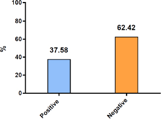
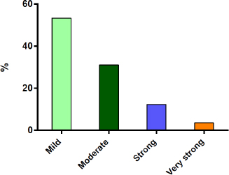
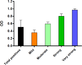
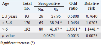
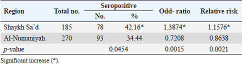
AbstractBackground: Since the first human case of Middle East Respiratory Syndrome (MERS) caused by Coronavirus (MERS-CoV) in 2012, several evidence bases have shown one-humped camels as the main reservoir host, from which infection is transmitted to humans. Aim: Serological investigation of MERS in dromedary camels in Wasit province (Iraq), detection severity of infection, and association to some risk factors. Methods: A total of 455 dromedary camels were selected randomly from two main districts in Wasit province, Iraq, during January and April (2023). Sera of all study camels were examined by enzyme-linked immunosorbent assay (ELISA), and titers of positive study animals were categorized according to their severity. Results: Serological testing yielded 37.58% positive animals for MERS infection. According to the severity of positive ODs (titer), a total of 53.22%, 30.99%, 12.28%, and 3.51% showed mild, moderate, strong, and very strong infections, respectively. Regarding risk factors, significant elevation in seropositivity was seen in camels of >3–6 and >6 years old and reduced in camels of £3 years old with an elevated risk of MERS with increased age. Regionally, seropositivity and relative risk were increased in the camels of Shaykh Sa’d when compared with Al-Numaniyah. Regarding sex, no significant variation was detected between seropositive females and males; however, male camels appeared at higher risk than females. Association between the severity of MERS infection and risk factors revealed that there was a significant increase in mild and moderate infections in female camels of >6 years old; whereas strong and very strong infections were seen in male camels of ³3–6 years old. Mild and very strong infections were recorded in Shaykh Sa’d; while moderate and strong infections in Al-Numaniyah. Conclusion: The study indicated a longstanding existence of MERS-CoV in camels of Wasit province; therefore, recent infections or active viral excretion are required for confirmation by molecular approaches. Keywords: MERS-CoV, Camelus dromedarius, Coronavirus, Serological test, ELISA. IntroductionMiddle East respiratory syndrome (MERS) is one of the most prevalent viral diseases in the last decade. It was first described in Saudi Arabia in 2012 and then reported in many countries in Africa, Asia, Europe, and America continents (Şenel and Topal, 2021; Islam et al., 2023). MERS-CoV belongs to Coronaviridae Family in Nidovirales Order of Orthornavirae Kingdom, which is an enveloped virus of single-stranded RNA genome and a nucleocapsid of helical symmetry with the club-shaped spikes that project from their surface (Li et al., 2019; Yan et al., 2020). Although, the origin of the virus is not fully understood, it is believed that it originated in wild animals, such as bats, and then spread to domestic animals, such as camel, cattle, goats, and sheep (Reusken et al., 2013a; Malik, 2020; Islam et al., 2023). However, the dromedary camels have been the only species so far to harbor specific antibodies against MERS-CoV, identifying them as the main potential reservoir that transmits the virus to humans (Hemida and Alnaeem, 2019). Notably, most MERS infections in camels appeared asymptomatic, but minor clinical signs could be seen after some field or experimental infections, which include fever, anorexia, respiratory discharge, sneezing, and coughing (Sharun et al., 2020). Traditional isolation of the virus is a great of challenge since the shedding of the virus occurs during 1–7 days after infection, and the virus can be detected in tissues of the upper respiratory system associated with its regional lymph nodes (Adney et al., 2014). Molecular studies of nasal swabs showed that the findings could remain positive after two weeks, and the next sampling after 1 or 2 months post-infection revealed negative results for viral RNA (Meyer et al., 2016; Muhairi et al., 2016; Ali et al., 2017a; Hemida et al., 2017). Different serological techniques were modified to identify MERS, such as neutralization test, protein microarray, western blot, immunofluorescence assay, and enzyme-linked immunosorbent assay (ELISA). Of these methods, ELISA is suitable for first-line screening as it is a simple operating procedure with short processing time and large throughput (Yan et al., 2020). Global epidemiological researchers have well documented that the virus circulates extensively in dromedary populations with a high seroprevalence varied between 80% and 100% in some areas, especially, in the Arabian Peninsula (Dighe et al., 2019; Pestil et al., 2022). In Iraq, there were only two serological reports that were carried out in Najaf, Muthana, and Basra (Thwiny et al., 2018) and Diyala (Hasan et al., 2022) provinces; therefore, this study represents a unique survey to investigate seroprevalence of MERS in one-humped camels of Wasit province (Iraq) by ELISA with detection the association of infection to some risk factors. Materials and MethodsStudy samplesA total of 455 camels (Camelus dromedarius) of variable ages and sexes were chosen from two main districts in Wasit province (Shaykh Sa’d and Al-Numaniyah) during January and April (2023). Each study animal was subjected to jugular venous blood sampling (5 ml), under aseptic conditions, into a labeled free-anticoagulant gel glass tube that was then centrifuged at 1000 ´ g for 5 minutes. Sera were kept in Eppendorf tubes at 4°C. Serological testingFollowing the manufacturers’ instructions of Qualitative MERS-COV ELISA Kit (MyBioSource, USA), the sera and ELISA kit reagents were prepared, processed and the reaction was read at an optical density (OD) 450 nm using the Automated Microplate Photometer (MicroTech, USA) after adding of Stop Solution. The averages of positive and negative controls were determined at 1.23 and 0.11, respectively; the Cut-Off value was calculated following the provided equal (Average of negative control + 0.15), and the positive samples were determined at ³0.26. According to the level of positive ODs (titer), the severity of infection was categorized into 4 groups: mild, moderate, strong, and very strong infections. Study limitationThe study investigates the seroprevalence of MERS in camels in two regions only, as these regions exhibited a high number of camels compared to other regions. Furthermore, the limited number of camels in the other regions associated with far distances and the difficulty of obtaining the consent of owners to take blood samples are the main reasons that the study depends on only two regions that represent the province. Statistical analysisThe t-test, one- and two-way ANOVA, as well as Odd-Ratio in the GraphPad Prism Software (GraphPad Inc, USA), was used to estimate differences between values of the study at p < 0.05. Values were represented as either percentage (%) or as Mean ± Standard deviation (AL-Shaeli et al., 2022). Ethical approvalThe study was licensed by the Scientific Committee of the College of Veterinary Medicine (University of Wasit, Wasit, Iraq). In contrast, the collection of blood samples and data was carried out after the oral agreement of the camels’ owners. ResultsSerological resultsSerological testing of 455 camels by indirect ELISA yielded 171 (37.58%) positive animals for MERS infection and 284 (62.42%) negative animals (Fig. 1). According to their positive OD levels, a total of 53.22% (91/171), 30.99% (53/171), 12.28% (21/171), and 3.51% (6/171) showed mild, moderate, strong, and very strong infections, respectively (Fig. 2). Subsequently, the OD values (M ± SD) were 0.355 ± 0.074, 0.594 ± 0.051, 0.799 ± 0.067, and 0.968 ± 0.036, respectively for mild, moderate, strong and very strong infections (Fig. 3). Risk factorsIn this study, values of positivity, odd ratio, and relative risk showed a significant variation among different groups of age (Table 1), region (Table 2), and sex (Table 3). For age, significant elevation in seropositivity was seen in camels of >3–6 (38.24%) and > 6 (41.67%) years old and reduced in those of £ 3 years old (27.96%). Also, the findings of Odd ratio and relative risk were increased significantly ( p < 0.05) with age, in particular in camels of >6 years old.
Fig. 1. Serological result for testing camel’s sera by indirect ELISA.
Fig. 2. Prevalence rate of positives according to severity of infection.
Fig. 3. Values of ODs of positives according to severity of infection. Table 1. Association between positivity, Odd ratio and relative risk with age groups.
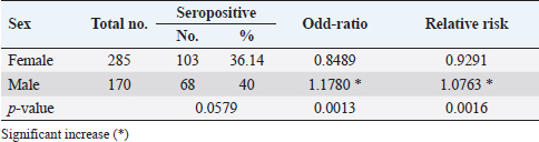
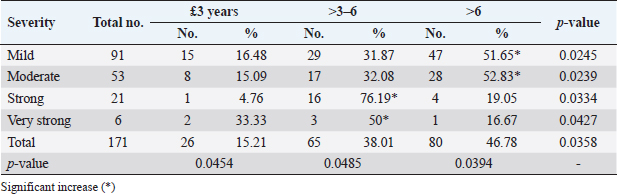
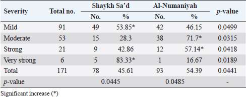
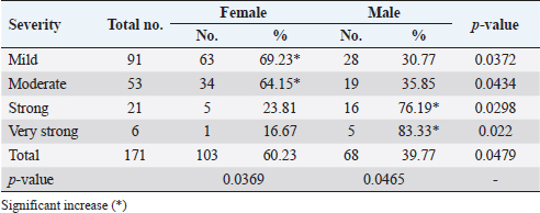
Concerning study regions, significant increases ( p < 0.05) in seropositivity were recorded in the camels of Shaykh Sa’d (42.16%) when compared to those of Al-Numaniyah (34.44%). In addition, values of Odd ratio and relative risk were in camels of Shaykh Sa’d than Al-Numaniyah. Regarding sex, no significant variation ( p < 0.0579) was detected between seropositive females and males; however, male camels have appeared at a significantly greater MERS risk than females ( p < 0.05). Association of severity of MERS infection to risk factorsAlthough the mild and moderate MERS infection increased significantly ( p < 0.05) in camels of >6 years old (51.65% and 52.83%, respectively), strong and very strong infections were seen significantly ( p < 0.05) in camels of ³3–6 years old (76.19% and 50%, respectively), (Table 4). Among both study regions, the findings of mild and very strong MERS-CoV infections were increased significantly ( p < 0.05) in Shaykh Sa’d (53.85% and 83.33%, respectively), whereas moderate and strong infections were significantly higher in Al-Numaniyah (71.7% and 57.14%, respectively), (Table 5). Regarding sex, mild and moderate MERS-CoV infections appeared significantly ( p < 0.05) in females (69.23% and 64.15%, respectively), while the strong and very strong seropositive infections were reported in males (76.19% and 83.33%, respectively), (Table 6). DiscussionThe seroprevalence of MERS infection in study camels (37.58%) was less than the reported prevalence in Najaf (85%), Muthana (84.7%), and Basra (85.7%) (Thwiny et al., 2018) and Diyala (90%) (Hasan et al., 2022) provinces. In comparison with seroprevalence of MERS in camels of other countries, there was 0.24% in Kazakhstan (Orynbayev et al., 2022), 4.12% in Canary Island (Gutiérrez et al., 2015), 19.44% in Nigeria (Gaddafi et al., 2020), 39.5% in Pakistan (Saqib et al., 2017), 51.05% in Jordan (Ababneh et al., 2021), and 52.7% in Turkey (Pestil et al., 2022). Furthermore, the prevalence is 59.8%–97.1% in United Arab Emirates (Meyer et al., 2014), 61.8% in Israel (David et al., 2018), 68.9% in Egypt (Kandeil et al., 2019), 70.81% in Saudi Arabia (Aljasim et al., 2020), 80.4% in Tunisia (Eckstein et al., 2021), 93% in Ethiopia (Fekadu et al., 2017), and 100% in Oman (Reusken et al., 2013b). The source of antigen is the main issue in different serological results; however, animal’s age and sex, geographic location, and sampling population characteristics, as the imported camels have a significantly higher seropositivity than local breed counterparts, and diagnostic assay serving in sero-survey are other factors. This study showed that mild infection was prevalent significantly among seropositive camels. Globally, few information is available about the longevity of antibody titers post-MERS-CoV infections. Meyer et al. (2016) found that neutralizing antibodies remain consistent during a year in camels following infection on a closed farm, whereas other researchers recognized that the titer of antibodies declines rapidly throughout 14 days (Ali et al., 2017b; Hemida et al., 2017). However, many of these studies that were conducted variously to dedicate the seroprevalence of MERS had taken the samples opportunistically from stored samples/sera of dromedary camels when there was an investigation of human outbreaks or from tested samples for other means. In addition, not all studies used serological assays to determine or confirm seropositivity, and between those that did, cut-off positivity titers varied. These findings are important since they supported the regional and global scientific consent as the one-humped camel is the only host species for MERS and, therefore, a potential source of infection in humans (Mirkena et al., 2018; Al-Salihi and Khalaf, 2021). Table 2. Association between positivity, odd ratio and relative risk with region groups.
Table 3. Association between positivity, odd ratio and relative risk with sex groups.
Table 4. Association of severity of MERS infection to age.
Table 5. Association of severity of MERS infection to region.
Table 6. Association of severity of MERS infection to sex.
Our results showed that seropositivity increased with age, as recorded by several studies (Ali et al., 2017a; Miguel et al., 2017; Kasem et al., 2018; Ommeh et al., 2018); however, in contrast to others (Deem et al., 2015; Harrath and Abu Duhier, 2018; Thwiny et al., 2018) that showed no effect of age in seropositivity results. Reusken et al. (2013a) mentioned that maternal immunity in dromedaries rapidly declined 14–35 days post-delivery, and a much higher titer of antibodies was generated against disease in adult dromedaries. Hence, alternative interpretations are that camels had just been infected and titers of antibodies were rising. In this study, increasing in seroprevalence and risk of MERS infection was reported in the camels of Shaykh Sa’d when compared to Al-Numaniyah. These findings were in contrast with those seen in other Iraqi locations by Thwiny et al. (2018), but it was similar to those reported in different countries in Africa and the Middle East, such as Egypt, Senegal, Tunisia, and Saudi Arabian (Kandeil et al., 2019). However, significant variation between study areas might be attributed to differences in camel management, herd size, exposure to wild animals, owners’ ethnic background, and facility of sampling site. Frequent transporting of camels appears to be another cause for the increasing regional seropositivity because transportation generates stress and lowers infection resistance, increasing the chance for direct contact with infected camels (Islam et al., 2023). Although, no significant variation was detected between seropositive females and males, male camels appeared at greater MERS risk than females. Some individual reports indicate a significantly high prevalence in females (Ali et al., 2017a; Miguel et al., 2017), while others found the opposite (Kasem et al., 2018) or do not record any significant difference (Thwiny et al., 2018). Sikkema et al. (2019) mentioned that most female camels are sampled at farms, whereas the majority of males were looked at quarantine areas, live animal markets, and slaughterhouses. Different studies indicated that positive rates of IgG antibodies increase gradually in dromedaries with age, whereas detection of MERS-CoV RNA decreases (Wernery et al., 2015; Wang et al., 2019; Loeffelholz and Tang, 2020). Much higher MERS seroprevalence could exist probably as a result of multiple MERS re-infections or long-lasting immune response toward MERS infections. However, immunity is not sterilizing as MERS infection and shedding have also been detected in adult camels that have MERS antibodies (Haagmans et al., 2016; Hemida et al., 2017; van Doremalen et al., 2017). ConclusionOur observations indicated that MERS might be circulated in dromedary camels of Wasit province, and the potential role of camels, as a reservoir, in the transmission of infection to humans is expected. Combining serological laboratory results of camels with sufficient background information (severity of infection, age, region, and sex) does result in valuable data. The sensitivity and specificity of ELISA, as well as comparative studying with other diagnostic techniques, are necessary in the detection of MERS infection. Mapping the movement of dromedaries is necessary to understand the underlying spatial transmission dynamics of MERS. AcknowledgmentsThanks to the College of Veterinary Medicine (University of Wasit), which allows us to use their laboratories throughout carrying out this study. Conflict of interestAll authors have no conflict of interest to disclose. Authors contributionsHAJG and AAS: Blood sampling and sera preparation. HSRA and HMAB: Serological testing of sera. HAJG: Statistical analysis of data. All authors read and approved the final copy of the manuscript. FundingThere is no external fund (private funding). Data availabilityAll data supporting the findings of this study are available within the manuscript. ReferencesAbabneh, M.M., Lafi, S.Q., Abutarbush, S.M., Khalifeh, M.S., Hijazeen, Z.S., Ramadneh, W.A. and von Dobschuetz, S. 2021. Longitudinal and abattoir-based surveillance of MERS-CoV in camels in Jordan, 2018–2020. Heliyon, 7(10), 1–8. Adney, D.R., van Doremalen, N., Brown, V.R., Bushmaker, T., Scott, D., de Wit, E. and Munster, V.J. 2014. Replication and shedding of MERS-CoV in upper respiratory tract of inoculated dromedary camels. Emerg. Infect. Dis. 20(12), 1999–2005. Ali, M.A., Shehata, M.M., Gomaa, M.R., Kandeil, A., El-Shesheny, R., Kayed, A.S. and Kayali, G. 2017a. Systematic, active surveillance for Middle East respiratory syndrome coronavirus in camels in Egypt. Emerg. Microb. Infect. 6(1), 1–7. Ali, M., El-Shesheny, R., Kandeil, A., Shehata, M., Elsokary, B., Gomaa, M. and Makonnen, Y.J. 2017b. Cross-sectional surveillance of Middle East respiratory syndrome coronavirus (MERS-CoV) in dromedary camels and other mammals in Egypt, August 2015 to January 2016. Eurosurveillance 22(11), 30487. Aljasim, T.A., Almasoud, A., Aljami, H.A., Alenazi, M.W., Alsagaby, S.A., Alsaleh, A.N. and Alharbi, N.K. 2020. High rate of circulating MERS-CoV in dromedary camels at slaughterhouses in Riyadh, 2019. Viruses 12(11), 1215–1224. Al-Salihi, K.A. and Khalaf, J.M. 2021. The emerging SARS-CoV, MERS-CoV, and SARS-CoV-2: an insight into the viruses zoonotic aspects. Vet. World, 14(1), 190–199. AL-Shaeli, S.J., Ethaeb, A.M. and Gharban, H.A. 2022. Determine the glucose regulatory role of decaffeinated Green Tea extract in reduces the metastasis and cell viability of MCF7 cell line. In AIP Conference Proceedings, pp: 1–8. David, D., Rotenberg, D., Khinich, E., Erster, O., Bardenstein, S., van Straten, M. and Davidson, I. 2018. Middle East respiratory syndrome coronavirus specific antibodies in naturally exposed Israeli llamas, alpacas and camels. One Health 5, 65–68. Deem, S.L., Fèvre, E.M., Kinnaird, M., Browne, A.S., Muloi, D., Godeke, G.J. and Reusken, C. B. 2015. Serological evidence of MERS-CoV antibodies in dromedary camels (Camelus dromedaries) in Laikipia County, Kenya. PLoS One, 10(10), e0140125. Dighe, A., Jombart, T., Van Kerkhove, M.D. and Ferguson, N. 2019. A systematic review of MERS-CoV seroprevalence and RNA prevalence in dromedary camels: implications for animal vaccination. Epidemics, 29, 100350. Eckstein, S., Ehmann, R., Gritli, A., Yahia, H.B., Diehl, M., Wölfel, R. and Moussa, M.B. 2021. Prevalence of Middle East respiratory syndrome coronavirus in dromedary camels, Tunisia. Emerg. Infect. Dis. 27(7), 1964–1968. Fekadu, G., Ayelet, G. and Abunna, F. 2017. Epidemiological investigation of Middle East respiratory syndrome corona virus (mers-cov) among dromedary camels in selected areas of Afar and Oromia region, Ethiopia. J. Vet. Med. Anim. Health 9(3), 47–54. Gaddafi, M.S., Faleke, O.O., Yakubu, Y., Garba, B., Musawa, I.A., Junaidu, A.U. and Aliyu, M.A. 2020. Seroprevalence of the Middle East respiratory syndrome coronavirus antibodies in camels from two international animal control-posts, Kebbi State, Nigeria. J. Anim. Health Prod. 8(2), 50–54. Gutiérrez, C., Tejedor-Junco, M.T., González, M., Lattwein, E. and Renneker, S. 2015. Presence of antibodies but no evidence for circulation of MERS-CoV in dromedaries on the Canary Islands, 2015. Eurosurveillance 20(37), 30019. Haagmans, B.L., Van Den Brand, J.M., Raj, V.S., Volz, A., Wohlsein, P., Smits, S.L. and Osterhaus, A.D. 2016. An orthopoxvirus-based vaccine reduces virus excretion after MERS-CoV infection in dromedary camels. Science 351(6268), 77–81. Harrath, R. and Abu Duhier, F.M. 2018. Sero-prevalence of Middle East respiratory syndrome coronavirus (MERS-CoV) specific antibodies in dromedary camels in Tabuk, Saudi Arabia. J. Med. Virol. 90(8), 1285–1289. Hasan, A., Ali, K. and Saleh, M. 2022. Cross-sectional study of Middle East respiratory syndrome coronavirus in humans and dromedary camels in Diyala, Iraq. Turkish J. Med. Sci. 52(4), 910–916. Hemida, M.G., Alnaeem, A., Chu, D.K., Perera, R.A., Chan, S.M., Almathen, F. and Peiris, M. 2017. Longitudinal study of Middle East respiratory syndrome coronavirus infection in dromedary camel herds in Saudi Arabia, 2014–2015. Emerg. Microb. Infect. 6(1), 1–7. Hemida, M.G. and Alnaeem, A. 2019. Some One Health based control strategies for the Middle East respiratory syndrome coronavirus. One Health 8, 100102. Islam, M.M., Khanom, H., Farag, E., Mim, Z.T., Naidoo, P., Mkhize-Kwitshana, Z.L. and Hassan, M.M. 2023. Global patterns of Middle East respiratory syndrome coronavirus (MERS-CoV) prevalence and seroprevalence in camels: a systematic review and meta-analysis. One Health 16, 100561. Kandeil, A., Gomaa, M., Nageh, A., Shehata, M.M., Kayed, A.E., Sabir, J.S. and Kayali, G. 2019. Middle East respiratory syndrome coronavirus (MERS-CoV) in dromedary camels in Africa and Middle East. Viruses 11(8), 1–16. Kasem, S., Qasim, I., Al-Hufofi, A., Hashim, O., Alkarar, A., Abu-Obeida, A. and Perera, R.A. 2018. Cross-sectional study of MERS-CoV-specific RNA and antibodies in animals that have had contact with MERS patients in Saudi Arabia. J. Infect. Public Health 11(3), 331–338. Li, Y.H., Hu, C.Y., Wu, N.P., Yao, H.P. and Li, L.J. 2019. Molecular characteristics, functions, and related pathogenicity of MERS-CoV proteins. Engineering 5(5), 940–947. Loeffelholz, M.J. and Tang, Y.W. 2020. Laboratory diagnosis of emerging human coronavirus infections–the state of the art. Emerg. Microb. Infect. 9(1), 747–756. Malik, Y.A. 2020. Properties of coronavirus and SARS-CoV-2. Malaysian J. Pathol. 42(1), 3–11. Meyer, B., Müller, M.A., Corman, V.M., Reusken, C.B., Ritz, D., Godeke, G.J. and Drosten, C. 2014. Antibodies against MERS coronavirus in dromedary camels, United Arab Emirates, 2003 and 2013. Emerg. Infect. Dis. 20(4), 552–559. Meyer, B., Juhasz, J., Barua, R., Gupta, A.D., Hakimuddin, F., Corman, V.M. and Nagy, P. 2016. Time course of MERS-CoV infection and immunity in dromedary camels. Emerg. Infect. Dis. 22(12), 2171–2173. Miguel, E., Chevalier, V., Ayelet, G., Bencheikh, M.N.B., Boussini, H., Chu, D.K. and Peiris, M. 2017. Risk factors for MERS coronavirus infection in dromedary camels in Burkina Faso, Ethiopia, and Morocco, 2015. Eurosurveillance 22(13), 30498. Mirkena, T., Walelign, E., Tewolde, N., Gari, G., Abebe, G. and Newman, S. 2018. Camel production systems in Ethiopia: a review of literature with notes on MERS-CoV risk factors. Pastoralism 8(1), 1–17. Muhairi, S.A., Hosani, F.A., Eltahir, Y.M., Mulla, M.A., Yusof, M.F., Serhan, W.S. and Abdelazim, A.S. 2016. Epidemiological investigation of Middle East respiratory syndrome coronavirus in dromedary camel farms linked with human infection in Abu Dhabi Emirate, United Arab Emirates. Virus Genes 52(6), 848–854. Ommeh, S., Zhang, W., Zohaib, A., Chen, J., Zhang, H., Hu, B. and Shi, Z.L. 2018. Genetic evidence of Middle East respiratory syndrome coronavirus (MERS-Cov) and widespread seroprevalence among camels in Kenya. Virologica Sinica 33, 484–492. Orynbayev, M.B., Hitch, A.T., Kerimbayev, A.A., Nissanova, R.K., Sultankulova, K.T., Rystayeva, R.A. and Mendenhall, I.H. 2022. Serological exposure in Bactrian and dromedary camels in Kazakhstan to a MERS or MERS-like coronavirus. Transbound. Emerg. Dis. 69(5), e1374–e1381. Pestil, Z., Ataseven, V.S., Ambarcıoğlu, P. and Gürel, K. 2022. First serologic evidence of Middle East Respiratory Syndrome (MERS-CoV) infection in camels (Camelus dromedarius), Turkey. https://doi.org/10.21203/rs.3.rs-1705625/v1. Reusken, C.B., Ababneh, M., Raj, V.S., Meyer, B., Eljarah, A., Abutarbush, S. and Koopmans, M.P. 2013a. Middle East Respiratory Syndrome coronavirus (MERS-CoV) serology in major livestock species in an affected region in Jordan, June to September 2013. Eurosurveillance 18(50), 1–7. Reusken, C.B., Haagmans, B.L., Müller, M.A., Gutierrez, C., Godeke, G.J., Meyer, B. and Koopmans, M.P. 2013b. Middle East respiratory syndrome coronavirus neutralising serum antibodies in dromedary camels: a comparative serological study. The Lancet Infect. Dis. 13(10), 859–866. Saqib, M., Sieberg, A., Hussain, M.H., Mansoor, M.K., Zohaib, A., Lattwein, E. and Corman, V.M. 2017. Serologic evidence for MERS-CoV infection in dromedary camels, Punjab, Pakistan, 2012–2015. Emerg. Infect. Dis. 23(3), 550–551. Şenel, E. and Topal, F.E. 2021. Holistic analysis of coronavirus literature: a scientometric study of the global publications relevant to sars-cov-2 (covid-19), mers-cov (mers) and sars-cov (sars). Disaster Med. Public Health Prepared. 15(6), e12–e19. Sharun, K., Sircar, S., Malik, Y.S., Singh, R.K. and Dhama, K. 2020. How close is SARS-CoV-2 to canine and feline coronaviruses? J. Small Anim. Pract. 61(8), 523–536. Sikkema, R.S., Farag, E.A.B.A., Islam, M., Atta, M., Reusken, C.B.E.M., Al-Hajri, M.M. and Koopmans, M.P.G. 2019. Global status of Middle East respiratory syndrome coronavirus in dromedary camels: a systematic review. Epidemiol. Infect. 147, 1–13. Thwiny, H.T., Al Hamed, T.A. and Nazzal, A.R. 2018. Seroepidemiological study of Middle East respiratory syndrome (MERS) virus infection in Iraqi dromedary camels. Veterinarski Arhiv 88(2), 191–200. van Doremalen, N., Hijazeen, Z.S., Holloway, P., Al Omari, B., McDowell, C., Adney, D. and Richt, J.A. 2017. High prevalence of Middle East respiratory coronavirus in young dromedary camels in Jordan. Vector-Borne Zoo. Dis. 17(2), 155–159. Wang, W., Wang, T., Deng, Y., Niu, P., Ruhan, A., Zhao, J. and Tan, W. 2019. A novel luciferase immunosorbent assay performs better than a commercial enzyme-linked immunosorbent assay to detect MERS-CoV specific IgG in humans and animals. Biosaf. Health 1(3), 134–143. Wernery, U., Rasoul, I.E., Wong, E.Y., Joseph, M., Chen, Y., Jose, S. and Woo, P.C. 2015. A phylogenetically distinct Middle East respiratory syndrome coronavirus detected in a dromedary calf from a closed dairy herd in Dubai with rising seroprevalence with age. Emerg. Microb. Infect. 4(1), 1–5. Yan, Y., Chang, L. and Wang, L. 2020. Laboratory testing of SARS-CoV, MERS-CoV, and SARS-CoV-2 (2019-nCoV): current status, challenges, and countermeasures. Rev. Med. Virol. 30(3), e2106. | ||
| How to Cite this Article |
| Pubmed Style AL-Taee HS, Sekhi AA, Gharban HA, Biati HM. Serological identification of MERS-CoV in camels of Wasit province, Iraq. Open Vet J. 2023; 13(10): 1283-1289. doi:10.5455/OVJ.2023.v13.i10.7 Web Style AL-Taee HS, Sekhi AA, Gharban HA, Biati HM. Serological identification of MERS-CoV in camels of Wasit province, Iraq. https://www.openveterinaryjournal.com/?mno=161536 [Access: July 27, 2024]. doi:10.5455/OVJ.2023.v13.i10.7 AMA (American Medical Association) Style AL-Taee HS, Sekhi AA, Gharban HA, Biati HM. Serological identification of MERS-CoV in camels of Wasit province, Iraq. Open Vet J. 2023; 13(10): 1283-1289. doi:10.5455/OVJ.2023.v13.i10.7 Vancouver/ICMJE Style AL-Taee HS, Sekhi AA, Gharban HA, Biati HM. Serological identification of MERS-CoV in camels of Wasit province, Iraq. Open Vet J. (2023), [cited July 27, 2024]; 13(10): 1283-1289. doi:10.5455/OVJ.2023.v13.i10.7 Harvard Style AL-Taee, H. S., Sekhi, . A. A., Gharban, . H. A. & Biati, . H. M. (2023) Serological identification of MERS-CoV in camels of Wasit province, Iraq. Open Vet J, 13 (10), 1283-1289. doi:10.5455/OVJ.2023.v13.i10.7 Turabian Style AL-Taee, Hala SR., Azhar Ali Sekhi, Hasanain A.J. Gharban, and Hussien M.A. Biati. 2023. Serological identification of MERS-CoV in camels of Wasit province, Iraq. Open Veterinary Journal, 13 (10), 1283-1289. doi:10.5455/OVJ.2023.v13.i10.7 Chicago Style AL-Taee, Hala SR., Azhar Ali Sekhi, Hasanain A.J. Gharban, and Hussien M.A. Biati. "Serological identification of MERS-CoV in camels of Wasit province, Iraq." Open Veterinary Journal 13 (2023), 1283-1289. doi:10.5455/OVJ.2023.v13.i10.7 MLA (The Modern Language Association) Style AL-Taee, Hala SR., Azhar Ali Sekhi, Hasanain A.J. Gharban, and Hussien M.A. Biati. "Serological identification of MERS-CoV in camels of Wasit province, Iraq." Open Veterinary Journal 13.10 (2023), 1283-1289. Print. doi:10.5455/OVJ.2023.v13.i10.7 APA (American Psychological Association) Style AL-Taee, H. S., Sekhi, . A. A., Gharban, . H. A. & Biati, . H. M. (2023) Serological identification of MERS-CoV in camels of Wasit province, Iraq. Open Veterinary Journal, 13 (10), 1283-1289. doi:10.5455/OVJ.2023.v13.i10.7 |

