


| Case Report | ||
Open Vet J. 2023; 13(11): 1485-1490 Open Veterinary Journal, (2023), Vol. 13(11): 1485-1490 Case Report Radiotherapy in combination with exenteration and partial orbitectomy for orbital multilobular tumor of bone in a Cocker SpanielNatthanet Sritrakoon1, Winutpuksinee Wibulchan2, Winyu Karntip1, Theerapol Sirinarumitr3 and Aree Thayananuphat4*1Ophthalmology Unit, Faculty of Veterinary Medicine, Kasetsart University Veterinary Teaching Hospital, Bangkok, Thailand 2Imaging and Radiotherapy Center, Faculty of Veterinary Medicine, Kasetsart University Veterinary Teaching Hospital, Bangkok, Thailand 3Department of Pathology, Faculty of Veterinary Medicine, Kasetsart University, Bangkok, Thailand 4Department of Companion Animal Clinical Sciences, Faculty of Veterinary Medicine, Kasetsart University, Bangkok, Thailand *Corresponding Author: Aree Thayananuphat. Department of Companion Animal Clinical Sciences, Faculty of Veterinary Medicine, Kasetsart University, Bangkok, Thailand. Email: areethaya [at] gmail.com Submitted: 19/07/2023 Accepted: 19/10/2023 Published: 30/11/2023 © 2023 Open Veterinary Journal
AbstractBackground: Multilobular tumor of bone or multilobular osteochondrosarcoma is a tumor of flat bone in the skull. The treatment of choice for a multilobular tumor of bone is local aggressive surgical excision. Case Description: A female Cocker Spaniel dog aged 11 years presented with a history of globe displacement of the right eye for 3 months. Ophthalmic examination revealed exophthalmos, third eyelid protrusion, and slightly increased intraocular pressure OD (oculus dexter; right eye). Computed tomography (CT) revealed a mass effect in the right retrobulbar, maxilla, zygomatic, and temporal areas. Right zygomatic and temporal bone lysis were observed. Physical examination, hematology, and blood chemistry results were within normal limits. Exenteration with zygomatic arch removal was performed. During surgery, a firm 2-lobed mass (4.8 × 3.7 and 1.6 × 1.4 cm) adhered to the mandible was found in the retrobulbar area OD. Histopathological findings revealed a multilobular tumor of bone. CT imaging was performed for the remaining tumor and an extended part of the right retrobulbar mass was found. Hypofractioned radiotherapy with 6 fractions of 6 Gy was performed on days 0, 7, 14, 21, 28, and 35. At 1-month and 4-month follow-up inspections, the mass gradually reduced in size. At 8 months and 11 months after radiotherapy, the mass was unremarkable. The dog was alert during all follow-up periods to 1 year and 8 months after hypofractioned radiotherapy combined with exenteration and partial orbitectomy. Conclusion: Hypofractioned radiotherapy combined with exenteration and partial orbitectomy extended the patient’s survival and decreased the size of the remaining tumor for the management of orbital multilobular tumor of bone in this dog for at least 1 year and 8 months. Keywords: Dog, Exenteration, Multilobular tumor of bone, Orbitectomy, Radiotherapy. IntroductionOrbital tumors are common in dogs (Flaherty et al ., 2020). A recent study of 112 cases, reported the tumors as 40% mesenchymal, 35% epithelial, 17% round cell, and 8% tumor of neural origin, with the most common tumor types being nasal adenocarcinoma, osteosarcoma, lymphoma, and meningioma (Flaherty et al., 2020). Orbital tumor types have been reported that have an osseous element, including osteosarcoma, chondrosarcoma, osteochondroma, chondroma rodens, and multilobular tumor of bone (Groff et al., 1992). The presence of orbital osteolysis has a poor prognosis (Hendrix and Gelatt, 2000). The average time of post-diagnosis living in dogs with and without orbital osteolytic lesions were 5.1 and 18.6 months, respectively (Hendrix and Gelatt, 2000). Multilobular tumors of bone or multilobular osteochondrosarcoma are bone tumors of the axial skeleton that frequently arise from the flat bones in the skull of dogs (Dernell et al., 1998; Chun, 2005; Boston, 2010). Dernell et al. (1998) studied 39 dogs with multilobular tumors of bone, having clinical signs of a firm, fixed mass (54%), swelling (10%), mass and exophthalmos (10%), neurologic signs (5%), exophthalmos and pain (5%), ocular signs (2.5%), mass and pain (2.5%), weight loss (2.5%), dyspnea (2.5%), and pain (2.5%). Reports of multilobular tumors of bone at the orbit have been limited (Pletcher et al., 1979; McCalla et al., 1989; Groff et el., 1992; Dernell et al., 1998; Garza et al., 2008; Leonardi et al., 2014; Raposa et al., 2016; Dent et al., 2019). In the recent largest study of canine orbital neoplasia (112 cases), only 1 case of multilobular tumor of bone was presented (Flaherty et al., 2020). Surgery is the treatment of choice for multilobular tumors of bone (Chun, 2005). In one study, orbitectomy for treatment of periorbital tumors in 24 dogs and 6 cats resulted in local recurrence for 64% of the animals, where all these animals had incomplete excision or close margins in histopathology (O’Brien et al., 1996). In invasive and extensive orbital tumors, orbitectomy or large resections are recommended to obtain complete margins (Dent et al., 2019). In general, after surgery, adjuvant therapy, such as radiation therapy, chemotherapy, or immunotherapy, may be recommended, depending on the tumor type (Betbeze, 2015). The current report investigated the success of hypofractioned radiotherapy in combination with exenteration and a partial orbitectomy for a canine orbital multilobular tumor of bone in a dog. Case DetailsHistory and ophthalmic examinationA female Cocker Spaniel dog aged 11 years was presented with a history of globe displacement of the right eye for 3 months. Ophthalmic examination revealed exophthalmos and third eyelid protrusion OD. Menace response, dazzle reflex, and pupillary light reflex were positive OU (oculus uterque; both eyes). Intraocular pressure (IOP) measured using a rebound tonometer (Icare®TonoVet; Icare Finland Oy; Helsinki, Finland) was slightly increased OD (IOP=29 mmHg) and within the normal limit OS (IOP=12 mmHg). Fundic examination was normal OU. Topical 1% brinzolamide (Azopt®; Alcon-Couvreur SA; Belgium) every 8 hours a day OD and artificial tears ointment with lanolin (Duratear: Alcon-Couvreur SA; Belgium) every 12 hours a day OU were prescribed. Computed tomography (CT; Optima CT600; GE Healthcare; USA) was planned to estimate the invasiveness of the lesion. CT demonstrated a mass effect with contrast enhancement in the right retrobulbar, maxilla, zygomatic, and temporal areas invading the right mandibular area with a periosteal reaction. Right zygomatic and temporal bone lysis was evident (Fig. 1). There was no detectable nodule metastasis in lung fields. The diagnosis was retrobulbar tumor OD. Cytologic examination at the retrobulbar area was sarcoma. Physical examination, hematology, and blood chemistry results were within normal limits. The planned treatment was tumor biopsy and surgical mass excision by exenteration combined with partial orbitectomy and zygomatic arch removal. Anesthesia and surgical managementThe pre-anesthetic medication was administered as 0.1 mg/kg midazolam (Midazolam Sandoz®; EVER Pharma Jena GmbH; Jena, Germany) IM, 0.5 mg/kg morphine (Morphine; M&H Manufacturing Co., Ltd; Samut Prakan, Thailand) IM, 20 mg/kg amoxicillin and clavulanic acid (Synulox®; Haupt Pharma S.r.l; Latina, Italy) SC, and a constant rate infusion with 0.24 mg/kg morphine. Anesthesia was induced using 3.5 mg/kg propofol (Anesvan; Chi Sheng Chemical Corporation; Hsinchu, Taiwan) IV and followed by maintenance with 2% isoflurane (Terrell®; Minrad Inc.; USA) inhalation. The periocular area and the incision area were prepared by shaving the hair over a wide area (Fig. 2a) and the skin was cleaned using a chlorhexidine scrub. A povidone-iodine solution was applied to the periocular area of the shaved skin. The incision line was made surrounding the mass with at least a 1–2 cm margin away from the tumor. The skin was incised along the dorsal part of the frontal bone to the caudal part of the zygomatic process of the frontal bone. The insertions of the temporalis and masseter muscles were incised to uncover the zygomatic arch. An osteotomy was performed by osteotome cutting the zygomatic process of the frontal bone to separate the dorsal part along the orbital rim of the frontal bone to the caudal part of the frontal bone. The zygomatic arch was incised at the caudal border area. A firm and irregular surface 2-lobed mass (4.8 × 3.7 and 1.6 × 1.4 cm) in the retrobulbar area was found adhesive to the right mandible. The small part of the tumor was located near the orbital muscle and the lateral orbital ligament. The large part of the tumor was located near the zygomatic arch. The tumor was removed including the globe, orbital muscle, retrobulbar muscle, and periorbital tissue (Fig. 2b). The temporalis muscle attached to the tumor was also removed. The temporalis muscle flap was attached to the massester muscle using 2/0 polypropylene (Prolene; Ethicon LLC; San Lorenzo, Puerto Rico). A Penrose drainage tube (URG®; Union Gloves Co.; Ltd.; Bangkok, Thailand) was placed beneath the flap and removed within 3 days. The subcutaneous layer was closed using 2/0 polydioxanone (PDS II; Ethicon LLC; Guaynabo, Puerto Rico). The skin was closed using 3/0 polyamide (Dafilon®; B. Braun Surgical; Rubi, Spain) using a simple interrupted suture pattern.
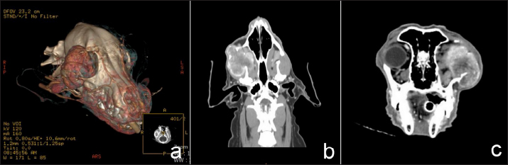
Fig. 1. Three-D (a), transverse (b), and dorsal (c) computed tomographic images in right retrobulbar, zygomatic, and temporal areas revealed a soft tissue mass involving periosteal bone with lysis of zygomatic and temporal bones. Postoperative management was orally prescribed with 20 mg/kg amoxicillin and clavulanic acid (Curam®; Sandoz GmbH; Kundl, Austria) every 12 hours a day and 2.2 mg/kg carprofen (Rimadyl®; Zoetis Inc.; Lincoln, NE) every 12 hours a day for 7 days, in addition to 3 mg/kg tramadol HCl (Tramada; Charoen Bhaesaj Lab Co. Ltd.; Bangkok, Thailand) every 12 hours a day for 3 days. Histopathology and postoperative planThe histopathological findings revealed a multilobulated neoplasm composed of various sizes of islands of bone and cartilage that were separated by diffuse anastomosed fibrovascular septa (Fig. 3a). The islands of cartilage were composed of anaplastic chondrocytes which were markedly anisocytosis and anisokaryosis (Fig. 3b). The histopathological diagnosis was orbital multilobular tumor of bone. Chemotherapy with cisplatin or radiotherapy has been reported as adjuvant therapy for multilobular tumors of bone (O’Brien et al., 1996). However, the owner denied chemotherapy due to the toxic side effects of cisplatin, including vomiting, anorexia, hemorrhagic diarrhea, nephrotoxicity, and bone marrow suppression (Knapp et al., 1988). Therefore, hypofractioned radiotherapy was further planned.
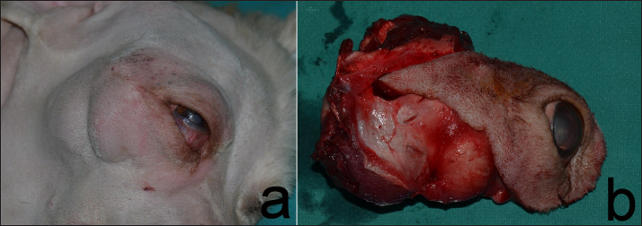
Fig. 2. Retrobular mass with exopthalmos and third eyelid protrusion OD in Cocker Spaniel dog before surgery (a). Firm 2-lobed mass in the retrobulbar area (b).
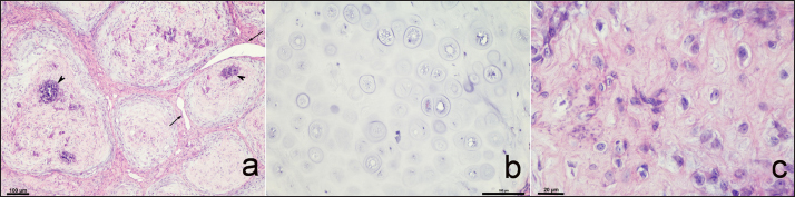
Fig. 3. Histopathology of retrobulbar mass from a female Cocker Spaniel aged 11 years with a multilobular tumor of bone. This mass showed multiple concentric islands of bone and cartilage surrounded by fibrovascular septa (arrow) and multiple foci of mineralization (arrowhead). Hematoxylin and eosin (H&E), 10× (a). Cartilaginous island composed of anaplastic chondrocytes. H&E, 20× (b). Osteocytes haphazardly arranged within lacunae. H&E, 60× (c). RadiotherapyCT for 4 weeks after the surgery demonstrated a heterogeneous mass effect (0.6 × 0.8 cm) in the right caudal maxilla area which extended to part of the right retrobulbar mass with hyper density contrast enhancement (Fig. 4). There was no detection of soft-tissue nodules in the lungs. The radiation treatment plan aimed to control retained mass and micro metastasis. The dog received hypofractioned radiation treatment with intensity-modulated radiation therapy using a linear accelerator (LINAC 2300C/D; Varian; USA) for six fractions. Each fraction used 6 Gy at an interval of 7 days. The total dose delivered was 36 Gy. The tumor size and clinical signs were determined before and after radiation treatment. After radiationAfter complete radiation, the dog had normal clinical signs with some hair loss at the radiation site. The CT images showed no detectable recurrent tumor or abnormal contrast enhancement in the right retrobulbar and zygomatic areas. After radiation treatment for 11 months, the CT images showed no detectable recurrent tumor or abnormal contrast enhancement in the right retrobulbar and zygomatic areas (Fig. 5). There were no complications from the hypofractioned radiotherapy. The dog was alert during all follow-up periods to 1 year and 8 months after the hypofractioned radiotherapy and surgical excision. At 1 year and 8 months following the radiotherapy, the dog was cleared of any local recurrence at the last follow-up time.
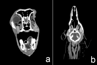
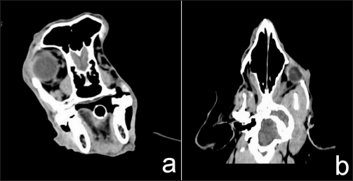
Fig. 4. Transverse (a) and dorsal (b) computed tomographic images in the right retrobulbar, maxilla, zygomatic, and temporal areas with mass effect at right caudal maxilla area, extending to part of right retrobulbar mass with hyper density contrast enhancement. DiscussionOrbital tumors in dogs can be primary tumors of the orbit or secondary tumors (either via metastastatic or multicentric invasion) or may invade into the orbit from local extension of tumors from any surrounding structures, such as the paranasal sinuses, oral cavity, or cranium (Hendrix and Gelatt, 2000; Betbeze, 2015; Flaherty et al., 2020). Ninety-five percent of orbital tumors have been reported as malignant (Hendrix and Gelatt, 2000; Attali-Soussay et al., 2001). Hendrix and Gelatt (2000) reported that 56% of dogs with orbital tumors were euthanized or had died within 6 months of diagnosis. The survival times of orbital tumors are short, resulting in the common practice of euthanasia at the time of diagnosis (Flaherty et al., 2020). Clinical signs of orbital tumor are exophthalmos, strabismus, conjunctival hyperemia, protrusion of the third eyelid, resistance to retropulsion, exposure keratitis, and periocular swelling with either normal vision or vision impairment (Hendrix and Gelatt, 2000; Attali-Soussay et al., 2001; Betbeze, 2015; Flaherty et al., 2020). General treatments are medication or surgery or their combination. Surgery of the orbital area is challenging because of the complex anatomy, the difficulty of exposing the affected area, and the tendency to bleed (Cho, 2008). Orbitotomy is a technique recommended for small and noninvasive periorbital tumors and for tissue biopsy, removal of small and encapsulated masses that do not warrant complete excision of a large and invasive tumor (O’Brien et al., 1996). The surgical technique indicated for a tumor that has invaded the orbital bone is total or partial orbitectomy (O’Brien et al., 1996; Boston, 2010). Where a tumor has invaded the vital structures of the eye or eyelids, exenteration has been suggested with orbitectomy (O’Brien et al., 1996; Betbeze, 2015). In a retrospective study of total and partial orbitectomy for the treatment of periorbital tumors with or without adjuvant therapy in 24 dogs, the most common tumor type (10/24) was a multilobular tumor of bone (O’Brien et al., 1996). Adjuvant therapy was applied in two dogs with multilobular tumors of bone. One dog was treated with preoperative radiation therapy and cisplatin, while the other dog was treated with only cisplatin after surgical resection. The dog treated with cisplatin and preoperative radiation therapy developed both local recurrence and metastasis, while the one treated with only cisplatin developed local recurrence. Both dogs had died before follow-up at 101 and 391 days, respectively. Adjuvant therapy has been recommended after surgical resection of multilobular tumors of bone because this type of tumor is predisposed to local recurrence and has a high potential for metastasis (O’Brien et al., 1996). Complications of total or partial orbitectomy are infection, strabismus, conjunctivitis, blindness in the contralateral eye, facial swelling, and hemorrhage and dehiscence of the wound (O’Brien et al., 1996; Corgozinho et al., 2015). However, no postoperative complication was observed in the dog in the current study.
Fig. 5. After 11 months of complete radiation treatment, transverse (a) and dorsal (b) computed tomographic images in the right retrobulbar, caudal maxilla, zygomatic, and temporal areas without mass effect or abnormal contrast enhancement. Dogs with multilobular tumors of bone have a slow, progressive, locally invasive, firm mass on the skull (Chun, 2005). This tumor can involve the mandible, maxilla, cranium, zygomatic bone, and tympanic bulla (McCalla et al., 1989; Straw et al., 1989; Dernell et al., 1998; Leonardi et al., 2014; Kim et al., 2017). The reported median survival time for untreated cases was 24 days (range 2–530 days). Of the treated cases, 47% (18/38) developed local recurrence, while 56% (19/34) developed metastasis. Ninety percent of metastasis was primarily in the lungs (Dernell et al., 1998). The median survival time for treated cases was 797 days (range 28–1,670 days) and the median time to death from the onset of recurrence or metastasis was 239 days (Dernell et al., 1998). The major prognostic factor regarding the survival time was the tumor location. The median survival time in dogs with the affected area being the mandible was 1,487 days, whereas it was 528 days for nonmandibular tumor sites, such as the maxilla, calvarium, and orbit (Dernell et al., 1998). The nonmandibular sites had decreased survival times approximately 12-fold worse compared to mandibular sites, perhaps because the tumors located on the mandible were easier to excise compared to other sites (Dernell et al., 1998). In the current study, the tumor invaded the orbit (a nonmandibular site) and the post-treatment disease-free interval for this dog was more than 1 year and 8 months, with no metastasis in any of the examinations. The survival time for this dog tended to be longer than for dogs (at 101 days) in other reports involving nonmandibular tumor sites that had been treated with surgery and radiation therapy (O’Brien et al ., 1996). Image scanning techniques such as CT or magnetic resonance imaging (MRI) should be performed before surgery to define the extent of the tumor (O’Brien et al., 1996; Boston, 2010). In the current study, a CT scan was chosen instead of MRI because the CT scan is faster, less expensive, and useful in the evaluation of neoplasia involving bone and lung metastasis at the same time (Boston, 2010). Furthermore, a CT scan can be used for radiation planning when radiation is chosen as adjuvant therapy (Boston, 2010). Hypofractioned radiotherapy has been suggested to relieve pain associated with bone metastasis (Siegel and Cronin, 1997). Hypofractioned radiotherapy has been used to improve the quality of life in advanced malignancies or in patients with a long-term uncontrolled tumor, even though the hypofractioned radiotherapy did not increase the survival time or eradicate the tumor (Siegel and Cronin, 1997). In veterinary medicine, commonly used protocols are 8–10 Gy fractions given on days 0, 7, and 21 or on a Monday–Wednesday–Friday schedule to total doses of 24–30 Gy (Siegel and Cronin, 1997). The reported maximum dose per fraction without any significant complications was 8.25 Gy (Siegel and Cronin, 1997). In multilobular tumors of bone, radiotherapy could probably prolong the disease-free interval (Chun, 2005). Complications of radiation therapy, such as conjunctivitis, keratitis, keratoconjunctivitis sicca, alopesia, erythema, and moist desquamation (LaDue and Klein, 2001), were not present in the dog treated in the current study and the dog had a good quality of life during the 1 year and 8 months follow-up period. In conclusion, hypofractioned radiotherapy, combined with exenteration and a partial orbitectomy, for an orbital multilobular tumor of bone in the dog in the current study resulted in no local recurrence and metastasis after surgery and adjuvant radiation for 1 year and 8 months. AcknowledgmentsThe authors thank Dr. Andrew Warner for the English copyediting of this manuscript. Authors contributionsNS performed the case management, assisted the surgical management, and wrote the manuscript. WW performed the radiotherapy and follow-up case after radiation and wrote the manuscript in part on radiation therapy and diagnostic imaging. WK performed the surgical management and wrote the manuscript in part of surgical procedures. TS was responsible for histopathological evaluation and results. AT has revised the manuscript. All authors contributed to the final manuscript. Conflict of interestThe authors declare that there is no conflict of interest. FundingThis study was supported by Kasetsart University Veterinary Teaching Hospital, Faculty of Veterinary Medicine, Kasetsart University, which provided surgical equipment and CT machine for this case. Data availabilityAll the data are presented in figures directly in this manuscript. ReferencesAttali-Soussay, K., Jegou, J. and Clerc, B. 2001. Retrobulbar tumors in dogs and cats: 25 cases. Vet. Ophthalmol. 4, 19–27. Betbeze, C. 2015. Management of orbital diseases. Top. Companion. Anim. Med. 30, 107–117. Boston, S.E. 2010. Craniectomy and orbitectomy in dogs and cats. Can. Vet. J. 51, 537–540. Cho, J. 2008. Surgery of the globe and orbit. Top. Companion. Anim. Med. 23, 23–37. Chun, R. 2005. Common malignant musculoskeletal neoplasms of dogs and cats. Vet. Clin. Small. Anim. 35, 1155–1167. Corgozinho, K.B., Cunha, S.C.S., Siqueira, R.S. and Souza, H.J.M. 2015. Successful subtotal orbitectomy in a cat with osteoma. JFMS. Open. Rep. 1, 1–4. Dent, B., Wavreille, V.A. and Selmic, L.E. 2019. Use of a temporalis fascia transposition flap for ventral orbital stabilization after ventral orbitectomy in a dog. Vet. Surg. 48, 1058–1063. Dernell, W.S., Straw, R.C., Cooper, M.F., Powers, B.E., LaRue, S.M. and Withrow, S.J. 1998. Multilobular osteochondrosarcoma in 39 dogs: 1979–1993. J. Am. Anim. Hosp. Assoc. 34, 11–18. Flaherty, E.H., Robinson, N.A., Pizzirani, S. and Pumphrey, S.A. 2020. Evaluation of cytology and histopathology for the diagnosis of canine orbital neoplasia: 112 cases (2004-2019) and review of the literature. Vet. Ophthalmol. 23, 259–268. Garza, A.M.N., Fernandez, E.M.A., Palacios, T.T., Tovar, L.E.R. and Romero, R.R. 2008. Multilobular tumor of bone: report of two cases in dogs. Vet. Mex. 39, 443–450. Groff, J.M., Murphy, C.J., Pool, R.R., Koblik, P. and Bellhorn, R. 1992. Orbital multilobulartumour of bone in a dog. J. Small. Anim. Pract. 33, 597–600. Hendrix, D.V.H. and Gelatt, K.N. 2000. Diagnosis, treatment and outcome of orbital neoplasia in dogs: a retrospective study of 44 cases. J. Small. Anim. Pract. 41, 105–108. Kim, S., Lee, J., Jeong, W., Song, H., Choi, S., Choi, H. and Lee, Y. 2017. Multilobular osteochondrosarcoma of the cranium in a Miniature Pinscher dog. J. Vet. Clin. 34, 470–473. Knapp, D.W., Richardson, R.C., Bonney, P.T. and Hahn, K. 1988. Cisplatin therapy in 41 dogs with malignant tumors. J. Vet. Intern. Med. 2, 41–46. LaDue, T. and Klein, M.K. 2001. Toxicity criteria of the veterinary radiation therapy oncology group. Vet. Radiol. Ultrasound. 42, 475–476. Leonardi, L., Carrano, A., Stoppini, L. and Floris, M. 2014. Multilobular tumor of the zygomatic bone in a dog. Open. Vet. J. 4, 9–11. McCalla, T.L., Moore, C.P., Turk, J., Collier, L.L. and Pore, E.R. 1989. Multilobular osteosarcoma of the mandible and orbit in a dog. Vet. Pathol. 26, 92–94. O’Brien, M.G., Withrow, S.J., Straw, R.C., Powers, B.E. and Kirpensteijn, J.K. 1996. Total and partial orbitectomy for treatment of periorbital tumors in 24 dogs and 6 cats: a retrospective study. Vet. Surg. 25, 471–479. Pletcher, J.M., Koch, S.A. and Stedham, M.A. 1979. Orbital chondroma rodents in a dog. J. Am. Vet. Med. Assoc. 175, 187–190. Raposa, A.C.S., Estrela-Lima, A., DóreaNeto, F.A. and Oriá, A.P. 2016. Orbital multilobular osteochondrosarcoma in a dog. Biosci. J. 32, 1567–1571. Siegel, S. and Cronin, K.L. 1997. Palliative radiotherapy. Vet. Clin. North. Am. Small. Anim. Pract. 27, 149–155. Straw, R.C., LeCouteur, R.A., Powers, B.E. and Withrow, S.J. 1989. Multilobular osteochondrosarcoma of the canine skull: 16 cases (1978-1988). J. Am. Vet. Med. Assoc. 195, 1764–1769. | ||
| How to Cite this Article |
| Pubmed Style Sritrakoon N, Wibulchan W, Karntip W, Sirinarumitr T, Thayananuphat A. Radiotherapy in combination with exenteration and partial orbitectomy for orbital multilobular tumor of bone in a Cocker Spaniel. Open Vet J. 2023; 13(11): 1485-1490. doi:10.5455/OVJ.2023.v13.i11.13 Web Style Sritrakoon N, Wibulchan W, Karntip W, Sirinarumitr T, Thayananuphat A. Radiotherapy in combination with exenteration and partial orbitectomy for orbital multilobular tumor of bone in a Cocker Spaniel. https://www.openveterinaryjournal.com/?mno=161732 [Access: May 13, 2024]. doi:10.5455/OVJ.2023.v13.i11.13 AMA (American Medical Association) Style Sritrakoon N, Wibulchan W, Karntip W, Sirinarumitr T, Thayananuphat A. Radiotherapy in combination with exenteration and partial orbitectomy for orbital multilobular tumor of bone in a Cocker Spaniel. Open Vet J. 2023; 13(11): 1485-1490. doi:10.5455/OVJ.2023.v13.i11.13 Vancouver/ICMJE Style Sritrakoon N, Wibulchan W, Karntip W, Sirinarumitr T, Thayananuphat A. Radiotherapy in combination with exenteration and partial orbitectomy for orbital multilobular tumor of bone in a Cocker Spaniel. Open Vet J. (2023), [cited May 13, 2024]; 13(11): 1485-1490. doi:10.5455/OVJ.2023.v13.i11.13 Harvard Style Sritrakoon, N., Wibulchan, . W., Karntip, . W., Sirinarumitr, . T. & Thayananuphat, . A. (2023) Radiotherapy in combination with exenteration and partial orbitectomy for orbital multilobular tumor of bone in a Cocker Spaniel. Open Vet J, 13 (11), 1485-1490. doi:10.5455/OVJ.2023.v13.i11.13 Turabian Style Sritrakoon, Natthanet, Winutpuksinee Wibulchan, Winyu Karntip, Theerapol Sirinarumitr, and Aree Thayananuphat. 2023. Radiotherapy in combination with exenteration and partial orbitectomy for orbital multilobular tumor of bone in a Cocker Spaniel. Open Veterinary Journal, 13 (11), 1485-1490. doi:10.5455/OVJ.2023.v13.i11.13 Chicago Style Sritrakoon, Natthanet, Winutpuksinee Wibulchan, Winyu Karntip, Theerapol Sirinarumitr, and Aree Thayananuphat. "Radiotherapy in combination with exenteration and partial orbitectomy for orbital multilobular tumor of bone in a Cocker Spaniel." Open Veterinary Journal 13 (2023), 1485-1490. doi:10.5455/OVJ.2023.v13.i11.13 MLA (The Modern Language Association) Style Sritrakoon, Natthanet, Winutpuksinee Wibulchan, Winyu Karntip, Theerapol Sirinarumitr, and Aree Thayananuphat. "Radiotherapy in combination with exenteration and partial orbitectomy for orbital multilobular tumor of bone in a Cocker Spaniel." Open Veterinary Journal 13.11 (2023), 1485-1490. Print. doi:10.5455/OVJ.2023.v13.i11.13 APA (American Psychological Association) Style Sritrakoon, N., Wibulchan, . W., Karntip, . W., Sirinarumitr, . T. & Thayananuphat, . A. (2023) Radiotherapy in combination with exenteration and partial orbitectomy for orbital multilobular tumor of bone in a Cocker Spaniel. Open Veterinary Journal, 13 (11), 1485-1490. doi:10.5455/OVJ.2023.v13.i11.13 |

