


| Case Report | ||
Open Vet J. 2023; 13(11): 1491-1497 Open Veterinary Journal, (2023), Vol. 13(11): 1491–1497 Case Report Right congestive heart failure in domestic kitten (Felis domesticus) by dilation of the pulmonary veins and cardiomegaliRatna Widyawati1, Wiwik Misaco Yuniarti2*, Annisa Tri Wardani3, Bima Satria Moekti3, Lalu Deni Mandala Putra3 and Maria Alfianti Clarista Janu31Faculty of Veterinary Medicine, Airlangga University, Surabaya, Indonesia 2Division of Veterinary Clinic, Faculty of Veterinary Medicine, Airlangga University, Surabaya, Indonesia 3Veterinary Surgery and Radiology Laboratory, Faculty of Veterinary Medicine, Wijaya Kusuma University, Surabaya, Indonesia *Corresponding Author: Wiwik Misaco Yuniarti. Division of Veterinary Clinic, Faculty of Veterinary Medicine, Airlangga University, Surabaya, Indonesia. Email: wiwikmisaco [at] yahoo.com Submitted: 28/07/2023 Accepted: 19/10/2023 Published: 30/11/2023 © 2023 Open Veterinary Journal
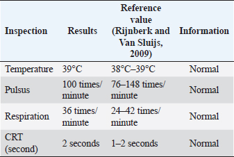
AbstractBackground: Congestive heart failure (CHF) is a pathological state characterized by the incapability of the heart to properly perform its essential function of delivering blood to meet the metabolic demands of the body. Case Description: The present case report concerns a 3-month-old male domestic kitten, displaying symptoms including an enlarged abdomen, emaciation, dehydration, dyspnea, rhinorrhea, and infestation with scabies. This animal, weighing 0.7 kg displays a tabby bicolor pattern. The findings gleaned from the clinical evaluation revealed the presence of a murmur upon auscultation of the cardiac region. Upon conducting an ultrasound examination, it was determined that the abdominal cavity contained a fluid accumulation known as ascites. Conclusion: The findings from the radiographic evaluation indicate that feline Hiro exhibits ascites alongside cardiomegaly, in conjunction with discernible vascular modifications characterized by both enlargements of the pulmonary arteries and veins. Keywords: Cardiac insufficiency, Corroborating medical evaluations, Diagnostic outcomes. IntroductionHeart failure is a discernable constellation of clinical manifestations arising from abnormalities in either the physiological capacity or anatomical integrity of the heart (Pilz et al., 2022). Heart failure is a pathological state that can be observed in felines regardless of their breed or age (Cote, 2017). Heart failure can be classified into three distinct categories, namely right ventricular failure, left ventricular failure, and bilateral ventricular failure, which encompasses both right and left ventricular dysfunction. The clinical manifestations of right-sided heart failure include systemic circulation congestion leading to a number of associated signs, such as hepatomegaly, elevated arterial pressure, and edema or called ascites (Dori et al., 2023). These complications may take the form of peripheral edema and ascites. The manifestation of left ventricular failure includes rapid breathing, the presence of rales or crackles that indicate the occurrence of pulmonary edema, muted percussion in the lungs, and decreased breath sounds, predominantly in the basal areas, suggestive of the presence of pleural effusion (Ferasin and Defrancesco, 2015; Abdul et al., 2016). This clinical presentation is indicative of the pathophysiological changes associated with the condition. Cyanosis arises due to reduced oxygen diffusion across pulmonary capillaries. Previous research on the management of acute heart failure in cats stated that, while diastolic dysfunction is the most common pathophysiological mechanism in cats with cardiomyopathy, systolic and diastolic dysfunction can coexist (Ferasin and Defrancesco, 2015). According to this explanation, the occurrence of heart failure may stem from systolic or diastolic dysfunction, or a combination thereof, where the heart’s pumping function is compromised and or its ability to adequately fill the ventricles is impaired. The effect of this phenomenon is an elevation in the magnitude of cardiac output which consequently leads to vasodilation, because studies regarding the enlargement of the pulmonary arteries and veins in CHF cases in cats are still rarely identified, this case can help provide a reference that the size of the pulmonary arteries and veins can be larger in CHF cases compared to other cardiac cases as mentioned in (Patata et al., 2020). Case DetailsA thorough physical assessment was conducted on a juvenile, domestic cat with an estimated age of 3 months and a recorded weight of 0.7 kg. The physical examination process requires the implementation of various stages including anamnesis, inspection, palpation, and auscultation (Abdisa, 2017, alongside supplementary assessments in the form of radiographic examination, namely X-ray, and ultrasonography (USG), specifically USG. The diagnostic modalities of ultrasound and X-ray examinations were employed in the dorsoventral and right lateral positional configurations, respectively (Meomartino et al., 2021). Signalement and anamnesisOn March 27, 2023, a juvenile male domestic cat exhibiting clinical symptoms including an enlarged abdomen, emaciation, dehydration, dyspnea, rhinorrhea, and scabies, and characterized by a tabby bicolor fur coat variety and a weight of 0.7 kg, underwent a comprehensive diagnostic evaluation at the Faculty of Veterinary Surgery and Radiology laboratory at UWKS. Physical examinationUpon conducting a physical evaluation (Table 1), it was determined that the abdomen exhibited distension (Fig. 1). In addition, symptomatic indicators such as scabies, dyspnea, dehydration, and rhinorrhea were noted. Furthermore, upon conducting an auscultatory examination, the presence of murmurs in the heart was identified. Upon palpation, the abdomen exhibits a pronounced enlargement without significant rigidity. The subsequent data pertains to the results obtained from the physical assessment conducted on Hiro’s feline. X-ray interpretationThe outcomes of the X-ray investigation revealed the occurrence of discoloration in the abdominal cavity, accompanied by a noticeable progression in opacity. This phenomenon is commonly attributed to the emergence of fluid in the abdominal cavity (Figs. 2 and 3). USG interpretationThe findings of USG performed in the dorsal recumbent position on the thoracic and abdominal regions revealed the existence of fluid accumulation in the peritoneal cavity (Fig. 4). Table 1. The results of the physical examination of the present status of Hiro’s cat.
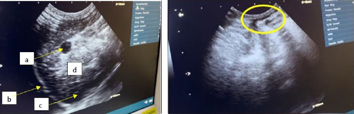
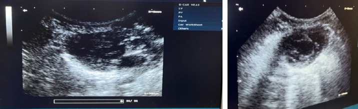
The abdominal cavity examination conducted through USG revealed certain structures appearing anechoic on the sonogram, indicating the presence of ascitic fluid in the abdominal cavity (Prajapati et al., 2022). The auscultation examination of Hiro’s heart revealed an abnormal murmur and the presence of ascites, prompting further investigation into the possible correlation between these findings. To determine whether there is a link between Hiro’s abnormal heart and ascites, an advanced examination of the heart’s organs through the use of ultrasound and X-ray was conducted. This was done in light of the fact, as detailed by Damara et al. (2023), that ascites can be caused by congestive heart failure (CHF). The findings from the ultrasound assessment revealed that the cardiac organ exhibited a normal structure (Fig. 5). The inability of researchers to utilize color flow Doppler imaging, a modality that permits direct visualization of blood flow velocity within the heart and related blood vessels, and consequent description of turbulent blood flow patterns, has precluded observation of blood flow in the heart of the Hiro cat. In examining the size of the heart via dorsoventral X-ray, the method used is a comparison of the width of the heart and the width between the right and left ribs and the comparison of the length of the boundaries of the right and left ribs with the heart. The measurement results in (Fig. 6) show a comparison of the length of the border of the right and left ribs with the heart RL, the length of the border between the ribs and the heart on the dexter and sinister parts is not the same (abnormal). In measuring the comparison of the width of the heart and the width between the right and left ribs, length B is greater than 2/3 A (abnormal), whereas normal is BA (Holland, 2020). When evaluating the cardiac dimensions through dorsoventral X-ray analysis (Birsan et al., 2017), a technique employed involves assessing the heart’s width relative to the space between the right and left ribs, as well as comparing the length of the right and left rib margins concerning the heart. The findings in Figure 6 indicate a comparison between the length of the border of the right and left ribs against the heart RL. Notably, there exists an abnormality in the length of the border between the ribs and the heart on the dexter and sinister parts, as they are not of equal dimensions. According to Holland (2020) findings, aberrant results were obtained when assessing the ratio of the width of the heart to the width between the right and left ribs, with length B being greater than 2/3 A. In contrast, a normal outcome was observed when the ratio of B to A was within typical parameters.
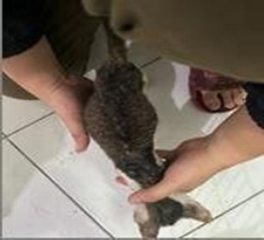
Fig. 1. Abdominal distension in the Hiro cat.
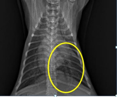
Fig. 2. Thoracic radiograph of Hiro’s cat in a dorso ventral (DV) changes in vascular patterns with dilatation of the pulmonary arteries and veins caudals.
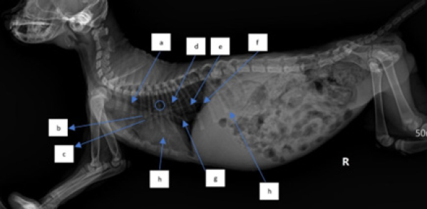
Fig. 3. Thoracic radiograph of Hiro’s cat in a lateral position with a dilated heart with an enlarged heart size. (a). Trachea, (b). cranial pulmonary artery, (c). pulmonary vein, (d). aorta, (e). pulmonary caudalis arteries and veins, (f). diaphragm, (g). vena cava caudalis, and (h). ascitic fluid. The way to measure the heart with the vertebrae heart size (VHS) method is by measuring it by drawing straight lines on the short axis and long axis (James, 2000), then the measurement results are aligned with the vertebrae and starting from the 4th rib (Marbella et al., 2023) then add up by looking at how long the vertebrae are. The normal size of VHS in the Buchanan and Bucheler method is 7.5 vertebrae (VT). In the case of Hiro’s cat (Fig. 6). VHS size using the Buchanan and Bucheler method (Litster and Buchanan, 2000) is 8 vertebrae with a long axis of 4.5 VT and a short axis of 3.5 VT so that the total becomes 8 VT (larger than normal size).
Fig. 4. The abdominal region of Hiro, a feline specimen, displays the presence of ascites as evidenced by medical imaging using USG.
Fig. 5. Ultrasound results of cat Hiro’s heart structure, (left: long axis, right: short axis) right parasternal view position.
Fig. 6. Method of measuring the heart in the DV position (Left). Heart measurement with the VHS method of the Buchanan and Bucheler methods (Right). Diagnosis and prognosisThe presence of ascites in the abdominal cavity, thickening of the ventricular wall (known as cardiomyopathy hypertrophy), cardiomegaly in the heart, abnormal heart sounds (referred to as murmurs), and dilatation of the caudal arteries and veins are typical manifestations and pathological alterations observed in patients experiencing right-sided congestive heart failure, as described by Damara et al. (2023). The prognosis for feline patients experiencing congestive heart failure is uncertain. A treatment plan involving the administration of pimobendane, diuretics, and angiotensin-converting enzyme inhibitors can enhance the quality of life of cats afflicted with heart failure. Despite a dubious prognosis, pets can enjoy a comfortable and satisfactory quality of life if an accurate diagnosis is promptly determined through early detection, as suggested by Damara et al. (2023) in their study. DiscussionCongestion Heart Failure (CHF) is a medical condition where the heart is unable to effectively pump blood to fulfill the body’s metabolic requirements such as oxygen and nutrients (Damara et al., 2023). CHF in cats can be diagnosed through clinical symptoms, physical examination, and supplementary tests (Suzuki et al., 2021). After conducting a physical examination, it was discovered that Hiro’s cat had a swollen abdomen. Therefore, additional tests such as X-ray and USG were performed to identify the reason behind the distended abdomen. The X-ray findings reveal a change in opacity in a section of the abdominal cavity, indicating an increase in radiopacity and the presence of ascites, or fluid buildup. Ultrasound imaging of the same area reveals anechoic regions between organs, which is a sign of fluid accumulation (Prajapati et al., 2022). Ascites are a symptomatic condition that occurs when fluid, both transudate and exudate, leaks into the space between the peritoneum and visceral layer of the abdominal cavity (Tilley et al., 2021). This is often a result of congestive heart failure or impaired venous flow and may cause cats to experience lethargy, abdominal distension, discomfort upon palpation, anorexia, vomiting, weight gain, scrotal or prepuce edema, and pain while lying down. Hypoalbuminemia, or low levels of albumin in the blood, is a contributing factor to ascites, as it reduces plasma osmotic pressure and increases vascular permeability, leading to fluid leakage from blood vessels into the abdominal cavity (Restijono et al., 2020). Ascites represent a clinical symptom manifestation of heart failure (Goh et al., 2022; Damara et al., 2023). The physical examination of the heart organ through auscultation revealed findings of a murmur (sound). Murmurs of the heart can be attributed to the narrowing (stenosis) of heart valves or the imperfect closing (insufficiency) of the heart valves, resulting in rapid blood flow to the heart (Santoso et al., 2021). Based on an unusual listening examination of the heart, a supplementary assessment is conducted to obtain more information about the heart’s condition. The additional tests carried out included a lateral and dorsoventral X-ray examination and an ultrasound examination. Unfortunately, due to the unavailability of certain tools, blood flow in the heart chambers could not be observed. During the X-ray examination, heart measurements were taken to identify any changes in the size of the heart organ. The lateral view measurements resulted in an intercostal measurement technique score of 4, with a normal range of 2.3–3.5. The height from the apex to the vertebral column was also measured compared to the carina, resulting in A being greater than 1/3 A + B and B being greater than 2/3 A + B (normal values are A < 1/3 A + B and B < 2/3 A + B). The VHS was also measured, and there was an increase in VHS results in CHF cases as mentioned in previous research in cats (Smith et al., 2004; antecedent corticosteroid administration has been noted in cats with congestive heart failure (CHFLaudhittirut et al., 2020), resulting in a score of 8 VT (the normal value is 7.5 VT). Measurements were then taken using the dorsoventral position, resulting in the heart size being different in R and L distances, with L being larger than R. The size of B was also greater than 2/3 (normally, R is equal to L, and B is less than 2/3). From these measurements, it was determined that the heart was enlarged or experiencing cardiomegaly. Cardiomegaly is the heart’s compensation for heart failure to meet the body’s circulatory needs, which can be caused by congestive heart failure (CHF), valvular heart disease, and cardiomyopathy (Triakoso, 2020). Following changes occur in the caudal pulmonary arteries and veins or vascular patterns observed in the dorsoventral view of the X-ray position, specifically alterations in size. The pulmonary veins and arteries caudalis exhibit dilation, resulting in a vessel diameter larger than the IXth rib diameter, this is consistent with previous research, which states that enlargement of the left atrium and pulmonary veins is one of the symptoms of CHF in cats (Guglielmini and Diana, 2015; Bernes et al., 2020). The dilation of pulmonary arteries and veins is due to augmented pressure from systemic veins toward the right ventricle (Berry, 2010). The upsurge of pressure from systemic veins toward the right ventricle is caused by the improper blood circulation process in the right atrium and ventricle, leading to a small output of blood from the ventricles and the retention of blood in the heart chambers at the end of systole, which increases. This condition increases hydrostatic pressure, leading to the dilation of pulmonary arteries and veins (Triakoso, 2020). The high hydrostatic pressure causes fluid leakage from veins into pleural and peritoneal spaces, and potentially into pericardium and peripheral interstitial tissues. Ascites occur when fluid leakage surpasses the lymphatic system’s capacity (Tilley et al., 2021). After evaluating clinical symptoms, physical examination, and supporting examinations, Hiro, the cat, was diagnosed with congestive heart failure right side (CHF-R), accompanied by ascites. The right heart chamber’s inability to pump blood caused by diseases of the right heart valves such as tricuspid insufficiency and tricuspid valve fibrosis, cardiomyopathy, congenital heart disease, and heartworm disease are some of the causes of this disease (Triakoso, 2020). The differential diagnosis in cases of CHF-R includes congenital heart disease in the form of tricuspid valve dysplasia, tetralogy of Fallot, patent ductus arteriosus, and cardiomyopathy (Jordan et al., 2016). AcknowledgmentsThe authors convey their thanks to Balai Pembiayaan Pendidikan Tinggi (BPPT) Ministry of Educational, Cultural, Research, and Technology of the Republic of Indonesia and Lembaga Pengelola Dana Pendidikan (LPDP) as Indonesia Endowment Fund for doctoral’s degree students with the scholarship program. Also to the Division of Veterinary Clinic, Faculty of Veterinary Medicine, Airlangga University who supported this study. Conflict of interestAll authors declare no conflicts of interest regarding the conduct of this study. FundingThis case report research was funded independently while carrying out doctoral studies. Authors contributionsWMY oversaw the research and did the work of analyzing and improving the writing. RW helped with planning, organizing, and checking data for accuracy. ATW, BSM, LDMP, and MACJ helped manage research projects (data processing, collection, and performing experiments). They also managed the project, conducted experiments, wrote the report, and made the first version of it. All writers have read, analyzed, and agreed with everything in the final version of their writing. Data availabilityThe data supporting the findings of this study are available within the article. ReferencesAbdisa, T. 2017. Review on practical guidance of veterinary clinical diagnostic approach. Int. J. Vet. Sci. Res. 3(1), 30–49. Abdul, A., Damanik, R. and Imawati, S. 2016. Hubungan Kejadian Efusi Pleura Pada Pasien Gagal Jantung Kongestif Berdasarkan Foto Thoraks Di RSUP Dr. Kariadi. JKD. 5(4), 393–402. Barnes, C.W., Ford, J.M., Harrington, M.A., Kedar, R.P., Tran, T.Q., Karlnoski, R.A. and Smith Jr, D.J. 2020. Radiographic comparison of superior and inferior gluteal vessels in jackknife versus prone position: a prospective, self-controlled trial. Plast Reconstr Surg. 146(4), 778–781. Berry, C.R. Thoracic radiographic interpretation. In Cvc In Kansas City Proceedings, The Mediastinum (Proceeding), Kansas, MO, 2010, pp 1–17 Birsan, O., Baisan, A.R., Paula, M. and Saftencu, V. 2017. Radiological changes of cardiac silhouette in cats with hypertrophic cardiomyopathy. Rev. Rom. Med. Vet. 27(1), 25–28. Cote, E. 2017. Feline congestive heart failure current diagnosis and management. Vet. Clin. Small. Anim. 47(5), 1055–1064. Damara, D., Batan, I.W. and Erawan, I.G.M.K. 2023. Laporan Kasus: Gagal Jantung Kongestif pada Anjing Shih-Tzu. Indones. Med. Veterinus. 12(1), 55–66. Dori, Y., Jeremy, M., Edo, B. and Christopher, S. 2023. Ascites in animals with right heart failure: correlation with lymphatic dysfunction. J. Am. Heart. Assoc. 12(7), 1–8. Ferasin, L. and Defrancesco, T. 2015. Management of acute heart failure in cats. J. Vet. Cardiol. 17, 173–189. Goh, Z.N.L., Roland, Y.L.T., Bui, K.C., Alexis, C.W. and Chen-June, S. 2022. At the heart of the problem: congestive cardiac failure as a cause of ascites A narrative review. Medicine 101(31), 1–3. Guglielmini, C. and Diana, A. 2015. Thoracic radiography in the cat: identification of cardiomegaly and congestive heart failure. J. Vet. Cardiol. 17, 87–101. Holland, M. 2020. Normal cardiovascular imaging. In Feline diagnostic imaging. Eds., Holland, M. and Hudson, J. First Edition, John Wiley & Sons, Inc, USA, pp: 161–177. James, W.B. 2000. Vertebral scale system to measure heart size in radiographs. Vet. Clin. North. Am. Small. Anim. Pract. 30(2), 379–393. Jordan, T.J., Scansen, B.A., Kent, A.M., Hitchcock, L.S. and Russell, D.S. 2016. Isolated unilateral absence of the right pulmonary artery in two cats visualized by computed tomography angiography. JFMS. Open. Rep. 2(2), 1–7. Laudhittirut, T., Natrada, R., Kornnicha, S., Peeraya, S., Teerawat, T. and Sirilak, D.S. 2020. Accuracy of methods for diagnosing heart diseases in cats. Vet. World. 13(5), 872–878. Litster, A.L., and Buchanan, J.W. 2000. Vertebral scale system to measure heart size in radiographs of cats. J. Am. Heart Assoc. 216(2), 210–214. Marbella, F.D., Veronica, G., Alexis, J.S. and Jose, A.A.M. 2023. The thoracic inlet heart size, a new approach to radiographic cardiac measurement. Animals 13(3), 1–14. Meomartino, L., Adelaide, G., Mauro, D.G., Arturo, B. and Giacomo, G. 2021. Imaging techniques in veterinary medicine. Part I: radiography and ultrasonography. Eur. J. Radiol. Open. 8, 1–12. Patata, V., Caivano, D., Porciello, F., Rishniw, M., Domenech, O., Marchesotti, F., Giorgi, M.E., Guglielmini, C., Poser, H., Spina, F. and Birettoni, F. 2020. Pulmonary vein to pulmonary artery ratio in healthy and cardiomyopathic cats. J. Vet. Cardiol. 27, 23–33. Pilz, P.M., Jennifer, E.W., Wei-Ting, C., Attila, K., Edward, B., Alokkumar, J., Sudeshna, F., Bruno, K. and Podesser, R.L. 2022. Large and small animal models of heart failure with reduced ejection fraction. Circ. Res. 130(12), 1888–1905. Prajapati, A.S., Suthar, A.N., Chauhan, P.M. and Patel, K.D. 2022. Retrospective study of asites in canines of north Gujarat region. IJBSM 13(9), 981–986. Restijono, E.H.M., Djazuli, P., Desiandura, K. and Wardhani, H.C.P. 2022. Ascites dan Hypoalbuinemia Pada Kucing Mix Domestic Long Hair. Acta. Vet. Indones. 2022, 65–71. Rijnberk, A. and Van Sluijs, F.J. 2009. Medical history and physical examination in companion snimals & accompanying CD, 2nd ed. Saunders, Netherlands, pp: 47–91. Santoso, S.W.H., Batan, I.W., Erawan, I.G.M.K. and Putra, I.P.C. 2021. Laporan Kasus: Pembesaran Jantung Pada Anjing Beagle Jantan Disertai dengan Asites. Indones. Med. Veterinus. 10(6), 936–945. Smith, S.A., Anthony, H.T., Deborah, M.F., Kristin, A.J. and Trasida, P. 2004. Corticosteroid-associated congestive heart failure in 12 cats. Intern. J. Appl. Res. Vet. Med. 2(3),159–170. Suzuki, R., Takahiro, S., Yunosuke,Y., Haruka, K., Takahiro, T., Hirotaka, M. and Hidekazu, K. 2021. Detection of congestive heart failure and myocardial dysfunction in cats with cardiomyopathy by using two-dimensional speckle-tracking echocardiography. Front. Vet. Sci. 8, 1–8. Tilley, L.P., Smith, F.W.K., Sloepr, M.M. and Brainard, B.M. 2021. Blackwell/s fiveminutes veterinary consult canine and feline. New York, NY: John Willey and Sons, Inc. Hoboken, NJ: Blackwell Publishing Press. Triakoso, N. 2020. Buku Ajar Ilmu Penyakit Dalam Veteriner Anjing dan Kucing, Airlangga University Press, pp: 43–44. | ||

| How to Cite this Article |
| Pubmed Style Widyawati R, Yuniarti WM, Wardani AT, Moekti BS, Putra LDM, Janu MAC. Right congestive heart failure in domestic kitten (Felis domesticus) by dilation of the pulmonary veins and cardiomegali. Open Vet J. 2023; 13(11): 1491-1497. doi:10.5455/OVJ.2023.v13.i11.14 Web Style Widyawati R, Yuniarti WM, Wardani AT, Moekti BS, Putra LDM, Janu MAC. Right congestive heart failure in domestic kitten (Felis domesticus) by dilation of the pulmonary veins and cardiomegali. https://www.openveterinaryjournal.com/?mno=162347 [Access: May 12, 2024]. doi:10.5455/OVJ.2023.v13.i11.14 AMA (American Medical Association) Style Widyawati R, Yuniarti WM, Wardani AT, Moekti BS, Putra LDM, Janu MAC. Right congestive heart failure in domestic kitten (Felis domesticus) by dilation of the pulmonary veins and cardiomegali. Open Vet J. 2023; 13(11): 1491-1497. doi:10.5455/OVJ.2023.v13.i11.14 Vancouver/ICMJE Style Widyawati R, Yuniarti WM, Wardani AT, Moekti BS, Putra LDM, Janu MAC. Right congestive heart failure in domestic kitten (Felis domesticus) by dilation of the pulmonary veins and cardiomegali. Open Vet J. (2023), [cited May 12, 2024]; 13(11): 1491-1497. doi:10.5455/OVJ.2023.v13.i11.14 Harvard Style Widyawati, R., Yuniarti, . W. M., Wardani, . A. T., Moekti, . B. S., Putra, . L. D. M. & Janu, . M. A. C. (2023) Right congestive heart failure in domestic kitten (Felis domesticus) by dilation of the pulmonary veins and cardiomegali. Open Vet J, 13 (11), 1491-1497. doi:10.5455/OVJ.2023.v13.i11.14 Turabian Style Widyawati, Ratna, Wiwik Misaco Yuniarti, Annisa Tri Wardani, Bima Satria Moekti, Lalu Deni Mandala Putra, and Maria Alfianti Clarista Janu. 2023. Right congestive heart failure in domestic kitten (Felis domesticus) by dilation of the pulmonary veins and cardiomegali. Open Veterinary Journal, 13 (11), 1491-1497. doi:10.5455/OVJ.2023.v13.i11.14 Chicago Style Widyawati, Ratna, Wiwik Misaco Yuniarti, Annisa Tri Wardani, Bima Satria Moekti, Lalu Deni Mandala Putra, and Maria Alfianti Clarista Janu. "Right congestive heart failure in domestic kitten (Felis domesticus) by dilation of the pulmonary veins and cardiomegali." Open Veterinary Journal 13 (2023), 1491-1497. doi:10.5455/OVJ.2023.v13.i11.14 MLA (The Modern Language Association) Style Widyawati, Ratna, Wiwik Misaco Yuniarti, Annisa Tri Wardani, Bima Satria Moekti, Lalu Deni Mandala Putra, and Maria Alfianti Clarista Janu. "Right congestive heart failure in domestic kitten (Felis domesticus) by dilation of the pulmonary veins and cardiomegali." Open Veterinary Journal 13.11 (2023), 1491-1497. Print. doi:10.5455/OVJ.2023.v13.i11.14 APA (American Psychological Association) Style Widyawati, R., Yuniarti, . W. M., Wardani, . A. T., Moekti, . B. S., Putra, . L. D. M. & Janu, . M. A. C. (2023) Right congestive heart failure in domestic kitten (Felis domesticus) by dilation of the pulmonary veins and cardiomegali. Open Veterinary Journal, 13 (11), 1491-1497. doi:10.5455/OVJ.2023.v13.i11.14 |

