| Original Article | ||
Open Vet J. 2021; 11(1): 174-179 Open Veterinary Journal, (2021), Vol. 11(1): 174–179 Original Research Isolation and identification of Mycoplasma mycoides subsp. mycoides in cattle from south-east NigeriaKingsley C. Anyika1*, Solomon O. Okaiyeto2, Anthony K. B. Sackey3, Clara N. Kwanashie4, Pam D. Luka5 and Paul I. Ankeli61Livestock Investigation Division, National Veterinary Research Institute Vom, Vom, Nigeria 2Veterinary Teaching Hospital, Ahmadu Bello University, Zaria, Nigeria 3Department of Veterinary Medicine, Ahmadu Bello University, Zaria, Nigeria 4Department of Veterinary Microbiology, Ahmadu Bello University, Zaria, Nigeria 5Molecular Biotechnology Division, National Veterinary Research Institute Vom, Vom, Nigeria 6Bacterial Vaccine Production Division, National Veterinary Reseasrch Institute Vom, Vom, Nigeria *Corresponding Author: Kingsley C. Anyika. Livestock Investigation Division, National Veterinary Research Institute Vom, Vom, Nigeria. Email: chinetoanyika [at] gmail.com Submitted: 10/12/2020 Accepted: 05/03/2021 Published: 19/03/2021 © 2021 Open Veterinary Journal
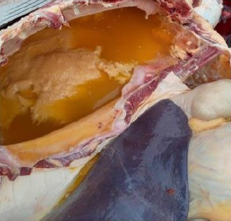
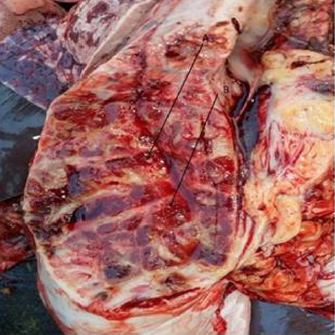
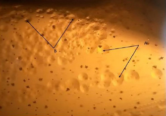
AbstractBackground: Mycoplasma mycoides subsp. mycoides is the causative organism of Contagious Bovine Pleuropneumonia (CBPP). It is a trans-boundary disease and an endemic in Nigeria having caused serious financial loss for the country’s economy. Aim: This study was undertaken to isolate and confirm the presence of M. mycoides subsp. mycoides (Mmm) in cattle, from three selected South-Eastern states of Nigeria. Method: A total of 90 bovine samples (25 pleural fluids and 65 lung tissues) suggestive of CBPP were collected from different abattoirs in the three selected South-eastern states of Nigeria (Anambra, Enugu, and Imo), for the isolation of Mmm by employing cultural method, whereas for confirmation polymerase chain reaction (PCR) approach was used. The collected samples were cultured on Pleuropneumonia like organism (PPLO) agar according to specific protocols. Results: Twenty five of the samples (lungs and pleural fluid) were positive for Mmm on PPLO agar giving an isolation rate of 27.7%. Only 21 of the isolates were further confirmed using PCR. The PCR amplification of the isolates produced a product of 1.1 kbp which is specific for Mmm. No positive isolates were recovered from Imo state. Conclusion: This study confirms the presence of Mmm as the causative organism of CBPP in Southeast Nigeria. It is recommended that active surveillance and vaccination protocol should be undertaken in the region for the control and prevention of this disease. Keywords: Cattle; CBPP; Mycoplasma mycoides subsp. mycoides; Polymerase chain reaction. IntroductionMycoplasma mycoides subsp. mycoides (Mmm) is the etiological agent of Contagious Bovine Pleuropneumonia (CBPP) (Thiaucourt et al., 2006; Campbell, 2015). It is a highly contagious and fatal disease of cattle, with serious financial implication in Nigeria (Fadiga et al., 2013). CBPP is a trans-boundary disease and has been classified as one of the bacterial diseases in OIE’s list A diseases (OIE, 1997; Litamoi et al., 2004). CBPP can manifest in hyper-acute form, where it is usually fatal and most times no clinical signs are observed (Thiaucourt et al., 2006). The incubatory period of this disease is relatively long with an inconsistent clinical course (Litamoi et al., 2004). Clinical signs of the disease include anorexia, fever, dyspnea, cough, and nasal discharges (Provost et al., 1987). Infections in adult cattle are most of the time restricted to the respiratory tract, while in calves, the disease usually manifests as arthritis (Litamoi et al., 2004; Campbell, 2015). Several molecular tools and technologies have been developed for the identification and confirmation of the causative organism (Dedieu et al., 1994; Lorenzon et al., 2002; Miles et al., 2006). Miles et al. (2006) developed a polymerase chain reaction (PCR) technique that could differentiate Mycoplasma species based on their origin. The PCR method is quite efficient and specific. The United States of America and Australia have eradicated CBPP through strict control of cattle movement, prohibiting large scale slaughter, and financial compensation to owners. This has, however, become difficult in Nigeria due to several factors, such as poor implementation of the test and slaughter policy, unrestricted movement of nomads across state boundaries which has made accurate monitoring of CBPP difficult (Egwu et al., 1996; Chima et al., 2001). Several authors have documented the outbreaks, prevalence, and economic importance of the disease in Nigeria (Fadiga et al., 2013; Ankeli et al., 2017; Jasini et al., 2016). In Africa, the application of the stamping out policy for the eradication of CBPP has faced so many challenges. Consequently, the control of the disease has depended heavily on the use of the vaccine (T1/44 or T1-SR) (Thiaucourt et al., 2000; Litamoi et al., 2004). National Veterinary Research Institute (NVRI) Vom, produces CBPP vaccine (T1/44) in Nigeria (NVRI, 2007). However, this vaccine has some drawbacks (Egwu et al., 1996; Thiaucourt et al., 2006). Presently, in Nigeria, there is little or no information on the status of this disease in South-East Nigeria as many studies have been centered within Northern Nigeria (Nwankpa, 2008; Danbirni et al., 2010; Okaiyeto et al., 2011; Jasini et al., 2016). Therefore, data generated from this research can serve as the baseline data for future studies in the region. Material and MethodsStudy areaThis study was undertaken in three selected South Eastern states of Nigeria (Anambra, Enugu and Imo states). The South-Eastern region is one of the geopolitical zones in the country. It consists of Anambra, Enugu, Imo, Ebonyi, and Abia states. Anambra state lies between latitude 5° 32′ and 6° 45′ N and longitude 6° 43′ and 7° 22′ E; Enugu state lies between latitude 5° 27′and 6° 33′ N and longitude 6° 28′ and 7° 32′ E, and Imo state is located between latitude 4° 45′ and 7° and 15′ N and longitude 6° 50′ and 7°25′ E. The region has an estimated cattle population of 4.5 million from a total of 16.3 million estimated cattle population in Nigeria (Ikhatua, 2011). Sample collection and processingA total of 90 bovine samples (65 lungs tissues and 25 pleural fluids) were collected from slaughtered animals showing classical signs of CBPP at the abattoir (Figs. 1 and 2). Samples were collected for 4 months between December 2019 and March 2020. The lung tissue samples were collected using sterile blades. Pleural fluids were collected using sterile 18G syringes (Fig. 1). Subsequently, all the collected samples were put in sterile sample bottles and transported in CBPP transport medium pleuropneumonia like organism (PPLO broth). They were properly labeled and conveyed to the Mycoplasma laboratory of NVRI, Vom for further processing. Culture and isolation of Mycoplasma specieThe lung tissues and pleural fluids collected from the suspected cattle at the abattoir were cultured in PPLO growth medium adhering to specific protocols by the OIE manual (OIE, 2014). Briefly, the suspected lung tissue and pleural fluid were incubated in PPLO broth for at least 48 hours at 37°C under anaerobic condition. After the incubation period, 20 μl of the overnight broth was added to 180 μl of PPLO broth and a ten-fold serial dilution (10−1–10−4) was carried out and finally sub-cultured onto PPLO agar. The agar plates were incubated for at least 72 hours at 37°C in a medium having 5% CO2and were monitored for colony growth. Small-to-medium sized colonies with the characteristic of a “fried egg” appearance under the stereomicroscope at 35× magnifications were considered presumptive of Mmm.
Fig. 1. CBPP infected animal at slaughter showing collection of pleural fluid.
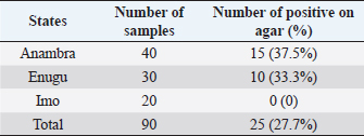
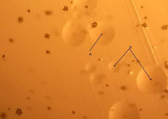
Fig. 2. Lung of CBPP infected cattle at post mortem showing thickening of the interlobular septa (marbled appearance) when cut open. Confirmation of Mmm isolates using PCRAll the positive Mycoplasma isolates after culture on growth medium (PPLO agar) were subjected to conventional PCR assay for the detection of Mmm according to protocols by Miles et al. (2006). Extraction of DNADNA was extracted from a 3 ml Mmm broth culture using QlAamp® DNA Mini kit. It was carried out according to the manufacturer’s instructions. Lyophilized T1/44 vaccine (NVRI, Vom Nigeria) was used as the positive control for this study. Briefly, Proteinase K (20 μl) was pipetted into 1.5 ml micro-centrifuge tube after which 200 μl of the sample (broth culture) was added. Subsequently, 200 μl Buffer AL was then added and mixed thoroughly by vortexing for 15 seconds. This mixture was incubated at 56°C for 10 minutes. Thereafter, 200 μl ethanol was added and mixed thoroughly. The mixture was centrifuged at 6,000 × g for 1 minute. The resultant pellets were then washed twice in Buffer AW1 and AW2 respectively. Finally, 200 μl of Buffer AE was added and incubated at room temperature for 1 minute and centrifuged at 8,000 × g for 1 minute to elute the DNA. Thereafter, 2.5 μl of the DNA extract was used as the template for all the reactions. Specific PCR protocol for the confirmation of M. mycoides subsp. mycoidesAll the PCR reactions were performed in a total volume of 25 μl, which contained dH2O, 5× FIREPol® master mix (12 mM MgCl, 1 mM dNTP mix, FIREPol® DNA polymerase and 1 μl IS1296F: Primer (5′–3′): CTA AAG AGC TTG GAG TTC AGT G and 1 μl R (all) (sequence 5′–3′): CCA GCT CAACCA GCT CCA G) (Miles et al., 2006). DNA amplification was performed using GeneAmp® PCR system 2,720 (Perkin Elmer, Courtaboeuf, France). It consisted of an initial denaturation step at 95°C for 5 minutes, followed by 32 cycles of denaturation at 95°C for 30 seconds, annealing at 62°C for 30 seconds and extension at 72°C for 1 minute and 20 seconds. The final extension stage was maintained at 72°C for 5 minutes. The PCR product was then running on 2% agarose gel impregnated with ethidium bromide (0.5 μg/ml) at 130 volts for 30 minutes. Subsequently, the DNA migration was viewed under ultra-violent light and photographs were taken. The production of a band equivalent to 1.1 kbp and at the same distance with the positive control (T1/44) was considered confirmatory for M. mycoides subsp mycoides. Ethical approvalThe consent of the Ahmadu Bello University Animal Care and Use was sought after for this study. Samples were collected from already slaughtered animals at registered abattoirs. The abattoirs abided by the government established guidelines for slaughter. ResultsCulture and isolation of M. mycoides subsp. mycoidesOut of the 90 bovine samples (25 pleural fluids and 65 lung tissues) collected, 25 (27.7%) were considered positive for M. mycoides subsp. mycoides based on their colonial morphology on PPLO agar (Table 1). A typical fried egg colonies with some of the colonies having dense centre while others were raised with nipple like appearance were observed from day 3 of culture on PPLO agar (Figs. 3 and 4). A total of 30 samples (10 pleural fluid and 20 lung tissues) were collected from Enugu state of which 10 (33.3%) were positive on PPLO agar (Table 1). Similarly, 40 tissue samples (15 pleural fluid and 25 lung tissues) were collected from Anambra state of which 15 (37.5%) were positive on PPLO agar. There were no positive isolates from the 20 lung tissue samples from Imo state (Table 1). Table 1. Number of Mycoplasma mycoides subsp. mycoides isolates positive on PPLO agar.
Fig. 3. Mycoplasma mycoides subsp. mycoides colonies on PPLO agar.
Fig. 4. Mycoplasma mycoides subsp. mycoides colonies (raised colonies with dark pinpoint centres) on PPLO agar.
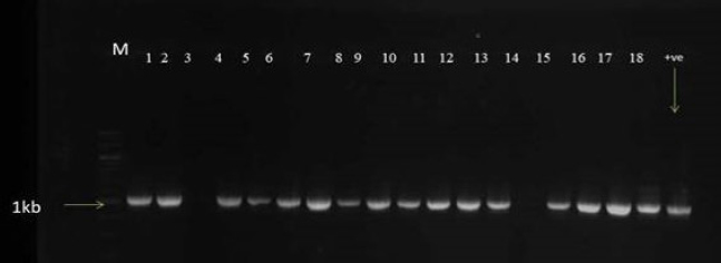
Fig. 5. Mmm specific PCR showing 1.1kb amplicon size. Lane M: 100 base pair ladder. Line 1-18: positive isolates. Lane +ve: for positive control.
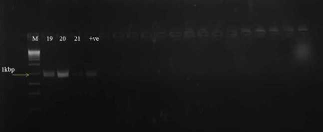
Fig. 6. Mmm specific PCR showing 1.1kb amplicon size. Lane M: 100 base pair ladder. Lane 19-21: positive isolates. Lane +ve: for positive control. Confirmation of Mmm by conventional PCRTwenty one out of the 25 Mycoplasma isolates cultured on PPLO agar were confirmed as M. mycoides subsp. mycoides using conventional PCR as described by Miles et al. (2006). Production of a band equivalent to 1.1 kbp and at the same distance with T1/44 positive control confirms the presence of Mmm (Figs. 5 and 6). DiscussionThe South east region of Nigeria is estimated to have a cattle population of 4.5 million (Ikhatua, 2011). There is an apparent successful settlement of pastoralist within the Southern region of Nigeria in the past four decades with thousands of zebu cattle (Blench, 1994). However, most of the studies on CBPP are centered within the Northern region, where most of the cattle population is located. In this study, the overall Mmm isolation rate was 27.75%. Twenty five of the isolates were positive on PPLO agar. This high isolation rate could be attributed to the fact that purposive sampling method was used for this study. Only apparently sick animals showing classical clinical signs and gross lesions suggestive of CBPP at the abattoirs were sampled. Samples such as pleural fluid and pneumonic lungs were collected from the abattoirs. Pleural fluid accumulation is one of the characteristic of CBPP and is considered as one of the best medium for the isolation of M. mycoides subsp. mycoides (Thiaucourt et al., 2006). Anambra state recorded the highest isolation rate of Mmm. Perhaps, more animals showing classical gross lesions of CBPP at the slaughter house were sampled in Anambra state followed by Enugu State. However, there was no isolate from Imo state. The failure to isolate Mmm in Imo state could be because most of the cattle sampled were apparently healthy, showing no classical gross lesions of CBPP at the abattoir. Similarly, the presence of antibiotics contamination in the collected samples may have also accounted for the inability to recover mycoplasma from the samples. The isolation rate in this study is in contrast to studies by Jasini et al. (2016), who recorded an overall isolation rate of 3.33% in the North eastern region of Nigeria. Similarly, Nwankpa (2008), recorded 6.27% from cattle within Northern Nigeria. Ikpa et al. (2020) also recorded a lower isolation rate of 4% in Nasarawa State, Nigeria. Several genomic and molecular tools have been developed for the identification and confirmation of M. mycoides subsp. mycoides (Bashiruddin et al., 1994: Miles et al., 2006). PCR is a robust technique for the detection, identification, and differentiation of members of the M. mycoides cluster (Miles et al., 2006). In this research, out of the total of 25 Mmm isolates positive on PPLO culture medium, 21 isolates were confirmed to be M. mycoides subsp. mycoides by PCR. The isolates yielded molecular size of 1.1 kbp specific for Mmm. This is in agreement with the findings by Miles et al. (2006). Generally, species within the M. mycoides cluster share many immunological, biochemical and genetic properties; which can result in major problems for diagnostic laboratories in the identification process of field strains (Cottew et al., 1987; Persson et al., 2002). Perhaps, some of the positive isolates on PPLO agar in this study were Mycoplasma species within the Mycoplasma cluster as the isolates were not subjected to biochemical tests. The use of PCR for the confirmation of Mycoplasma species from several clinical samples has shown a higher efficiency, specificity, and sensitivity for laboratory diagnosis when compared with conventional culture-based diagnostic procedures (Bashiruddin et al., 1994). ConclusionThis study confirms the presence of M. mycoides subsp. mycoides in South eastern Nigeria. This is the first report on the isolation and confirmation of Mmm in South-eastern region of Nigeria. We, therefore, recommend active surveillance and vaccination programs within the region for the control and prevention of Contagious Bovine Pleuropneumonia. AcknowledgementsWe sincerely thank the Executive Director of NVRI, Dr David Shamaki, for the kind approval of some of the CBPP vaccines used for this research. Conflict of interestThe authors declare no conflict of interest. Authors contributionSOO, CNK, and AKBS supervised the study. KCA, PDL, and PIA collected and analyzed the samples. KCA wrote the manuscript. All authors read and approved the manuscript for submission. ReferencesAnkeli, P.I, Raji, M.A., Kazeem, H.M., Tambuwal, F.M., Francis, M.I., Ikpa, L.T., Fagbamila, I.O., Luka, P.D. and Nwakpa, N.D 2017. Seroprevalence of Contagious Bovine pleuropneumonia in plateau state, North-Central Nigeria. Bull. Anim. Hlth. Prod. 65, 357–366. Bashiruddin, J.B., Taylor, T.K. and Gould, A.R. 1994. A PCR-based test for the specific identification of Mycoplasma mycoides subspecies mycoides. J. Vet. Diagn. Invest. 6(4), 428–434. Blench, R. 1994. The expansion and adaptation of Fulbe Pastoralism to sub-humid and humid conditions in Nigeria. Afican studies Centre Leiden. Cahier d’ Etudes Africaines, pp: 197–212. Campbell, J. 2015. Contagious Bovine pleuropneumonia. In The Merck’s veterinary manual. Eds., Kahn, C.M., Line, S., Aiello, S.E. Whitehouse Station, NJ: Merck and Co. Available via www.merckvetmanual.com (Accessed 04 November 2020). Chima, J.C., Lombin, L.H., Molokwu, J.H., Abiaye, E.A. and Majiyagbe, K.A. 2001. Current situation of contagious bovine pleuropneumonia in Nigeria and the relevance of c-ELISA in the control of the disease. Proceedings of the Research coordination meeting of the FAO/IAEA coordinated research programmeheld in Nairobi, Kenya. Cottew, G.S., Breard, A., Damassa, A.J., Erne, H., Leach, R.H., Lefevre, P.C., Rodwell, A.W. and Smith, G.R. 1987. Taxonomy of the Mycoplasma mycoides cluster. Isr. J. Med. Sci. 23, 632–635. Danbirni, S., Okaiyeto, S.O., Pewan, S.B. and Kudi, A.C. 2010. Concurent infection of Contagious Bovine pleuropneumonia and Bovine tuberculosis in Bunaji nomadic cows. Res. J. Anim. Sci. 4(1), 23–25. Dedieu, L., Mady, V. and Lefevre, P.C. 1994. Development of a selective polymerase chain reaction assay for the detection of Mycoplasma mycoides subsp. mycoides S.C. (contagious bovine pleuropneumonia agent). Vet. Microbiol. 42(4), 327–339. Egwu, G.O., Nicholas, R.A.J., Ameh, J.A. and Bashiruddin, J.B. 1996. Contagious bovine pleuropneumonia: an update. Vet. Bull. 66, 875–888. Fadiga, M.L., Lost, C. and Ihedioha, J. 2013. Financial cost of disease burden, morbidity and mortality from priority livestock diseases in Nigeria: disease burden and cost-benefit analysis of targeted interventions. International livestock Research Institute, Nairobi, Kenya. Ikhatua, U.J. 2011. Nigerian institute of animal science (NIAS) beef cattle production report. Timestamp: 2011-02-18. Available via http://repository.fuoye.edu.ng. (Accessed 08 September 2020). Ikpa, L., Bwala, D., Ankeli, P., Kaikabo, A., Maichibi, M., Muraina, I., Abenga, J., Nwannkpa, N. and Adah, M. 2020. Isolation and molecular characterization of mycoplasma mycoides mycoides in 3 agro ecological zones of Nasarawa State, Nigeria. Open J. Vet. Med. 10, 15–26. Jasini, A.M., James, O.O., Haruna, M.K., Nicholas, N.D., Andrea, D.P., Flavio, S., Katiusia, Z., Ann, A., Massimo, S. and Attilio, P. 2016. Molecular detection of Nigerian Field isolates of Mycoplasma mycoides subsp. mycoides as causative agents of Contagious Bovine Pleuropneumonia. Int. J. Vet. Sci. 4(2), 46–53. Litamoi, J.K., Ayelet, G. and Rweyemamu, M.M. 2004. Evaluation of the xerovac process for the preparation of heat tolerant contagious bovine pleuropneumonia (CBPP) vaccine. Vaccine 23(20), 2573–2579. Lorenzon, S., Arzul, I., Peyraud, A., Hendrikx, F. and Thiaucourt, F. 2003. Molecular epidemiology of contagious bovine pleuropneumonia by multilocus sequence analysis of Mycoplasma mycoides subspecies mycoides biotype SC strains. Vet. Microbiol. 93, 319–333. Miles, K., Churchward, C.P., McAuliffe, L., Ayling, R.D. and Nicholas, R.A.J. 2006. Identification and differentiation of European and African/Australian strains of Mycoplasma mycoides subspecies mycoides small-colony type using polymerase chain reaction analysis. J. Vet. Diagn. Invest. 18(2), 168–171. NVRI. 2007. Its activities and opportunities to veterinary professionals. A presentation to the 44 annual congress of the Nigerian veterinary medical association, Conference Centre, Petroleum Training Institute Effurun, National Veterinary Research Institute, Vom, Nigeria 2007 Oct 22nd–26th. Nwankpa, N.D. 2008. Serological and molecular studies of Mycoplasma mycoides mycoides small colony in Northern Nigeria. PhD Thesis. Department of Veterinary Pathology and Microbiology, University of Nigeria Nsukka, Nsukka, Nigeria. Nwanta, D.R. and Umoh, J.U. 1992. Epidemiology of Contagious Bovine pleuropneumonia (CBPP) in Northern states of Nigeria. An update. Rev. Eleven Pays. Trop. 45(1), 17–20. OIE. 1997. Contagious Bovine pleuropneumonia. Man. Stand. 2, 16. OIE. 2014. World Animal Health Information Database - Version: 1.4. World Animal Health Information Database. Paris, France: World Organisation for Animal Health. http://www.oie.int (Accessed 17 October 2020). Okaiyeto, S.O., Danbirni, S., Allam, L., Akam, E., Pewan, S.B. and Kudi, A.C. 2011. On farm diagnosis of Contagious Bovine pleuropneumonia in nomadic herds using latex agglutination test (LAT). J. Vet. Med. Anim. Health 3(5), 62–66. Persson, A., Pettersson, B., Bölske, G. and Johansson, K.E. 2002. Diagnosis of Contagious Bovine pleuropneumonia by PCR-laser-induced fluorescence and PCR-restriction endonuclease analysis based on the 16S rRNA genes of Mycoplasma mycoides subsp. mycoides SC. J. Clin. Microbiol. 37(12), 3815–3821. Provost, A., Perreau, P., Beard, Goff, L.E. and Cottew, G.S. 1987. Contagious Bovine pleuropneumonia. Rev. Sci. Tech. 6, 625–679. Thiaucourt, F., Lorenzon, S., David, A., Tulasne, J.J. and Domenech, J. 2006. Vaccination against Contagious Bovine pleuropneumonia and the use of molecular tools in epidemiology 1998. Ann N Y Acad Sci 849, 146–151. Thiaucourt, F., Yaya, A., Wesonga, H., Huebschle, O.J.B., Tulasne, J.J. and Provost, A. 2000. Contagious Bovine pleuropneumonia. Ann N Y Acad Sci 916, 71–80. | ||
| How to Cite this Article |
| Pubmed Style Anyika KC, Okaiyeto SO, Sackey AK, Kwanashie CN, Luka PD, Ankeli P. Isolation and Identification of Mycoplasma mycoides subsp. mycoides in Cattle from South-East Nigeria.. Open Vet J. 2021; 11(1): 174-179. doi:10.5455/OVJ.v11.i1.25 Web Style Anyika KC, Okaiyeto SO, Sackey AK, Kwanashie CN, Luka PD, Ankeli P. Isolation and Identification of Mycoplasma mycoides subsp. mycoides in Cattle from South-East Nigeria.. https://www.openveterinaryjournal.com/?mno=21657 [Access: September 01, 2024]. doi:10.5455/OVJ.v11.i1.25 AMA (American Medical Association) Style Anyika KC, Okaiyeto SO, Sackey AK, Kwanashie CN, Luka PD, Ankeli P. Isolation and Identification of Mycoplasma mycoides subsp. mycoides in Cattle from South-East Nigeria.. Open Vet J. 2021; 11(1): 174-179. doi:10.5455/OVJ.v11.i1.25 Vancouver/ICMJE Style Anyika KC, Okaiyeto SO, Sackey AK, Kwanashie CN, Luka PD, Ankeli P. Isolation and Identification of Mycoplasma mycoides subsp. mycoides in Cattle from South-East Nigeria.. Open Vet J. (2021), [cited September 01, 2024]; 11(1): 174-179. doi:10.5455/OVJ.v11.i1.25 Harvard Style Anyika, K. C., Okaiyeto, . S. O., Sackey, . A. K., Kwanashie, . C. N., Luka, . P. D. & Ankeli, . P. (2021) Isolation and Identification of Mycoplasma mycoides subsp. mycoides in Cattle from South-East Nigeria.. Open Vet J, 11 (1), 174-179. doi:10.5455/OVJ.v11.i1.25 Turabian Style Anyika, Kingsley Chineto, Solomon Olu Okaiyeto, Anthony KB Sackey, Clara Nna Kwanashie, Pam D Luka, and Paul Ankeli. 2021. Isolation and Identification of Mycoplasma mycoides subsp. mycoides in Cattle from South-East Nigeria.. Open Veterinary Journal, 11 (1), 174-179. doi:10.5455/OVJ.v11.i1.25 Chicago Style Anyika, Kingsley Chineto, Solomon Olu Okaiyeto, Anthony KB Sackey, Clara Nna Kwanashie, Pam D Luka, and Paul Ankeli. "Isolation and Identification of Mycoplasma mycoides subsp. mycoides in Cattle from South-East Nigeria.." Open Veterinary Journal 11 (2021), 174-179. doi:10.5455/OVJ.v11.i1.25 MLA (The Modern Language Association) Style Anyika, Kingsley Chineto, Solomon Olu Okaiyeto, Anthony KB Sackey, Clara Nna Kwanashie, Pam D Luka, and Paul Ankeli. "Isolation and Identification of Mycoplasma mycoides subsp. mycoides in Cattle from South-East Nigeria.." Open Veterinary Journal 11.1 (2021), 174-179. Print. doi:10.5455/OVJ.v11.i1.25 APA (American Psychological Association) Style Anyika, K. C., Okaiyeto, . S. O., Sackey, . A. K., Kwanashie, . C. N., Luka, . P. D. & Ankeli, . P. (2021) Isolation and Identification of Mycoplasma mycoides subsp. mycoides in Cattle from South-East Nigeria.. Open Veterinary Journal, 11 (1), 174-179. doi:10.5455/OVJ.v11.i1.25 |


