


| Case Report | ||
Open Vet J. 2022; 12(6): 855-858 Open Veterinary Journal, (2022), Vol. 12(6): 855–858 Case Report Esophageal obstruction due to trichobezoar in a she-camel (Camelus dromedarius)Isam Eljalii, Mohamed K. Zabady Ayman Elnahas and Turke Shawaf*Department of Clinical Sciences, College of Veterinary Medicine, King Faisal University, Al-Ahsa, Saudi Arabia Submitted: 15/05/2022 Accepted: 14/10/2022 Published: 16/11/2022 *Corresponding Author: Turke Shawaf. Department of Clinical Sciences, College of Veterinary Medicine, King Faisal University, Al-Ahsa, Saudi Arabia. Email: tshawaf [at] kfu.edu.sa © 2022 Open Veterinary Journal
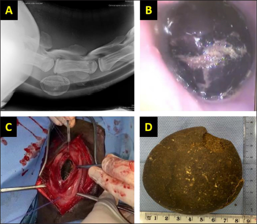
AbstractBackground: Occurrences of esophageal foreign bodies are common in camels. Esophageal obstruction in camels due to bezoars is rare. Case Description: This report describes esophageal obstruction in camel due to trichobezoar. A 2-year-old she-camel presented with a history of inability to swallow and there was food and water regurgitation for one day before. Radiography and endoscopic examination revealed an oval-shaped foreign body embedded in the esophageal lumen in the level distal third of the neck. The foreign body was successfully removed using cervical esophagotomy under general anesthesia. Successful esophagostomy revealed trichobezoar weighing 45 g and measuring 85 mm × 75 mm × 42 mm. The trichobezoar removed from the esophagus was the cause of esophageal obstruction. Conclusion: Esophageal obstruction in camel could be due to trichobezoar. Radiography and endoscopy are valuable diagnostic methods to determine the position and nature of an obstructive object. Keywords: Camel, Esophageal obstruction, Trichobezoar. IntroductionBezoars are ball-like materials that formed in the gastrointestinal tract of humans (Pitiakoudis et al., 2003; Zhang et al., 2020) and animals (Eljalii et al., 2014). Bezoars may be of plant fibers origin (phytobezoars), wool or hair origin (trichobezoars), or mixed (trichophytobezoars) (Cardoso et al., 2017). Occurrences of esophageal foreign bodies are common in camels (Ramadan et al., 1986; Shawaf et al., 2017). The varieties of foreign bodies include bezoars, grass seeds, twigs, pieces of wood, and thorns passages (Rush, 2004). However, esophageal bezoars are rare (Zhang et al., 2020). In camels, bezoars were reported in different ages and in different gastrointestinal compartments (Fahmy et al., 1995). In animals, esophageal obstruction due to a trichobezoar was reported in cows (Patel and Brace, 1995). Esophageal obstruction in camels due to bezoars is rare but obstruction due to other foreign bodies is frequently reported especially in young calves (Ramadan and Abdi-Bey, 1990; Shawaf et al., 2017). Eljalii et al. (2014) reported the presence of trichobezoars in the intestine of a camel calf. Some foreign bodies are visible on raising the alar margins but others are identified only during endoscopic examination (Shawaf, 2017). Radiographic examination may be used in the diagnosis of radio-opaque or metallic foreign bodies (Rush, 2004). There are few published studies describing the clinical findings and treatment in camels with esophageal obstruction and these mainly included smaller numbers of camels (Ramadan and Abdi-Bey, 1990; Ramadan, 2016; Shawaf et al., 2017). Available literature lacks information about esophageal obstruction in young camels. The objective of the present case report was to document obstruction of the esophagus in a she-camel with a massive trichobezoar. Case DetailsA 2-year-old she-camel weighing 150 kg was presented to the Veterinary Teaching Hospital, King Faisal University. On presentation, the she-camel had normal body condition, normothermic, heart rate was 48 beats/minute, slightly tachypneic with respiratory rate 19 breaths/minute, and had a normal blood profile. The main complaint was the inability to swallow and there was food and water regurgitation for one day before. The animal had a history of bad vices of self-licking and licking other camels. On palpation, there was a clear visible mass on the neck region about 105 cm caudal to the base of the head. Insertion of stomach tube in the esophagus stopped at the area of swelling. Lateral radiograph for the distal third of the neck showed a round to ovoid, radio-opaque foreign body mass located in the esophagus at the level of the sixth and seventh cervical vertebrae (Fig. 1A). An endoscopy according to Shawaf et al. (2017), using a flexible endoscope with 8 mm diameter and 110 cm length (VetVu, a unit of Swiss Precision Products), after the camel was sedated with xylazine (Rompun® 2%, Bayer) at dose 0.1 mg/kg, identified a black foreign body mass lodged in the third part of the esophagus (Fig. 1B). The mass was found about 105 cm from oral opening and embedded in the lumen of the esophagus. Depending on the history, palpation, X-rays, and endoscopy presence of the foreign body in the esophagus leading to obstruction was confirmed.
Fig. 1. (A) Lateral radiograph showing radio-opaque foreign body mass at the level of intervertebral disc between sixth and seventh cervical vertebrae. (B) Endoscopic view reveals the foreign body mass lodged in the esophageal lumen with accumulated saliva and redness in the esophageal mucosa. (C) Surgical esophageal incision to expose the large trichobezoar. (D) large, rounded to ovoid trichobezoar (85 mm × 75 mm × 42 mm). Surgical management (esophagotomy)Cervical esophagotomy was performed for the removal of the foreign body mass. The anesthetic regimen comprised sedation with xylazine 2% (0.2 mg/kg IV), followed by general anesthesia using ketamine hydrochloride 10% (2 mg/kg IV). The she-camel was placed on the right lateral recumbency and a stomach tube was introduced to the level of obstruction. The distal third of the neck was prepared for surgery. A 10 cm long incision was made at the ventrolateral aspect at skin and fascia, and then sternocephalicus and sternothyrohyoideus muscles were bluntly separated while preserving the jugular vein that lies under the sternocephalicus muscle (Fig. 1C). The esophagus that is located dorsolateral to the trachea and under the jugular vein was held with two stay stitches (Fig. 1C). A 5 cm longitudinal incision was made in its dorsolateral aspect through the outer fibrous and muscular layer and then through the inner submucosal and mucosal layers, to expose the lumen. Rochester-Carmalt forceps were used to extract the obstructing bezoar (Fig. 1C and D). The stomach tube was then pushed toward the chest to check the patency of the esophagus and the surgical field was flushed using a sterile normal saline solution to remove any food debris. The esophageal mucosa and submucosa were sutured using a simple interrupted pattern (in out – out in) using 1–0 Surgicryl® (Smi AG, HÜnningen 37 Belgium). The outer muscular and fibrous layers were opposed with a simple continuous pattern using the same suture material. Benzylpenicillin 3 g powder was dusted into the depth of the wound. The fascia over the muscles was opposed using 1–0 Surgicryl® (Smi AG, HÜnningen 37 Belgium) in a simple continuous suture pattern. The skin was closed using 2 silk (Lukens Medical, USA) sutures in an interrupted horizontal mattress suture pattern. Post-operative careFood and water were withheld for 48 hours after surgery and maintenance intravenous fluid therapy (Lactate Ringer’s solution) was administered at a rate of 10 ml/kg/hour, over approximately 3 hours. Camel was only fed milk or soft food subsequently. The animal was injected with Penstrept® (IM at a dose of 20 mg/kg) every 72 hours, and anti-inflammatory flunixin meglumine (Flunixin)® at a dose of 1.1 mg/kg for 7 days. The skin sutures were removed 14 days post-operatively. Post-operative follow-up information via telephone contact with the owner revealed general improvement with a lack of any gastrointestinal disorders. The animal was inspected after a 4-week-follow-up period. DiscussionThis study showed that the weight of the bezoar removed from the esophagus was 45 g and its dimensions were 85 mm × 75 mm × 42 mm (Fig. 1D). In our study, the bezoar that was removed from the animal’s esophagus consisted of hair, most likely due to self-licking and licking other animals. Trichobezoars were reported in the rumen and intestines of camel calves (Gameel et al., 2000; Eljalii et al., 2014). However, trichobezoars are extremely rare in camels (Ramadan and Abdi-Bey, 1990; Gameel et al., 2000). In camel calves, usually esophageal obstruction occurs due to the ingestion of foreign materials like plastic bags, ropes, metallic objects, and plastic boltless usually found in the environment of the animal (Ramadan and Abdi-Bey, 1990; Gameel et al., 2000; Ahmed, 2011; Shawaf et al., 2017). Esophageal obstruction due to bezoars is generally caused by reverse migration from the stomach leading to acute esophageal obstruction. It is very rare and to the best of our knowledge, there was no report of esophageal obstruction due to trichobezoar in camels. Also, esophageal obstruction due to phytobezoar (wheat straw) was reported by Singh et al. (2021) in two cases of camels. The mechanism of how this bezoar moved from the rumen upwards to the esophagus in camel is questionable, but similar previous studies reported the probable causes for regurgitation of bezoars from the lower digestive tract into the esophagus when severe retching or nausea occur and become lodged in the esophagus causing an acute esophageal obstruction (Pitiakoudis et al., 2003; Zhang et al., 2020), which could the most likely the reason in this case. Clinical signs, radiography, and endoscopy could identify the type and size of the foreign body and its location (Archer, 2013). Three methods were tried in this case to diagnose the esophageal obstruction. Oro-pharyngeal insertion of the stomach tube into the esophagus gave an indication of the presence of the obstruction (Fowler, 1996). The esophageal obstruction was fully confirmed by the experimental administration of water to the animal using a gastric tube and the impossibility of its passage into the rumen. Plain radiography succeeded in visualizing the radio opacity of obstructive mass (Pitiakoudis et al., 2003; Ramadan, 2016; Zhang et al., 2020). Endoscopic examination revealed the nature of the obstructive mass and the condition of the esophageal lumen and mucosa (Fig. 1B) as previously reported (Ahmed, 2011; Shawaf et al., 2017; Zhang et al., 2020). In this case, we had difficulty withdrawing the trichobezoar with endoscopic vision using special forceps as described previously (Shawaf et al., 2017), because the foreign body was large and its oval smooth shape made it difficult to grasp the bezoar. ConclusionThis case represented the first report of esophageal obstruction by a trichobezoar in a camel. Radiography and endoscopy are valuable diagnostic methods to determine the position and nature of obstructive objects. Conflict of interestThe authors declare that there is no conflict of interest. Author contributionsIE, TS, and AE did the examination; MZ and AE did surgical work. TS, IE drafted the manuscript. The final manuscript was approved by all of the authors. ReferencesAhmed, A. 2011. Esophageal obstruction in young camel calves (Camelus dromedaries). Res. J. Vet. Sci. 41, 20–26. Archer, D. 2013. Handbook of equine emergencies, Oxford, UK: Elsevier Ltd. Cardoso, F., Silva, N.C., Silva, Y., Pereira, A.M., Mendonça, W.D.S., Feitosa Junior, F. and Tenório, T. 2017. Intestinal obstruction by phytobezoar in cattle: review. Pubvet. 11, 610–615. Eljalii, I., Ramadan, R. and Almubarak, A. 2014. Trichbezoars associated with intestinal obstruction in a she-camel (Camelus dromedarius). J. Camel Pract. Res. 21, 1–3. Fahmy, L., El-Zomor, S., Mostafa, M.A. and Hegazy, A. 1995. An abattoir survey of presence of foreign body in the stomach of the camel (Camelus dromedarius). J. Camel Pract. Res. 2(2), 139–141. Fowler, E. 1996. Concretions in camelids. J. Camel Pract. Res. 3, 107–113. Gameel, A., Alhendi, B., Ramadan, R.O. and Dafalla, A. 2000. The incidence of foreign bodies in the stomach of camels (Camelus dromedarius). J. Camel Pract. Res. 7, 159–161. Patel, J.H. and Brace, D.M. 1995. Esophageal obstruction due to a trichobezoar in a cow. Can. Vet. J. 36, 774–775. Pitiakoudis, M., Tsaroucha, A., Mimidis, K., Constantinidis, T., Anagnostoulis, S., Stathopoulos, G. and Simopoulos, C. 2003. Esophageal and small bowel obstruction by occupational bezoar: report of a case. BMC Gastroenterol. 3, 13. Ramadan, R.O. 2016. Advances in surgery and diagnostic imaging of the dromedary camel, King Faisal University, Saudi Arabia. Ramadan, R.O. and Abdi-Bey, M.R. 1990. Obstruction of the oesophagus in camels. Indian Vet. J. 67, 363–364. Ramadan, R.O., Kock, R.A. and Higgins, A.J. 1986. Observations on the diagnosis and treatment of surgical conditions in the camel. Br. Vet. J. 142, 75–89. Rush, B. 2004. Equine respiratory diseases Blackwell Science Ltd. Oxford, UK: Blackwell Publishing Company. Shawaf, T., Ramadan, R., Al Nehas, A. and Al Salman, M. 2017. Oesophagoscopy and endoscopic aided removal of oesophageal foreign bodies in camel calves (Camelus dromedarius). J. Camel Pract. Res. 25, 35–39. Shawaf, T.M. 2017. Endoscopic diagnosis and management of an unusual nasal foreign body in a mare. Int. J. Vet. Sci. Med. 5, 81–83. Singh, S., Palecha, S., Bishnoi, P. and Gahlot, T.K. 2021. Oesophageal obstruction in dromedary camels: report of 4 cases. J. Camel Pract. Res. 28, 197–200. Zhang, F.H., Ding, X.P., Zhang, J.H., Miao, L.S., Bai, L.Y., Ge, H.L. and Zhou, Y.N. 2020. Acute esophageal obstruction caused by reverse migration of gastric bezoars: a case report. World J. Clin. Cases. 8, 3130–3135. | ||
| How to Cite this Article |
| Pubmed Style Eljalii I, Zabady M, Elnahas A, Shawaf T. Esophageal obstruction due to trichobezoar in a she-camel (Camelus dromedarius). Open Vet J. 2022; 12(6): 855-858. doi:10.5455/OVJ.2022.v12.i6.10 Web Style Eljalii I, Zabady M, Elnahas A, Shawaf T. Esophageal obstruction due to trichobezoar in a she-camel (Camelus dromedarius). https://www.openveterinaryjournal.com/?mno=40056 [Access: September 01, 2024]. doi:10.5455/OVJ.2022.v12.i6.10 AMA (American Medical Association) Style Eljalii I, Zabady M, Elnahas A, Shawaf T. Esophageal obstruction due to trichobezoar in a she-camel (Camelus dromedarius). Open Vet J. 2022; 12(6): 855-858. doi:10.5455/OVJ.2022.v12.i6.10 Vancouver/ICMJE Style Eljalii I, Zabady M, Elnahas A, Shawaf T. Esophageal obstruction due to trichobezoar in a she-camel (Camelus dromedarius). Open Vet J. (2022), [cited September 01, 2024]; 12(6): 855-858. doi:10.5455/OVJ.2022.v12.i6.10 Harvard Style Eljalii, I., Zabady, . M., Elnahas, . A. & Shawaf, . T. (2022) Esophageal obstruction due to trichobezoar in a she-camel (Camelus dromedarius). Open Vet J, 12 (6), 855-858. doi:10.5455/OVJ.2022.v12.i6.10 Turabian Style Eljalii, Isam, Mohamed Zabady, Ayman Elnahas, and Turke Shawaf. 2022. Esophageal obstruction due to trichobezoar in a she-camel (Camelus dromedarius). Open Veterinary Journal, 12 (6), 855-858. doi:10.5455/OVJ.2022.v12.i6.10 Chicago Style Eljalii, Isam, Mohamed Zabady, Ayman Elnahas, and Turke Shawaf. "Esophageal obstruction due to trichobezoar in a she-camel (Camelus dromedarius)." Open Veterinary Journal 12 (2022), 855-858. doi:10.5455/OVJ.2022.v12.i6.10 MLA (The Modern Language Association) Style Eljalii, Isam, Mohamed Zabady, Ayman Elnahas, and Turke Shawaf. "Esophageal obstruction due to trichobezoar in a she-camel (Camelus dromedarius)." Open Veterinary Journal 12.6 (2022), 855-858. Print. doi:10.5455/OVJ.2022.v12.i6.10 APA (American Psychological Association) Style Eljalii, I., Zabady, . M., Elnahas, . A. & Shawaf, . T. (2022) Esophageal obstruction due to trichobezoar in a she-camel (Camelus dromedarius). Open Veterinary Journal, 12 (6), 855-858. doi:10.5455/OVJ.2022.v12.i6.10 |

