| Original Article | ||
Open Vet J. 2020; 10(2): 144-156 doi: 10.4314/ovj.v10i2.4 Open Veterinary Journal, (2020), Vol. 10(2): 144–156 Original Research DOI: http://dx.doi.org/10.4314/ovj.v10i2.4 More cell culture passaged Camelpox virus sequences found resembling those of vaccinia virusAbdelmalik I. Khalafalla1,2*, Mohamed A. Al Hosani1, Hassan Zackaria Ali Ishag1 and Salama S. Al Muhairi11Veterinary Laboratories Division, Animal Health Sector, Abu Dhabi Agriculture and Food Safety Authority (ADAFSA), Abu Dhabi, UAE 2Department of Microbiology, Faculty of Veterinary Medicine, University of Khartoum, Shambat, Khartoum North Sudan *Corresponding Author: Abdelmalik Ibrahim Khalafalla. Veterinary Laboratories Division, Animal Health Sector, Abu Dhabi Agriculture and Food Safety Authority (ADAFSA), Abu Dhabi, UAE. Email: Abdelmalik.khalafalla [at] adafsa.gov.ae Submitted: 18/09/2019 Accepted: 24/03/2020 Published: 23/04/2020 © 2020 Open Veterinary Journal
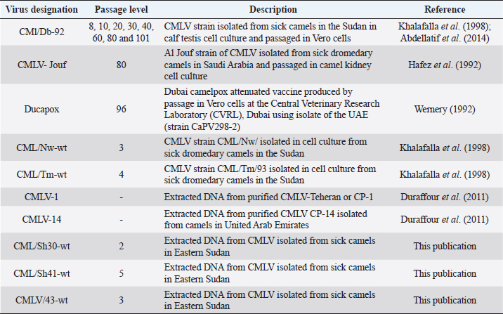
AbstractBackground: Camelpox is the most infectious and economically important disease of camelids that causes significant morbidity and mortality rates. Several live attenuated vaccines against Camelpox virus (CMLV) are produced worldwide by passaging field isolates in cell culture. Sequence of a high passage Saudi isolate of CMLV was previously found closely resembled Vaccinia virus (VACV). Aim: To determine whether other high cell culture passage CMLV isolates are genetically resemble VACV and further to explore the possible mechanism of the resemblance. Methods: We performed polymerase chain reaction and DNA sequence analysis of A-type inclusion body protein (ATIP), L1R, and open reading frame (ORF) 185 genes on different cell culture passage levels of a field isolate, two high passage vaccines, wild-type, and reference strains of CMLV. Results: We demonstrate that additional two high passage attenuated vaccine candidate from Sudan and UAE likewise contain sequences resembling VACV more than CMLV. Furthermore, sequence analysis of the ATIP gene of selected virus passages in cell culture revealed that the shift to VACV-like occurred between passage 11 and 20 and up to the 10th passage the genome still resembles wild-type virus. This observation was further confirmed by recombination analysis which indicated recombination events at ATIP and ORF185 genes occurred at higher passages. Conclusion: We confirmed that the cell culture passage CMLV turns to resemble VACV after cell culture passage and concluded that the resemblance may not be a result of contamination or misidentification as previously thought but could be due to recombination events that occurred during the passage process. Keywords: Camelpox virus, cell culture passage, phylogenetic analysis. IntroductionCamelpox virus (CMLV), the causative agent of camelpox is a member of the genus Orthopoxvirus (OPXV), subfamily Chordopoxvirinae in the family Poxviridae which at present contain 12 genera (ICTV, 2018). CMLV have recently received much attention due to the emergence of infections in humans (Bera et al., 2011; Khalafalla and Abdelazim, 2017). Camelpox is routinely diagnosed based on clinical signs, pathological findings and cellular and molecular assays. The currently available polymerase chain reaction (PCR) assays to identify CMLV are based on the detection of sequences encoding for the A-type inclusion body protein (ATIP), the hemagglutinin (HA), the ankyrin repeat protein (C18L), or the DNA polymerase (DNA pol) genes (Meyer et al., 1994; Ropp et al., 1995; Balamurugan et al., 2009; Venkatesan et al., 2012). ATIP gene-based PCR has been performed with a single set of primers, which enables the differentiation of OPXV species by producing amplicons of different sizes (Meyer et al., 1997). The used primer pairs, common to all OPXV species flanked a region exhibiting distinct and specific DNA deletions in the corresponding sequences of vaccinia virus (VACV), Ectromelia virus (ECTV), monkey pox (MPXV), and CMLV. For this reason, PCR resulted in DNA fragments of different sizes. The method yields 881 bp from CMLV, 1,219 bp from ECTV (strain Munich), 1,500 bp from MPXV (strain Copenhagen), 1,575 bp from the VARV (strain Bangladesh), 1,603 bp from VACV (VACV WR), and 1,673 bp from CPXV (strain Brighton) genomes (Meyer et al., 1997). Yousif and Al Ali (2012) surprisingly obtained a PCR fragment longer than 881 bp by using the consensus ATIP gene primers to identify CMLV in Jouf CMLV vaccine produced in Saudi Arabia. They sequenced the ATIP, two extracellular enveloped viruses-specific (A33R and B5R), and two intracellular mature virus (IMV) (L1R and A27L) orthologue genes and these sequences were found like VACV Lister strain. They concluded that the tissue culture attenuated CMLV vaccine based on the isolate Jouf-78 strain produced in Saudi Arabia (Hafez et al., 1992) contains a Lister-like strain of VACV rather than CMLV and a possible contamination event during production may have caused this mistaken identity. Previously, the development of a live attenuated CMLV vaccine by passage of a Sudanese isolate in Vero cells has been reported by Abdellatif et al. (2014). The present study was prompted by the interesting findings of Yousif and Al Ali (2012) on the mistaken identity of Al Jouf-78 CMLV vaccine. Table 1. Details on viruses and DNAs used in the study.
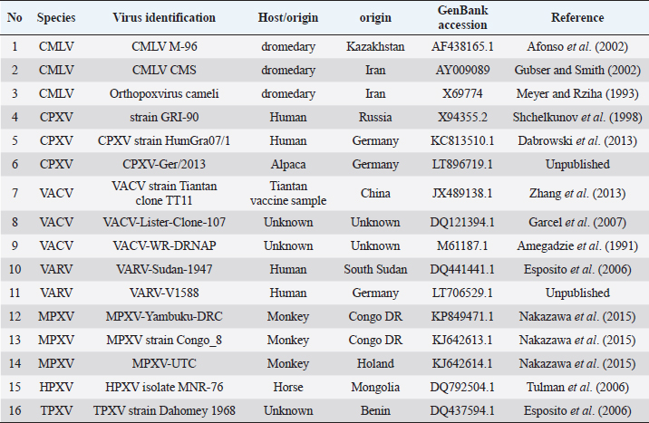
Materials and MethodsVirusesDifferent passage levels of cell culture attenuated CMLV strain CMl/Db-92 from the available stored samples at Department of Microbiology, faculty of Veterinary medicine, University of Khartoum were used in this study (Table 1). Commercial live attenuated camelpox vaccines and wild-type CMLV isolates previously isolated in cell culture from sick dromedary camels as well as two extracted DNAs from scabs collected from field cases of the disease are detailed in Table 1. PCRWe selected three genes to represent different regions on the CMLV genome; the ATIP gene region which, was used to determine the species of the OPXV via PCR by producing amplicons of different sizes (Meyer et al., 1994) or VACV similar sequence (Yousif and Al Ali, 2012), second, we targeted the L1R gene IMV gene of ORXV also used by Yousif and Al Ali (2012), additionally, a unique region in the CMLV genome which encode open reading frame (ORF) 185 claimed absent in other OPXVs (Afonso et al., 2002) was used. ATIP geneTo exclude other pox like diseases, used viruses were first identified as CMLV by using a multiplex gel-based PCR (Khalafalla et al., 2015). Secondly, the extracted DNAs were subjected to PCR using the consensus ATIP gene primers (Meyer et al., 1997). L1R and ORF 185 genesTo corroborate the ATIP gene results, we amplified two more CMLV genes. First, IMV gene (L1R) was followed as previously described (Yousif and Al Ali, 2012). Second, a unique region in the CMLV genome which encode ORF 185 absent in other OPXVs (Afonso et al., 2002) was used for PCR primer design, namely 185F forward sequence 5′ CTCAATGAGAGTTCCTGACCATCC 3′ and 185R reverse 5′ ACACTCTAATACAACACAGGCAACA 3′ that amplify a 923 bp product. PCR condition in a total volume of 25 μl containing 2.5 μl Go Taq® Green (Promega) master mix, 0.2 mM each primer, 2 μl template DNA. With 1 U of Taq polymerase. Cycling conditions were set to first denaturation at 95°C for 5 m followed by 30 cycles of 94°C/ 30 seconds, 52.4/30 seconds, and 72°C/3 minutes and then an extension period of 5 minutes at 72°C. Table 2. Information on OPXV sequences used for the phylogenetic analysis in the present study.
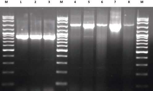
SequencingThe PCR products were then checked in agarose gel, purified using the QIAquick PCR Purification Kit (Qiagen, Hilden, Germany) and then were sequenced. Sequencing was completed using the BigDye® Terminator v3.1 cycle sequencing chemistry kit for each primer pairs. Nucleotide positions were confirmed based on two independent sequencing reactions in both directions. Phylogenetic analysisThe terminal unreliable nucleotides of the obtained gene sequences were first trimmed-off automatically and both forward and reverse sequences were aligned in the Geneious version 8.1 created by Biomatters, available from http://www.geneious.com. The forward and the reverse sequences were used to obtain the correct sequence of the fragment. The biologically correct sequences were subjected to basic local alignment search and comparison with nucleotide sequence in the GenBank database using the online BLASTN program on the NCBI website (Altschul et al., 1997). Phylogenetic tree based on nucleotide sequences was constructed using the Neighbor-Joining method in MEGA X (Kumar et al., 2018). Nucleotide sequences of CMLV investigated in the present study were compared with analogous sequences from other CMLV isolates and related OPXVs available in the GenBank database (Table 2). Recombination analysis using RDP5The Recombination Detection Program v.3 (RDP5) was used for recombination analysis. Recombination events, likely parental isolates of recombinants, and recombination break points were analyzed using the RDP, GENECONV, Chimaera, MaxChi, BOOTSCAN, and SISCAN methods implemented in the RDP4 program with default settings (Martin et al., 2015). Ethical approvalThis article does not contain any studies with human or animal subjects performed by any of the authors. ResultsPCRTwo sizes of PCR products were obtained: 1) band of ~880 bp in wild-type CMLV isolates (CML-Nw-SD, CML/Sh41), passages 8 and 10 of CML/Db/92 (ML/Db-P8 and P10) (Fig. 1) as well as the reference CMLV strains 1 and 14 (not shown), and 2) band of ~1,250 bp in passage 20 and higher of CML/Db/92 (Fig. 1) and the two vaccine strains Al-Jouf 78 and Ducapox vaccine (not shown). To support results of the ATIP gene, PCR was performed to amplify L1R and ORF 185 genes. DNAs from the CMLV positive control and the various passages produced amplicons of expected size (765 bp and for L1R and ORF 185), but there is no size difference in CMLV passages (passages 10 to 89) (data not shown).
Fig. 1. Agarose gel electrophoresis of PCR products amplified from the ATIP gene of CMLVs strains. Lane M; 100 bp marker, Lanes 1, 2, and 3: CML/Nw-wt, CML/Sh41 and CML/Db-P10, respectively. Lanes, 4, 5, 6, 7 and 8: CML/Db-P20, P40, P60, P80, and P101, respectively.
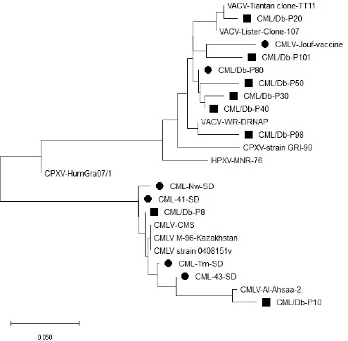
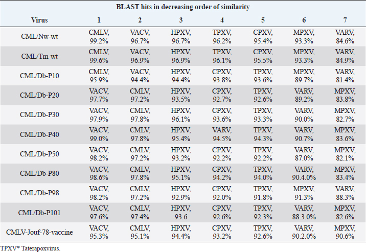
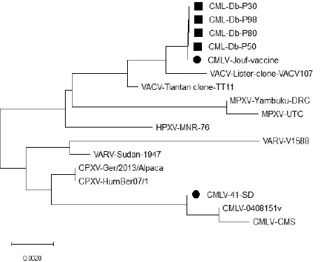
Fig. 2. Phylogenetic tree of the ATIP gene nucleotide sequences of wild type and cell culture passaged CMLV strains in comparison with selected OPXV sequences inferred using the Neighbor-Joining method (Saitou and Nei, 1987). Evolutionary analyses were conducted in MEGA X. The black squares refer to high passage CMLVs, black dots to wild-type CMLVs and vaccine strains, both sequenced in this work. Details on origin of the OPX sequences is shown in Table 1. Table 3. Results of BLAST search using CMLV ATIP gene sequences investigated in this work for best hit with published CMLV, VACV, CPXV, VARV, HPXV, MPXV, and TPXV* sequences. Shown are virus names and percent identity (%) in order of decreasing percent identity (1–7).
SequencingOverall, 26 partial genome sequences representing the three investigated genes ATIP (n=10), L1R (n=6), and ORF 185 (n=10 were obtained and are being deposited in the GenBank database. We failed to amplify ATIP gene sequences from CMLV-Ducapox vaccine and L1R sequence from some passages of CMLV strain CMl/Db-92 (data not shown). Nucleotide sequences obtained in the present study were deposited in the GenBank under accession numbers MN970175–MN970184 for ATIP gene of CML-Nw-SD, CML/Db-P8, CML/Db-P10, CML/Db-P20, CML/Db-P30, CML/Db-P40, CML/Db-P50, CML/Db-P80, CML/Db-P98, and CML/Db-P101, respectively; MN970185–MN970190 for LIR gene of CMLV-41-SD, CML-Db-P30, CML-Db-P50, CML-Db-P80, CML-Db-P98, and CMLV-Jouf-vaccine; MN970165–MN970174 for ORF 185 of CML-Nw-SD, CML-Tm-SD, CML-Db-P10, CML-Db-P20, CML-Db-P50, CML-Db-P98, CML-Db-P101, CMLV-41-SD, CMLV Ducapox-vaccine, CMLV-Jouf-vaccine, respectively. Phylogenetic analysisA BLAST search revealed that the wild-type or low-passage CMLV strains are closely related to the previously sequenced strains of CMLV [CMLV M-96 from Kazakhstan (Gen-Bank ID: AF438165.1; Afonso et al. (2002) (Sequences 138104 to 138920)], CMLV CMS from Iran [GenBank ID: AY009089; Gubser and Smith (2002) (Sequences 136224 to 137040) and Orthopoxvirus cameli, CMLV Teheran (GenBank ID: X69774; Meyer and Rziha (1993)]. These include passages 8 and 10 of the strain CMLV/Db (CMLV/Db-P8 and P10). Starting from passage 20 (CML/Db-P20), the ATI sequences become closer to VACV rather than CMLV. Passage 20 of this strain (CMLV/Db-P20), which has an added block of nucleotides that gave a larger (than the 881 bp produced by wild-type CMLVs) PCR product of ~1,250 bp showed 98% nt identity with VACVs and less than 93% (not shown in the BLAST) with CMLV sequences. Accordingly, the phylogeny analysis of the cell culture passaged virus showed three groups 1) Passage 8 clustered with wild type CMLVs; 2) Passage 10 forms a closely related branch to that of CMLVs cluster, and 3) Passage 20 to 101 forms a uniform group within VACV cluster (Fig. 2, Table 3). Based on BLAST search, passage 8 (and several wild type isolates) showed 99% nucleotide sequence homology with wild type such as CMLV M-96 (GB# AF438165.1), but 97% with VACVs including Lister clone VACV107. Passage 10 demonstrated 98% homology with CMLV strain Al Ahsa JQ901104.1, 96% with wild type such as CMLV M-96, but 94% with VACV, Rabbitpox virus, (AY484669.1). Passage 20 (and higher passages) showed 98% homology with VACV Tiantan clone TT11 (JX489138.1), VACV Lister clone VACV107 (DQ121394.1), VACV Lister (AY678276.1), VACV TianTan clone TP5 (KC207811.1) and VACV TianTan clone TP3 (KC207810.1) and 97% with other VACV and Rabbitpox virus (AY484669.1). 95%, 94% with other VACV and Horsepox virus (HPXV) isolate MNR-76 (DQ792504.1); and less than 93% (not shown in the BLAST) with CMLV sequences. Table 4. Results of BLAST search using CMLV L1R gene sequences investigated in this work for best hit with published CMLV, VACV, CPXV, VARV, HPXV, MPXV, and TPXV sequences. Shown are virus name, GenBank accession # and percent identity (%) in order of decreasing percent identity (1–7).
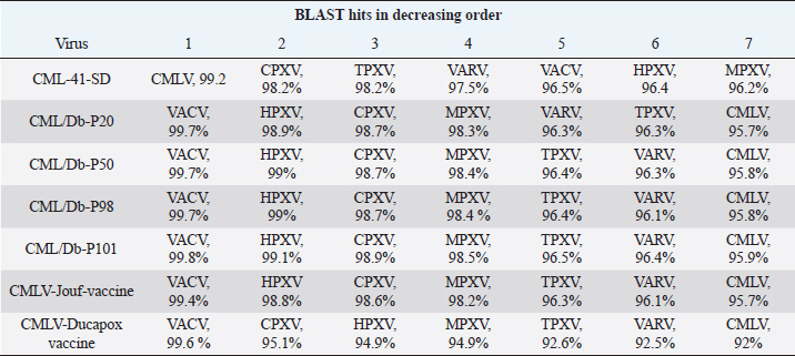
Fig. 3. Phylogenetic tree of the L1R gene nucleotide sequences of wild type and cell culture passaged CMLV strains in comparison with selected OPXV sequences inferred using the Neighbor-Joining method (Saitou and Nei, 1987). Evolutionary analyses were conducted in MEGA X. The black squares refer to high passage CMLVs, black dots to wild-type CMLVs and vaccine strains, both sequenced in this work. Details on origin of the OPX sequences are shown in Table 1. Alignment by Muscle (MEGA 6.06) of passages 8, 10, and 20 with CMLV-M-96; (Position 138,127–138,907; GB# NC_003391) and vaccinia virus sequence (position 135,622–136,649; GB# NC_006998) indicated that sequence 136,059–136,649 is almost identical in all the five sequences and sequence 136,622–136,059 is variable with several insertions of 10–50 nucleotides. On the other hand, testing the long insertion (216 nt) for the presence of tandem repeat using online EMBOSS tools (einverted, palindrome, and equicktandem) was found negative. Table 5. Results of BLAST search using CMLV ORF185 sequences investigated in this work for best hit with published CMLV, VACV, CPXV, VARV, HPXV, MPXV, and TPXV sequences. Shown are virus name and percent identity (%) in order of decreasing percent identity (1–7).
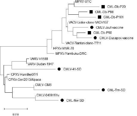
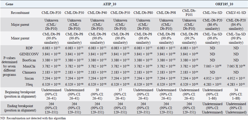
Fig. 4. Phylogenetic tree of the ORF 185 nucleotide sequences of wild-type and cell culture passaged CMLV strains in comparison with selected OPXV sequences inferred using the Neighbor-Joining method (Saitou and Nei, 1987). Evolutionary analyses were conducted in MEGA X. The black squares refer to high passage CMLVs, black dots to wild-type CMLVs and vaccine strains, both sequenced in this work. Details on origin of the OPX sequences are shown in Table 2. Results of BLAST and phylogenetic analysis for L1R gene are shown in Table 4 and Figure 3 and those of ORF 185 in Table 5 and Figure 4. Table 6. Summary of unique recombination events identified by the Recombination Detection Program v.3 (RDP5).
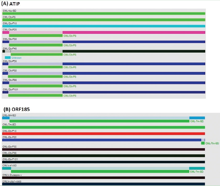
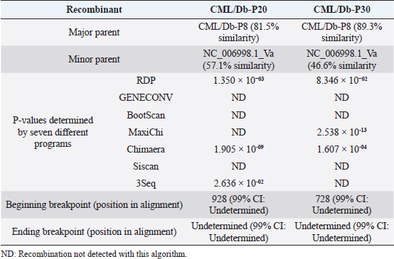
Fig. 5. Analysis of possible recombination events in full-length of ATIP and ORF185 genes. (a) Seven recombination events were detected in ATIP gene resulted in CML/Db-P20, CML/Db-P40, CML/Db-P50, CML/Db-P60, CML/Db-P80, CML/Db-P98, and CML/Db-P101 recombinants. (b) Two recombination events (possibly reassortment) were detected in ORF185 that generated CML-Nw-SD and CMLV-41-SD recombinants. Detailed information for recombination was also provided in Table 4. Each segment is indicated by different color bar. Recombination analysisTo identify the recombination events, we performed recombination detection analysis using the RDP5 program, which contains various recombination detection algorithms, such as RDP, GENECONV, Chimaera, MaxChi, BOOTSCAN, SISCAN, and 3Seq. All these seven algorithms can predict the seven recombination events in the ATIP gene (Table 6 and Fig. 5). In all recombinants, the minor parent is CML/Db-P8 (99.6% similarity), however, the major parent was unknown, but possibly be (CML/Db-P10). For all recombinant, the Beginning breakpoint (position in alignment) was undetermined except for CML/Db-P40, CML/Db-P50 (ATIP gene) and CMLV-41-SD (ORF185) which begin at position 39, 37, and 34, respectively. For all ATIP recombinant, the end breakpoint (position in alignment) was at 264. The end breakpoint (position in alignment) for ORF185 recombinants was undetermined. Another recombination event in OERF185 gene was observed (CMLV-Db-P20) but possibly due to misalignment artifact. To further evaluate whether sequence from Vaccinia virus had inserted into CMLV genome, we carried recombination analysis using aligned sequences of full-length of ATIP (passage 8, 10, 20, and 30), Vaccinia virus (NC_006998.1_Va) and CMLV (NC_003391.1_CM). Five recombination events have been identified (Fig. 6), of which two events clearly generated recombinant having fragments of Vaccinia virus (CML/Db-P20 and CML/Db-P30 recombinants) (Fig. 6 and Table 7).
Fig. 6. Analysis of possible recombination events in aligned full-length of ATIP (passage 8, 10, 20 and 30), Vaccinia virus (NC_006998.1_Va) and CMLV (NC_003391.1_CM). The most important events showing insertion of fragment of Vaccinia virus into CMLV at passage 20 and 30 (shown by yellow arrow (CML/Db-P20 and CML/Db-P30 recombinants). Detailed information for recombination was also provided in Table 1. Each segment is indicated by different color bar. Table 7. Summary of unique recombination events identified by the Recombination Detection Program v.3 (RDP5).
DiscussionSequence of the ATIP gene region could determine the species of the OPXV via PCR by producing amplicons of different sizes due to the heterogeneity of the primer binding sites (Meyer et al., 1994). Our results indicated two different banding patterns 1) the CMLV-typical band in wild-type CMLV isolates, cell culture passages 8 and 10 of the virus as well as the reference CMLV strains and 2) band of ~1250 bp in passage 20 and higher of the virus and the two vaccine strains; Al-Jouf 78 and Ducapox vaccine. Sequence analysis of ATIP gene region as well as of two other OPXV genes indicated that high cell culture passage CMLV turn to resemble VACV after cell culture passage. Of note, PCR revealed no differences in product size of L1R and ORF 185 genes between the wild type and the passaged CMLV. Yousif and Al-Ali (2012) likewise observed no size difference in four other OPXV genes including the L1R. These findings of the present study corroborated results obtained by Yousif and Al Ali (2012), who had identified VACV-like sequences in Al-Jouf-78 vaccine strain. The authors claimed that a conceivable contamination event during production may have caused the mistaken identity as VACV Elstree (Lister) which was used during initial testing of the developed vaccine (Hafez et al., 1992) supporting the previous studies which have shown a high recombination rate during a poxvirus co-infection (Oliveira et al., 2015). How a contamination of such scale remained undetected could be explained by the use of serologic techniques, which due to the cross-reactivity of OPXVs cannot differentiate between CMLV and VACV, and not gene-based assays in the process of virus identification and quality control. Since VACV Lister was successfully used in neighboring country Bahrain to control outbreaks of camelpox in 1987, it would be interesting, if available, to amplify the ATIP gene of the wild type Al Jouf isolate to see if it is a true CMLV or not, owing to its broad host range, a VACV Lister that had spread from vaccinated camels in Bahrain to unvaccinated herds in Saudi Arabia. It could be also a VACV Lister that probably have had emerged and circulated in dromedary camels in a pattern like the recent emergence of vaccinia-like viruses in Brazil and India (Singh et al., 2012; Oliveira et al., 2015). The recombination in OPXVs is well documented (Bourke and Dumbell, 1972; Takahashi-Nishimaki et al., 1987; Okeke et al., 2012) and early investigations indicated that homologous recombination could occur between the genomes of two replicating poxviruses and later, marker rescue studies demonstrated that fragments of genomic and cloned DNA could recombine with the genome of VACV in infected cells (Sánchez-Sampedro et al., 2015). The present study shows that other two attenuated strains of CMVs which undergone several passages in cell culture are also genetically like VACV. It is unlikely that events of contamination by VACV have occurred in the three laboratories (Saudi Arabia, UAE and Sudan) at different time span resulting in attenuation. In the present study, a band of ~1,250 bp was detected in passage 20 and higher of CML/Db/92 which is less than that of the VACV Western Reserve (~1,603 bp) as depicted by Meyer et al. (1997). In our study, we concluded that high passage CMLVs from Sudan and UAE contain sequences resembling VACV and speculated that this event is unlikely due to contamination with VACV as proposed in a previous publication, otherwise the size would be comparable or near ~1,603 bp. However, there is a need to suggest or test other possibilities that could disclose what happened to the virus. A multiple passage in a different host without any other factor though can change some of the genetic makeup of the CMLV, but probably not to the level of more than 90%. For instance, this could have been due to modified vaccinia virus Angara, which was passaged more than 570 times in primary chick embryo fibroblast cells but lost only 15% (around 30 kb) of its parental genome (Sánchez-Sampedro et al., 2015). The cell culture system used to attenuate the three CMLVs is different (camel kidney cell culture/Vero), but all were supplemented with fetal bovine serum. There is the possibility that a recombination even has occurred between passages of CMLVs and genomic fragments of some OPXVs present in the serum, which was used as a nutrient supplement for the cell culture as previous studies have detected OPXV DNA in serum of infected hosts (Savona et al., 2006; Cohen et al., 2007; Abrahão et al., 2010). Further research is needed to prove the hypothesis of VACV contamination by whole genome sequencing and studying more high passage CMLV isolates to examine a possible event of genetic recombination between vaccine strains and some genetic CMLV elements during attenuation in the cell culture or other possibilities such as the non-genetic genome reactivation which can generate infectious virus within and between members of genera in the subfamily Chordopoxvirinae (ICTV, 2009). Moreover, our findings highlighted the importance of studying the genetic variability among CMLV strains circulating in the environment using the ATIP gene PCR and next generation sequencing. The present study confirmed that high cell culture passage CMLV turn to resemble VACV after cell culture passage and concluded that the resemblance may not be a result of contamination or misidentification as previously thought but probably involve molecular events that are resultant from the passage process. The results of this investigation can add to the understanding of the molecular bases of poxvirus attenuation and give genetic markers to differentiate between wild type and vaccine strains. AcknowledgmentsThe author thanks Dr. Anwar Ghaleb for valuable suggestions and Dr. Javed Rashid for reading the manuscript. Authors contributionAIK and SSA designed and supervised the study. AIK performed the experiments and drafted the manuscript. MAA analyzed and interpreted the data, HZAI performed the recombination analysis and reviewed the manuscript, and SSA revised and approved the manuscript. Conflict of interestThe authors declare that they have no conflict of interest. ReferencesAbdellatif, M.M., Ibrahim, A.A. and Khalafalla, A.I. 2014. Development and evaluation of a live attenuated camelpox vaccine from a local field isolate of the virus.Rev. Sci. Tech. 33, 831–838. Abrahão, J.S., Silva-Fernandes, A.T., Lima, L.S., Campos, R.K., Guedes, M.I., Cota, M.M., Assis, F.L., Borges, I.A., Souza-Júnior, M.F., Lobato, Z.I, Bonjardim, C.A., Ferreira, P.C., Trindade, G.S. and Kroon, E.G. 2010. Vaccinia virus infection in monkeys, Brazilian Amazon. Emerg. Infect. Dis. 16, 976–979. Afonso, C.L., Tulman, E.R., Lu, Z., Zsak, L., Sandybaev, N.T., Kerembekova, U.Z., Zaitsev, V.L., Kutish, G.F. and Rock, D.L. 2002. The genome of Camelpox virus. Virology 295, 1–9. Amegadzie, B.Y., Ahn, B.Y., Moss, B. 1991. Identification, sequence, and expression of the gene encoding a Mr 35,000 subunit of the vaccinia virus DNA-dependent RNA polymerase. J. Biol. Chem. 266, 13712–8. Altschul, S.F., Madden, T.L., Schäffer, A.A., Zhang, J., Zhang, Z., Miller, W. and Lipman, D.J. 1997. Gapped BLAST and PSI-BLAST: a new generation of protein database search programs. Nucleic Acids Res. 25, 3389–3402. Balamurugan, V., Bhanuprakash, V., Hosamani, M., Jayappa, K.D., Venkatesan, G., Chauhan, B. and Singh, R.K. 2009. A polymerase chain reaction strategy for the diagnosis of camelpox. J. Vet. Diagn. Invest. 21, 231–237. Bera, B.C., Shanmugasundaram, K., Barua, S., Venkatesan, G., Virmani, N., Riyesh, T., Gulati, B.R., Bhanuprakash, V., Vaid, R.K., Kakker, N.K., Malik, P., Bansal, M., Gadvi, S., Singh, R.V., Yadav, V., Sardarilal., Nagarajan, G., Balamurugan, V., Hosamani, M., Pathak, K.M. and Singh, R.K. 2011. Zoonotic cases of camelpox infection in India. Vet. Microbiol. 152, 29–38. Bourke, A. T. and Dumbell, K.R. 1972. An unusual poxvirus from Nigeria. Bull. World Health Organ. 46, 621–623. Cohen, J.I., Hohman, P., Preuss, J.C., Li, L., Fischer, S.H. and Fedorko, D.P. 2007. Detection of vaccinia virus DNA, but not infectious virus, in the blood of smallpox vaccine recipients. Vaccine 25, 4571–4574. Dabrowski, P.W., Radonić, A., Kurth, A., Nitsche, A. 2013. Genome-wide comparison of cowpox viruses reveals a new clade related to Variola virus. PLoS One 8(12), e79953. Duraffour, S., Meyer, H., Andrei, G. and Snoeck, R. 2011. Camelpox virus. Antiviral Res. 92, (2), 167–186. Esposito, J.J., Sammons, S.A., Frace, A.M., Osborne, J.D., Olsen-Rasmussen, M., Zhang, M., Govil, D., Damon, I.K., Kline, R., Laker, M., Li, Y., Smith, G.L., Meyer, H., Leduc, J.W., Wohlhueter, R.M. 2006. Genome sequence diversity and clues to the evolution of variola (smallpox) virus. Science 313, 807–812. Garcel, A., Crance, J.M., Drillien, R., Garin, D., Favier, A.L. 2007. Genomic sequence of a clonal isolate of the vaccinia virus Lister strain employed for smallpox vaccination in France and its comparison to other orthopoxviruses. J. Gen. Virol. 88, 1906–1916. Gubser, C. and Smith, G.L. 2002. The sequence of Camelpox virus shows it is most closely related to variola virus, the cause of smallpox. J. Gen. Virol. 83, 855–872. Hafez, S.M., Al-Sukayran, A., Dela-Cruz, D.M., Mazloum, K.S., Al-Bokmy, A.M., Al-Mukayel, A. and Amjad, A.M. 1992. Development of a live cell culture camelpox vaccine. Vaccine 10, 533–537. ICTV International Committee on Taxonomy of Viruses (2018). Available via https://talk.ictvonline.org/taxonomy/ (accessed 23 September 2018). ICTV. 2009. Ninth Report. Family: Poxviridae Chapter Version Taxonomy Release. Available via https://talk.ictvonline.org/ictv-reports/ictv_9th_report/dsdna-viruses2011/w/dsdna_viruses/74/poxviridae) (accessed 13 July 2017). Khalafalla, A.I. and Abdelazim, F. 2017. Human and Dromedary Camel Infection with Camelpox virus in Eastern Sudan. Vector Borne Zoonotic Dis. 17, 281–284. Khalafalla, A.I., Al-Busada, K.A. and El-Sabagh, I.M. 2015. Multiplex PCR for rapid diagnosis and differentiation of pox and pox-like diseases in dromedary Camels. Virol. J. 12, 102–110. Khalafalla, A.I., Mohamed, M.E.M. and Ali, B.H. 1998. Camel pox in the Sudan: I – Isolation and identification of the causative virus. J. Camel Pract. Res. 5, 229–233. Kumar, S., Stecher, G., Li, M., Knyaz, C. and Tamura, K. 2018. MEGA X: Molecular Evolutionary Genetics Analysis across computing platforms. Mol. Biol. Evol. 35, 1547–1549. Martin, D.P., Murrell, B., Golden, M., Khoosal, A. and Muhire, B. 2015. RDP4: Detection and analysis of recombination patterns in virus genomes. Virus Evol. 1(1), vev003; doi.org/10.1093/ve/vev003 Meyer, H., Pfeffer, M. and Rziha, H.J. 1994. Sequence alterations within and downstream of the A-type inclusion protein genes allows differentiation of Orthopoxvirus species by polymerase chain reaction. J. Gen. Virol. 75, 1975–1981. Meyer, H., Ropp, S.L. and Esposito, J.J. 1997. Gene for A-type inclusion body protein is useful for a polymerase chain reaction assay to differentiate orthopoxviruses. J. Virol. Methods 64, 217–221. Meyer, H. and Rziha, H.J. 1993. Characterization of the gene encoding the A-type inclusion protein of Camelpox virus and sequence comparison with other orthopoxviruses. J. Gen. Virol. 74, 1679–1684. Nakazawa, Y., Mauldin, M.R., Emerson, G.L., Reynolds, M.G/, Lash, R.R/, Gao. J/, Zhao. H/, Li, Y., Muyembe, J.J., Kingebeni, P.M., Wemakoy, O., Malekani, J., Karem, K.L., Damon, I.K., Carroll, D.S. 2015. A phylogeographic investigation of African monkeypox. Viruses 7, 2168–2184. Okeke, M.I., Hansen, H. and Traavik, T. 2012. A naturally occurring cowpox virus with an ectromelia virus A-type inclusion protein gene displays atypical A-type inclusions. Infect. Genet. Evol. 12, 160–168. Oliveira, G., Assis, F., Almeida, G., Albarnaz, J., Lima, M., Andrade, A.C., Calixto, R., Oliveira, C., Diomedes-Neto, J., Trindade, G., Ferreira, P.C., Kroon, E.G. and Abrahão, J. 2015. From lesions to viral clones: biological and molecular diversity amongst autochthonous Brazilian vaccinia virus. Viruses 7, 1218–1237. Ropp, S.L., Jin, Q., Knight, J.C., Massung, R.F. and Esposito, J.J. 1995. Polymerase chain reaction strategy for identification differentiation of smallpox and other orthopox viruses. J. Clin. Microbiol. 33, 2069–2076. Saitou, N. and Nei, M. 1987. The neighbour-joining method: A new method for reconstructing phylogenetic trees. Mol. Biol. Evol. 4, 406–425. Sánchez-Sampedro, L., Perdiguero, B., Mejías-Pérez, E., García-Arriaza, J., Di Pilato, M. and Esteban, M. 2015. The evolution of poxvirus vaccines. Viruses 7, 1726–1803. Savona, M.R., Dela-Cruz, W.P., Jones. M.S., Thornton, J.A., Xia, D., Hadfield, T.L. and Danaher, P.J. 2006. Detection of vaccinia DNA in the blood following smallpox vaccination. JAMA 295, 1898–1900. Shchelkunov, S.N., Safronov, P.F., Totmenin, A.V., Petrov, N.A., Ryazankina, O.I., Gutorov, V.V., Kotwal, G.J. 1998. The genomic sequence analysis of the left and right species-specific terminal region of a cowpox virus strain reveals unique sequences and a cluster of intact ORFs for immunomodulatory and host range proteins. Virology. 243, 432–460. Singh, R.K., Balamurugan, V., Bhanuprakash, V., Venkatesan, G. and Hosamani, M. 2012. Emergence and reemergence of vaccinia-like viruses: global scenario and perspectives. Indian J. Virol. 23, 1–11. Takahashi-Nishimaki, F., Suzuki, K., Morita, M., Maruyam, T., Miki, K., Hashizume, S. and Sugimoto, M. 1987. Genetic analysis of vaccinia virus Lister strain and its attenuated mutant LC16m8: production of intermediate variants by homologous recombination. J. Gen. Virol. 68, 2705–2710. Tulman, E.R., Delhon, G., Afonso, C.L., Lu,. Z, Zsak, L., Sandybaev, N.T., Kerembekova, U.Z., Zaitsev, V.L., Kutish, G.F., Rock, D.L. 2006. Genome of horsepox virus. J Virol 80:9244–9258. Yousif, A.A., Al-Ali, A.M. 2012. A case of mistaken identity? Vaccinia virus in a live camelpox vaccine. Biologicals 40, 495–498. Venkatesan, G., Bhanuprakash, V., Balamurugan, V., Prabhu, M. and Pandey, A.B. 2012. TaqMan hydrolysis probe based real time PCR for detection and quantitation of Camelpox virus in skin scabs. J. Virol. Methods 181, 192–196. Wernery, U. 1992. Production of an attenuated camelpox vaccine (Ducapox). Camel Pract. Res. 7, 117–119. Zhang, Q., Tian, M., Feng, Y., Zhao, K., Xu, J., Liu, Y., Shao, Y. 2013. Genomic sequence and virulence of clonal isolates of vaccinia virus Tiantan, the Chinese smallpox vaccine strain. PLoS One 8, e60557. | ||
| How to Cite this Article |
| Pubmed Style Khalafalla AI, Muhairi SSA, Ishag HZA, Hosani MAA. More Cell Culture Passaged Camelpox virus Sequences Found Resembling those of Vaccinia Virus. Open Vet J. 2020; 10(2): 144-156. doi:10.4314/ovj.v10i2.4 Web Style Khalafalla AI, Muhairi SSA, Ishag HZA, Hosani MAA. More Cell Culture Passaged Camelpox virus Sequences Found Resembling those of Vaccinia Virus. https://www.openveterinaryjournal.com/?mno=66095 [Access: November 08, 2024]. doi:10.4314/ovj.v10i2.4 AMA (American Medical Association) Style Khalafalla AI, Muhairi SSA, Ishag HZA, Hosani MAA. More Cell Culture Passaged Camelpox virus Sequences Found Resembling those of Vaccinia Virus. Open Vet J. 2020; 10(2): 144-156. doi:10.4314/ovj.v10i2.4 Vancouver/ICMJE Style Khalafalla AI, Muhairi SSA, Ishag HZA, Hosani MAA. More Cell Culture Passaged Camelpox virus Sequences Found Resembling those of Vaccinia Virus. Open Vet J. (2020), [cited November 08, 2024]; 10(2): 144-156. doi:10.4314/ovj.v10i2.4 Harvard Style Khalafalla, A. I., Muhairi, . S. S. A., Ishag, . H. Z. A. & Hosani, . M. A. A. (2020) More Cell Culture Passaged Camelpox virus Sequences Found Resembling those of Vaccinia Virus. Open Vet J, 10 (2), 144-156. doi:10.4314/ovj.v10i2.4 Turabian Style Khalafalla, Abdelmalik Ibrahim, Salama S Al Muhairi, Hassan Zackaria Ali Ishag, and Mohamed A Al Hosani. 2020. More Cell Culture Passaged Camelpox virus Sequences Found Resembling those of Vaccinia Virus. Open Veterinary Journal, 10 (2), 144-156. doi:10.4314/ovj.v10i2.4 Chicago Style Khalafalla, Abdelmalik Ibrahim, Salama S Al Muhairi, Hassan Zackaria Ali Ishag, and Mohamed A Al Hosani. "More Cell Culture Passaged Camelpox virus Sequences Found Resembling those of Vaccinia Virus." Open Veterinary Journal 10 (2020), 144-156. doi:10.4314/ovj.v10i2.4 MLA (The Modern Language Association) Style Khalafalla, Abdelmalik Ibrahim, Salama S Al Muhairi, Hassan Zackaria Ali Ishag, and Mohamed A Al Hosani. "More Cell Culture Passaged Camelpox virus Sequences Found Resembling those of Vaccinia Virus." Open Veterinary Journal 10.2 (2020), 144-156. Print. doi:10.4314/ovj.v10i2.4 APA (American Psychological Association) Style Khalafalla, A. I., Muhairi, . S. S. A., Ishag, . H. Z. A. & Hosani, . M. A. A. (2020) More Cell Culture Passaged Camelpox virus Sequences Found Resembling those of Vaccinia Virus. Open Veterinary Journal, 10 (2), 144-156. doi:10.4314/ovj.v10i2.4 |


