


| Original Article | ||
Open Vet J. 2021; 11(2): 295-300 Open Veterinary Journal, (2021), Vol. 11(2): 295–300 Original Research Patellar luxation in Hejazi goatsMohamed H. Abushhiwa1*, Adulrhman M. Alrtib2, Taher N. Elmeshreghi1, Mouna A. Abdunnabi3, Mansur E. Shmela3 and Emad M. Bennour41Department of Surgery and Theriogenology, Faculty of Veterinary Medicine, University of Tripoli, Tripoli, Libya 2Department of Anatomy, Histology and Embryology, Faculty of Veterinary Medicine, University of Tripoli, Tripoli, Libya 3Department of Preventive Medicine (Genetics & Animal Breeding), Faculty of Veterinary Medicine, University of Tripoli, Tripoli, Libya 4Department of Internal Medicine, Faculty of Veterinary Medicine, University of Tripoli, Tripoli, Libya *Corresponding Author:Mohamed H. Abushhiwa. Department of Surgery and Theriogenology, Faculty of Veterinary Medicine, University of Tripoli, Tripoli, Libya. Email: m.abushhiwa [at] uot.edu.ly Submitted: 01/04/2021 Accepted: 21/05/2021 Published: 18/06/2021 © 2021 Open Veterinary Journal
AbstractBackground: Patellar luxation (PL) is a common orthopedic affection among farm and pet animals with mostly congenital (environmental and/or genetic) background. Aim: We report here the first observation of lateral PL in Hejazi goats bred in Libya. Methods: Five Hejazi goats aged between 4 months and 2 years with severe hind limb lameness were admitted to Al-Sorouh veterinary clinic in Tripoli during the period from 2016 to 2018. The goats were thoroughly examined clinically and radiographically. Two goats were surgically treated, and the other three cases were not because of either the cost limitation or expected poor prognosis. The surgical intervention involved femoral trochlear sulcoplasty, medial joint capsule imbrication, and tibial tuberosity transposition. Results: The clinical examination showed grade III–IV lateral PL. Radiologically, there were unilateral or bilateral, ventrocaudal, and dorsal PLs. Two cases were referred to surgical correction. One case almost restored the normal movement of stifle joint together with a good general status 1 year postsurgery. However, the surgical treatment was not effective in correcting the luxated patella in the second case. Conclusion: Lateral PL is common among orthopedic affections in Hejazi goats in Libya, and its surgical treatment provided a quite convenient approach. An association between inbreeding and the PL was suggested in those cases. Keywords: Clinical and radiological findings, Hejazi goat breed, Inbreeding, Patellar luxation, Surgical treatment. IntroductionPatellar luxation (PL) is a musculoskeletal affection characterized by deviation of patella from its normal sliding movement on the trochlear groove (Mostafa et al., 2008; Di Dona et al., 2018). The patella either deviates in dorsal, lateral, medial, or rarely ventral direction (Burnei et al., 2020). The cause of PL in different species has been suggested to be a multi-causal type of disorders; congenital in immature animals and acquired in adults due to traumatic incidences (Fowler, 1998; Van Hoogmoed et al., 1998; Shettko and Trostle, 2000; Vidoni et al., 2006; Abuja et al., 2014; Di Dona et al., 2018). The patella is a large sesamoid bone embedded into the inserted tendon of the quadriceps muscle of the thigh (Frandson et al., 2009). The femeropatellar joint formed between the trochlear groove of the femur and the patella (Dyce et al., 2009). The trochlear groove is formed from two oblique medial and lateral trochleae (trochlear ridges) (Dyce et al., 2009). The medial one is higher than the lateral one and continued with the medial condyle of the femur (Fathi et al., 2016). During PL, one or both of the trochlear ridge(s) flatten(s), allowing the patella to slide from its normal pathway (Luxation) (Di Dona et al., 2018). The fixation and motion mechanisms of the patella are mainly controlled by femeropatellar and patellar ligaments (Dyce et al., 2009). This mechanism is lost during PL leading to the characteristic stifle joint lock (Fossum et al., 2007). Additionally, it has been found that the direction of luxated patellae is variable among animal species because of some anatomical differences (Fossum et al., 2007). Medial and lateral PLs have been reported in sheep (Shettko and Trostle, 2000), goat (Baron, 1987), alpaca and llamas (Kaneps et al., 1989; Abuja et al., 2014), miniature horses (Arighi and Wilson, 1993), and deer (Sundberg and Nielsen, 1981), and are the most common orthopedic problems encountered in dogs’ stifle joint (Arthurs and Langley-Hobbs, 2006; Stanke et al., 2014). This orthopedic problem is also reported in humans (Gao et al., 2018; Sanders et al., 2018). PL is corrected surgically in different animal species using various surgical procedures based on the underlying causes (Shettko and Trostle, 2000; Arthurs and Langley-Hobbs, 2006; Abuja et al., 2014; Stanke et al., 2014). These surgical techniques include trochlear sulcoplasty, tibial tuberosity transposition (TTT), retinacular imbrication, trochlear block recession, trochlear wedge recession, abrasion trochleoplasty, and in adult dogs anti-rotational suture placement (fabella-patella or fabella-tibial). Mostly two or more surgical techniques are used in combination (Shettko and Trostle, 2000; Arthurs and Langley-Hobbs, 2006; Abuja et al., 2014; Stanke et al., 2014). Breeding of Hejazi goats has been recently adopted in Libya. Hejazi goats are cross-breed goats thought to have originated from breeding Kapla breed and local goats of the Arabian Peninsula (Amills et al., 2017; Mahmoud et al., 2020). This breed belongs to long-eared goats, a feature that makes them popular among Libyan breeders (Amills et al., 2017). For that, and because of their limited number in Libya, Hejazi goats were subjected to inbreeding practice, which might be responsible for the emergence of some genetic disorders like PL (Cecchi et al., 2020). To the best of the authors’ knowledge, no reports are available about the prevalence and management of PL in Hejazi goats. Therefore, the purpose of this study was to present the clinical, anatomical, and radiological aspects of PL in this particular breed of goats. The present report also aims to document the surgical intervention as the most appropriate approach to correct this orthopedic condition. Materials and MethodsFive Hejazi goats from both genders, suffering from either unilateral or bilateral hind limb lameness, were enrolled in the current study. Four goats were young (between 4 and 6 months of age), and one case was older (2-year-old). A complete examination of the affected limb was carried out, particularly the stifle area. In addition, a clinical examination was carried out, including vital signs, mucous membranes, and other general status indicators for investigating any other abnormalities and as a pre-operative measure. When PL was noticed, it was graded according to the grading system previously published (Roush, 1993; Arthurs and Langley-Hobbs, 2006). Two orthogonal radiographic views were taken to each case’s effected stifles, including mediolateral and craniocaudal views. The surgical intervention was elected for only two from the presented cases. Surgical treatment was decided upon owner’s consent. General anesthesia was administered using 0.1 mg/kg xylazine hydrochloride 2% intramuscular (Adwia, Egypt), as a premedication. The anesthesia was then induced using ketamine hydrochloride 10% (Bremer-Pharma, Warburg, Germany) at a dose of 2.5 mg/kg intravenously. The anesthesia was maintained using a double drip of xylazine 0.1 mg/ml and ketamine 2 mg/ml at a 2.5 ml/kg/hour rate. The animal was then placed on lateral recumbency, and the stifle area was shaved and aseptically prepared. A surgical incision was made over the craniolateral aspect of the stifle joint, which was fully extended. The subcutaneous tissue and joint capsule were cut, and the joint was exposed. Sulcoplasty on the trochlear groove, joint capsule imbrication, joint capsule release, and TTT were carried out following the standard procedures (Baron, 1987; Kobluk, 1993; Roush, 1993; Shettko and Trostle, 2000; Arthurs and Langley-Hobbs, 2006; Abuja et al., 2014; Di Dona et al., 2018). The skin and subcutaneous tissues were finally sutured. Clinical examination was carried out 10 days post-treatment. Phone call follow-up was possible with the successfully operated case more than 1 year later. Ethical approvalThis study was carried out as an investigation based on the routine diagnostic procedures and did not involve any new materials or protocols requiring special approval. In addition, the researchers were committed to doing all interventions ensuring that they meet the animal welfare protocols, such as shortening the time of manipulation and minimizing the pain and stress during animal handling and surgical procedures. ResultsThe clinical examination showed that three cases suffered from a severe unilateral hind limb lameness with locked stifle correlated with grade IV lateral PL (Fig. 1). The fourth case showed the same clinical picture with bilateral PLs. The fifth case was a buck that showed a grade-III lateral PL. Radiographically, the stifle areas of the affected goats showed ventrocaudal PL in three cases (Figs. 2 and 3) and dorsal PL in the other two cases, for which surgical treatment was performed (Fig. 4A). Surgical incision of the stifle joint revealed an almost flat trochlear groove suggesting trochlear ridge hypoplasia. Surgical correction of luxated patella showed satisfactory results in only one case but not with the other. This doe was 6 months old with grade IV lateral PL. Immediately after surgery, the stifle joint was moving in a normal way, and the doe was able to bear weight on the treated leg. Ten days postsurgery, the doe walks normally, and the wound made complete healing. Phone call follow-up 1 year later revealed that the doe did well, gave birth once and got pregnant again. The other goat was a 2-year-old buck with approximately 50 kg body weight. In this case, the surgical incision revealed a normal trochlear groove and only three surgical procedures were performed; namely, joint capsule imbrication, joint capsule release, and TTT. The correction of the luxated patella was difficult in this case. The surgical suture was ruptured, and the pin fixation had broken once the buck stood on the operated leg. DiscussionPL is an orthopedic affection, which was described in many animal species (Kobluk, 1993; Shettko and Trostle, 2000; Mostafa et al., 2008; Abuja et al., 2014; Di Dona et al., 2018) but with only one report in goats (Baron, 1987). Of particular note, PL was not reported in Hejazi goats. This study presents a number of cases of lateral PL in Hejazi goats, a particular crossbred breed. This breed is one of the popular imported fair animals in Libya, bred in small herds with an inbreeding practice. This practice may have contributed to the emergence of such musculoskeletal disorders. Cases with congenital luxation with a suspected hereditary component have been reported and are thought to be inherited in different animal breeds (Kaneps et al., 1989; Vidoni et al., 2006; Gangwar et al., 2014; O’Neill et al., 2016). In addition, PL was suggested to result from a transmission of recessive genes in Shetland ponies and sheepdogs (Hermans et al., 1987; Solanti et al., 2002). Interestingly, genomic analysis has shown that loci on some canine chromosomes are involved in PL in the Flat-Coated Retriever breed (Lavrijsen et al., 2014). A relationship was recently pointed out between inbreeding and the PL disorder in dogs (Cecchi et al., 2020). Despite the above data, the heritability for PL has shown to be moderate, indicating that environmental factors play a large role in the manifestation of this disorder (Lavrijsen et al., 2013; Zanders, 2014). However, there are neither epidemiological general population data nor information regarding such orthopedic defects in Hejazi breed.
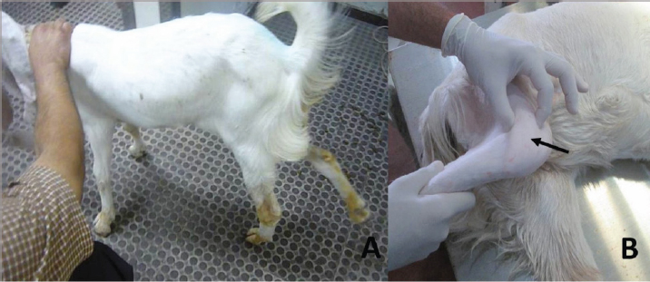
Fig. 1. Clinical presentation of PL in Hejazi goat. (A) Non-weight bearing lameness and locked stifle joint in the right hind limb. (B) Laterally luxated patella (arrow).
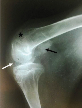
Fig. 2. Mediolateral radiograph of the right stifle of a 12-month-old goat with a unilateral PL. The radiograph shows a severely ventrocaudally luxated patella (black arrow) with loss of trabecular pattern of the distal extremity of femur (star), probably due to trochlear ridge hypoplasia. The infra-patellar fat bad is superimposed by a joint fluid due to stifle joint effusion (white arrow). The patella deviated laterally in all presented cases with a ventrocaudal or dorsal direction. The degree of luxation was severe in most cases (80% of cases). Lateral PL was reported in a goat (Baron, 1987), while in sheep, medial PL was found to be more frequent (Shettko and Trostle, 2000).
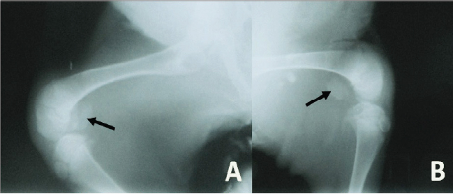
Fig. 3. Mediolateral radiograph of the right (A) and left (B) stifle of a 3-month-old goat with bilateral PLs. The radiograph shows severely ventrocaudally luxated patellae (arrows).
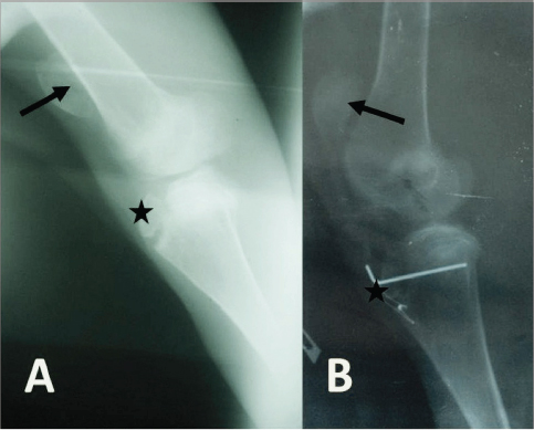
Fig. 4. (A) Mediolateral radiograph of the right stifle of a 10-month-old goat showing a dorsal PL (arrow) with no signs of trochlear ridge hypoplasia. There is also an avulsion fracture of the tibial tuberosity (star). (B) Mediolateral radiograph of the right stifle of the same goat immediately postsurgery showing that the patella (arrow) and the tibial tuberosity (star) are placed almost at their normal anatomical locations. Among the presented PL cases, surgical intervention was possible with two cases with diverse outcomes. While the surgery was beneficial to one case with a complete correction of luxation and normal leg movement for a long period after surgery, it was harder and with different results in the other. The younger age with more flaccidity of the anatomical structures may be attributed to the good outcome in the former case as compared with the heavier weight and denser and harder anatomical structures of the latter mature buck. In line with these findings, postsurgical PL outcome was also variable in the literature. Surgical intervention of PL in sheep showed a successful correction in all cases, but persistent femeropatellar instability was reported by owners (Shettko and Trostle, 2000). In addition, in dogs, it has been reported that in medial PL cases, almost half of the repaired cases had persistent palpable PL (Willauer and Vasseur, 1987; Di Dona et al., 2018), and the frequency of postoperative complications represented 18% of treated dogs (Arthurs and Langley-Hobbs, 2006). Furthermore, the higher grade of PL and heavier body weight were accountable for the higher percentage of postoperative complications in the latter report (Arthurs and Langley-Hobbs, 2006). ConclusionThe present report highlights the clinical presentation and the surgical outcome of PL in Hejazi goats. This disorder could be attributed to a genetic basis resulting from inbreeding practice, considering that this disorder was only found in this breed but not in local goats. PL was always lateral with either ventrocaudal or dorsal directions. The surgical intervention was with variable results but with a probable favorable prognosis in younger goats. Conflict of interestThe authors declare that there is no conflict of interest. Authors’ contributionMohamed Abushhiwa, Adulrhman Alrtib, Ataher Almashregi, and Emad Bennour performed the clinical examination and surgical interventions. Mansur Shmela and Mouna Abdunnabi contributed in the genetic and breeding interpretation of findings. All authors contributed equally in the manuscript writing. AcknowledgmentThe authors thank the staff of Al-Sorouh veterinary clinic and the Department of Surgery and Theriogenoloy for their cooperation. ReferencesAbuja, G.A., Kowaleski, M.P. and García-López, J.M. 2014. Management of bilateral patellar luxation in an alpaca. Vet. Surg. 43, 459–464. Amills, M., Capote, J. and Tosser-Klopp, G. 2017. Goat domestication and breeding: a jigsaw of historical, biological and molecular data with missing pieces. Anim. Genet. 48, 631–644. Arighi, M. and Wilson, J.W. 1993. Surgical correction of medial luxation of the patella in a Miniature Horse. Can. Vet. J. 34, 499–501. Arthurs, G.I. and Langley-Hobbs, S.J. 2006. Complications associated with corrective surgery for patellar luxation in 109 dogs. Vet. Surg. 35, 559–566. Baron, R. 1987. Laterally luxating patella in a goat. J. Am. Vet. Med. Assoc. 191, 1471–1472. Burnei, G., Răducan, I., Lală, C., Klinaku, I., Marti, T. and Burnei, C. 2020. Patellar dislocation: etiopathogenic diagnosis and treatment methods. Clin. Surg. 5, 1–8. Cecchi, F., Vannucchi, I., Carlini, G. and Macchioni, F. 2020. Inbreeding, phenotypic traits, coat colours and prevalence of health problems in a population of English Cocker Spaniels: the first survey in Italy. Rend. Fis. Acc. Lincei. 31, 873–880. Di Dona, F., Della Valle, G. and Fatone, G. 2018. Patellar luxation in dogs. Vet. Med. (Auckl). 9, 23–32. Dyce, K.M., Sack, W.O. and Wensing, C.J.G. 2009. Textbook of veterinary anatomy-E-book. London, UK: Elsevier Health Sciences. Fathi, N., Elbakary, R.A., Karkoura, A.A., El-Gendy, S.A. and Abumandour, M.A. 2016. Advanced morphological and radiological studies on the stifle joint of Egyptian Baladi goat (Capra hircus). AJVS. 51, 199–210. Fossum, T.W., Fossum, T.W., Duprey, L.P. and O’Connor, D. 2007. Small animal surgery. St. Louis, MO: Mosby Elsevier St Louis. Fowler, M.E. 1998. Medicine and surgery of South American camelids: llama, alpaca, vicuna, guanaco. Iowa City, IA: Iowa State University Press. Frandson, R.D., Lee Wilke, W. and Dee Fails, A. 2009. Anatomy and physiology of farm animals. Hoboken, NJ: Wiley-Blackwell: A John Wiley & Sons. Gangwar, A.K., Devi, K.S., Singh, A.K., Katiyar, N., Patel, G. and Srivastava, S. 2014. Congenital anomalies and their surgical correction in ruminants. Adv. Anim. Vet. Sci. 2, 369–376. Gao, S., Liu, X., Zhang, F., Gao, T., Zhang, Z. and Dai, M. 2018. A novel technique of using a miniature plate in combination with tension band wiring to treat comminuted patellar fractures. Medicine 97, 1–5. Hermans, W., Kersjes, A., Van Der Mey, G. and Dik, K. 1987. Investigation into the heredity of congenital lateral patellar (sub) luxation in the Shetland pony. Vet. Q. 9, 1–8. Kaneps, A., Riebold, T., Schmotzer, W., Watrous, B. and Huber, M. 1989. Surgical correction of congenital medial patellar luxation in a llama. J. Am. Vet. Med. Assoc. 194, 547–548. Kobluk, C.N. 1993. Correction of patellar luxation by recession sulcoplasty in three foals. Vet. Surg. 22, 298–300. Lavrijsen, I., Heuven, H., Breur, G., Leegwater, P., Meutstege, F. and Hazewinkel, H. 2013. Phenotypic and genetic trends of patellar luxation in D utch F lat-C oated R etrievers. Anim. Genet. 44, 736–741. Lavrijsen, I.C., Leegwater, P.A., Wangdee, C., van Steenbeek, F.G., Schwencke, M., Breur, G.J., Meutstege, F.J., Nijman, I.J., Cuppen, E. and Heuven, H.C. 2014. Genome-wide survey indicates involvement of loci on canine chromosomes 7 and 31 in patellar luxation in flat-coated retrievers. BMC Genet. 15, 1–9. Mahmoud, A.H., Farah, M.A., Rady, A., Alanazi, K.M., Mohammed, O., Amor, N. and Alarjani, K.M. 2020. Molecular characterization of goats from Saudi Arabia using microsatellite markers. J. King Saud. Univ. Sci. 32, 1681–1686. Mostafa, A.A., Griffon, D.J., Thomas, M.W. and Constable, P.D. 2008. Proximodistal alignment of the canine patella: radiographic evaluation and association with medial and lateral patellar luxation. Vet. Surg. 37, 201–211. O’Neill, D.G., Meeson, R.L., Sheridan, A., Church, D.B. and Brodbelt, D.C. 2016. The epidemiology of patellar luxation in dogs attending primary-care veterinary practices in England. Canine Genet. Epidemiol. 3, 1–12. Roush, J.K. 1993. Canine patellar luxation. Vet. Clin. North Am. Small Anim. Pract. 23, 855–868. Sanders, T.L. Pareek, A., Hewett, T.E., Stuart, M.J., Dahm, D.L. and Krych, A.J. 2018. High rate of recurrent patellar dislocation in skeletally immature patients: a long-term population-based study. Knee Surg. Sports Traumatol. Arthrosc. 26, 1037–1043. Shettko, D.L. and Trostle, S.S. 2000. Diagnosis and surgical repair of patellar luxations in a flock of sheep. J. Am. Vet. Med. Assoc. 216, 564–566. Solanti, S., Laitinen, O. and Atroshi, F. 2002. Hereditary and clinical characteristics of lateral luxation of the superficial digital flexor tendon in Shetland sheepdogs. Vet. Ther. 3, 97–103. Stanke, N.J., Stephenson, N. and Hayashi, K. 2014. Retrospective risk factor assessment for complication following tibial tuberosity transposition in 137 canine stifles with medial patellar luxation. Can. Vet. J. 55, 349. Sundberg, J. and Nielsen, S. 1981. Deer fibroma: a review. Can. Vet. J. 22, 385. Van Hoogmoed, L., Snyder, J. and Vasseur, P. 1998. Surgical repair of patellar luxation in llamas: 7 cases (1980–1996). J. Am. Vet. Med. Assoc. 212, 860–865. Vidoni, B., Sommerfeld-Stur, I. and Eisenmenger, E. 2006. Diagnostic and genetic aspects of patellar luxation in small and miniature breed dogs in Austria. EJCAP 16, 149–158. Willauer, C. and Vasseur, P. 1987. Clinical results of surgical correction of medial luxation of the patella in dogs. Vet. Surg. 16, 31–36. Zanders, S. 2014. Patellar luxation-a genetic study. M. S. thesis, Swedish University of Agricultural Sciences, Uppsala, Sweden. | ||
| How to Cite this Article |
| Pubmed Style MHA, AMA, TNE, MAA, Shmela ME, EMB, . Patellar Luxation in Hejazi Goats. Open Vet J. 2021; 11(2): 295-300. doi:10.5455/OVJ.2021.v11.i2.14 Web Style MHA, AMA, TNE, MAA, Shmela ME, EMB, . Patellar Luxation in Hejazi Goats. https://www.openveterinaryjournal.com/?mno=68365 [Access: July 27, 2024]. doi:10.5455/OVJ.2021.v11.i2.14 AMA (American Medical Association) Style MHA, AMA, TNE, MAA, Shmela ME, EMB, . Patellar Luxation in Hejazi Goats. Open Vet J. 2021; 11(2): 295-300. doi:10.5455/OVJ.2021.v11.i2.14 Vancouver/ICMJE Style MHA, AMA, TNE, MAA, Shmela ME, EMB, . Patellar Luxation in Hejazi Goats. Open Vet J. (2021), [cited July 27, 2024]; 11(2): 295-300. doi:10.5455/OVJ.2021.v11.i2.14 Harvard Style , M. H. A., , A. M. A., , T. N. E., , M. A. A., Shmela, M. E., , E. M. B. & (2021) Patellar Luxation in Hejazi Goats. Open Vet J, 11 (2), 295-300. doi:10.5455/OVJ.2021.v11.i2.14 Turabian Style , Mohamed H. Abushhiwa, Adulrhman M. Alrtib, Taher N. Elmeshregh, Mouna A Abdunnabi, Mansur E. Shmela, Emad M Bennour, and . 2021. Patellar Luxation in Hejazi Goats. Open Veterinary Journal, 11 (2), 295-300. doi:10.5455/OVJ.2021.v11.i2.14 Chicago Style , Mohamed H. Abushhiwa, Adulrhman M. Alrtib, Taher N. Elmeshregh, Mouna A Abdunnabi, Mansur E. Shmela, Emad M Bennour, and . "Patellar Luxation in Hejazi Goats." Open Veterinary Journal 11 (2021), 295-300. doi:10.5455/OVJ.2021.v11.i2.14 MLA (The Modern Language Association) Style , Mohamed H. Abushhiwa, Adulrhman M. Alrtib, Taher N. Elmeshregh, Mouna A Abdunnabi, Mansur E. Shmela, Emad M Bennour, and . "Patellar Luxation in Hejazi Goats." Open Veterinary Journal 11.2 (2021), 295-300. Print. doi:10.5455/OVJ.2021.v11.i2.14 APA (American Psychological Association) Style , M. H. A., , A. M. A., , T. N. E., , M. A. A., Shmela, M. E., , E. M. B. & (2021) Patellar Luxation in Hejazi Goats. Open Veterinary Journal, 11 (2), 295-300. doi:10.5455/OVJ.2021.v11.i2.14 |

