


| Case Report | ||
Open Vet J. 2021; 11(3): 379-384 Open Veterinary Journal, (2021), Vol. 11(3): 379–384 Case Report Surgical management of bilateral ear pinna lesions associated with traumatic aural hematoma in a 3-day-old goat kidPanagiotis D. Katsoulos1* and Anna Dedousi21Clinic of Farm Animals, School of Veterinary Medicine, Aristotle University of Thessaloniki, Thessaloniki, Greece 2Veterinary Research Institute, Hellenic Agricultural Organization DIMITRA, Thermi, Thessaloniki, Greece *Corresponding Author: Panagiotis D. Katsoulos. Clinic of Farm Animals, School of Veterinary Medicine, Aristotle University of Thessaloniki, Thessaloniki, Greece. Email: katsoulo [at] vet.auth.gr Submitted: 07/04/2021 Accepted: 02/07/2021 Published: 25/07/2021 © 2021 Open Veterinary Journal
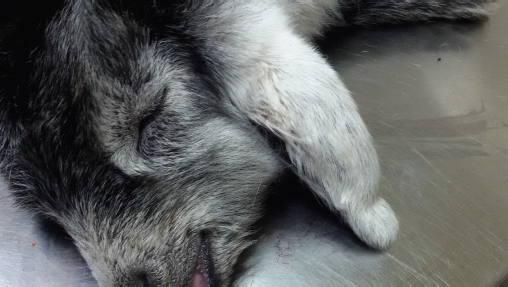
AbstractBackground: Aural hematomas are not uncommon in ruminants’ clinical practice; however, there is a lack of information regarding their management in newborn ruminants, especially for complicated cases with rupture of the hematoma and secondary ear pinna necrosis. Case Description: A 3-day-old orphan goat kid was admitted due to swelling on the left ear pinna and trauma on the right pina caused by biting by other goats. The swelling on the left ear which was located at the convex surface was soft, painless, and fluid-filled, suggestive of aural hematoma located at the convex surface. The right pinna was swollen, bleeding, and extremely painful at palpation. The skin was necrotized at the distal 2/3rd of the convex surface and the 1/2 of the concave surface. Underneath the necrotized skin of the convex surface, blood, and blood clots were trapped, and there was a pocket between the remaining normal skin and the cartilage indicating possible rupture of aural hematoma. The kid was surgically treated under general anesthesia with xylazine and ketamine. The aural hematoma was drained by the convex surface using a Penrose tube after flushing the cavity with 2 mg dexamethasone. The trauma of the right pinna was left to heal by secondary intention after resection of all necrotized, edematous tissues, and blood clots. Post-surgery, the animal was treated with parenteral antibiotic administration and daily application of a topical antiseptic solution. The Penrose tube was removed after 5 days, and the animal recovered uneventfully. Conclusions: The present case indicates that aural hematoma can occur in newborn goat kids secondary to ear pinna biting and might evolve to pinna necrosis. In addition, tube drainage after flushing the cavity with corticosteroids appears to be an effective treatment approach without requiring bandaging post-operatively. Keywords: Aural hematoma, Ear pinna necrosis, Goat kid, Penrose drainage, Surgery. IntroductionAural or auricular hematoma is characterized by blood accumulation between the pinnae’s dermal surface and the underlying perichondrium (Lanz and Wood, 2004; Macphail, 2016) or within the scaphal cartilage (Pavletic, 2015). Typically, it is located at the concave surface of the pinna and is usually presented as a fluctuant swelling. Aural hematomas have been described in detail in dogs and cats. They commonly result from self-induced traumas due to head shaking or scratching secondary to otitis externa (Pavletic, 2015; Macphail, 2016). Although aural hematomas are not uncommon in clinical practice, few detailed reports in ruminants have been reported (Tsioli et al., 2013; Dewangan et al., 2016; Lamani et al., 2019). Similar to dogs and cats, the cases resulted from self-induced traumas; intense head shaking because of sarcoptic mange (Tsioli et al., 2013) or ticks (Lamani et al., 2019) in sheep cases, and scratching secondary to ear injury of thorny bush in a goat case (Dewangan et al., 2016). Based on these reports, the most effective treatment option for aural hematomas in ruminants is incisional drainage. Although tube drainage is a common treatment option for aural hematomas in dogs and cats (Hall et al., 2016), this technique was associated with adverse post-operative complications when it was applied in sheep based on a report (Tsioli et al., 2013). According to the authors’ knowledge, there is a lack of information in the available literature regarding their management in newborn ruminants, especially for complicated cases with rupture of the hematoma and secondary ear pinna necrosis. The objective of the present report was to describe the surgical management of traumatic lesions in both ear pinnae, associated with an aural hematoma on the convex surface of the left ear and necrosis on the right one, which was attributed to a ruptured hematoma, in a 3-day-old goat kid. The hematoma was successfully treated by tube drainage technique from the convex surface without post-surgical bandaging and the necrosis by secondary healing after removing the necrotic tissues. Case DetailsA 3-day-old mixed breed female goat kid was admitted due to swelling on the left ear pinna and wound on the right pinna. According to the owner, the kid’s mother died 3 hours after parturition, and the kid was fed by nourishing from other goats after that. The lesion on the left ear pinna was the first sign observed on the day of admission. After closer inspection, the trauma at the other ear pinna was also detected. Regarding the possible cause of this condition, the owner claimed that the goats who refused to accept this kid were biting its ears to discourage it from nursing. At the clinical examination, the kid was 3.6 kg, moderately depressed, had rectal temperature 38.2°C, heart and respiratory rate at 120 beats/minutes and 30 breaths/minutes, respectively, and normal color of mucus membranes. The auscultation of the heart and lungs did not reveal anything abnormal, and there was no ocular or nasal discharge. Blood glucose was 76 mg/dl as determined by a portal glucometer (Precision Xceed®; Abbott, Abbott Diabetes Care Ltd., Witney, UK) The clinical examination of the left ear revealed a soft, painless, and fluid-filled swelling at the convex surface (Fig. 1), and no sign of otitis was detected after the otoscopic examination. Based on these findings, a diagnosis of auricular hematoma was made. The right pinna was bleeding, was swollen, and extremely painful at palpation. After clipping of the hair, it was observed that the skin was necrotized at the distal 2/3 of the convex surface (Fig. 2) and the 1/2 of the concave surface. Underneath the necrotized skin of the convex surface, blood and blood clots were trapped. It was also observed that there was a pocket between the remaining normal skin and the cartilage at the same surface. The kid was anaesthetized with xylazine hydrochloride (Xylazine, Neocell, Hellas) at the dose of 0.1 mg/kg and ketamine hydrochloride (Imalgene 1000, Merial, France) at the dose of 4 mg/kg body weight intramuscularly. Before the onset of surgical procedures, meloxicam (0.5 mg/kg BW SC; Melovem 5 mg/ml, Dophrma, The Netherlands) and lincomycin-streptomycin (LINCO-SPECTIN® Pharmacia & Upjohn Company, USA; dosage: 1 ml/10 kg BW IM) was administered. Throughout the procedures and until the recovery from anesthesia, the kid was kept on an electric heat pad covered with double layer cotton surgical drapes and received IV 2.5% dextrose added in Lactated Ringers solution at the rate of 10 ml/kg/hours. Hemoglobin oxygen saturation (SpO2) and heart rate were monitored using a pulse oximetry monitor (HS-10 Veterinary Handheld Pulse oximeter, Hunan Accurate Bio-Medical Co., Ltd.) with the reflective probe placed on the tongue and rectal temperature was measured every 15 minutes.
Fig. 1. Swelling at the convex surface of the left ear pinna at a 3-day-old goat kid with aural hematoma.
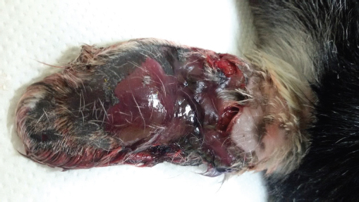
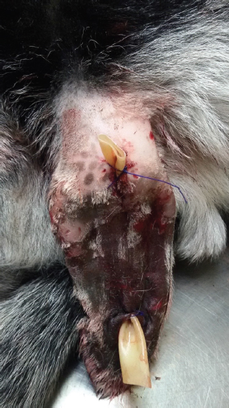
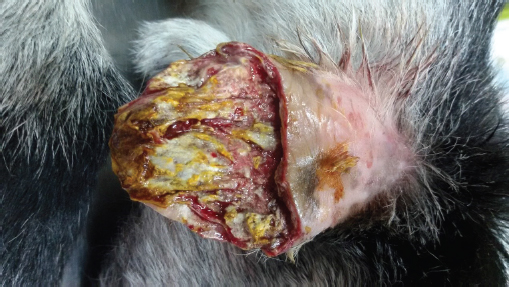
Fig. 2. Skin necrosis at the distal 2/3 of the convex surface of the right ear pinna of the 3-day-old goat kid of Figure 1. After clipping the hair of the left pinna, it was observed that the skin of the convex surface was bruised; the epidermis was easily detached at some parts, but the skin underneath was intact (Fig. 3). The convex surface of the pinna was surgically prepared, and the hematoma was drained by using a Penrose tube. At first, an incision was made at the distal border of the hematoma using a scalpel with a No 10 surgical blade. After removing the hematoma content, the cavity was flushed with 1ml of dexamethasone 2 mg/ml solution diluted in 4 ml of normal saline. After that, a Pean forceps was inserted in the cavity until the proximal border of the hematoma, and a second incision was made. A Penrose drain (Kendall Tyco, Mansfield, MA, USA) (6 mm) was inserted from the proximal incision through the cavity and was exited from the distal one (Fig. 3). The drain was sutured to the skin with two simple interrupted sutures using absorbable sutures (PGA 2/0; Medipac, Greece). The right ear pinna was surgically prepared, and all necrotized, edematous tissues and blood clots were removed. After a thorough inspection, it was observed that the pocked was extended to the base of the ear pinna. In addition, the ear cartilage was thickened throughout. Therefore, it was decided not to resect the pinna completely but to be left to heal by secondary intention (Fig. 4). All the procedures were completed within 30 minutes, and the kid was able to stand within 1 hour after the administration of anesthesia. After the complete restoration of suckling reflex, the kid was bottle fed with 200 ml of warmed (39oC) goat milk. When the animal was released, 2 hours after standing, the rectal temperature was 39.2oC and blood glucose was 98 mg/dl. Postoperative treatment consisted of: (a) intramuscular administration of lincomycin-spectinomycin once per day for 5 days, (b) twice a day lavage of the right pinna with a chlorhexidine solution 1% until granulation, and (c) daily cleansing of the incision sites of the left pinna with chlorhexidine solution 1%. It was suggested the Penrose drain be removed after 5 days. It was also advised that the kid be kept separated from the rest of the flock and be bottle-fed 3–4 times per day.
Fig. 3. The left ear pinna of the goat kid of Figure 1 after drainage of the aural hematoma with a Penrose tube. Two months after surgery, the kid was re-evaluated. It was observed that the left pinna had complete hair coverage without any malformations except from two small scars at the incision sites (Fig. 5). The right pinna was healed completely and the tip was covered by scar tissue (Fig. 6). According to the farmer, the recovery was uneventful; the drain was removed after 5 days, and the right pinna was healed within approximately 3 weeks. Written informed consent for the publication of the clinical details and images of this case was obtained from the farmer. DiscussionAuricular hematomas and necrosis of the ear pinna are rare in small newborn ruminants in clinical practice. To the authors’ knowledge, there is no relevant reference in the available literature. According to the farmer, this condition was caused due to biting from adult goats; the observed skin necrosis of the right pinna and the necrosis of the epidermis of the left one are both consistent with such traumas.
Fig. 4. Post-operative picture of the right ear pinna of the goat kid of Figure 2 after the resection of necrotizing tissues.
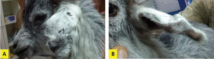
Fig. 5. Lateral (A) and frontal (B) view of the left ear pinna of the goat kid of Figure 1 2 months after surgery.
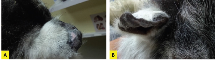
Fig. 6. Lateral (A) and frontal (B) view of the right ear pinna of the goat kid of Figure 2 2 months after surgery. At admission, the kid was generally in good condition. Apart from the observed lesions at both ears, the only remarkable findings at the clinical examination were moderate depression and mild hypothermia. The latter is attributed to the low blood glucose concentration (Abdolvahabi et al., 2016), probably because of reduced appetite. On the other hand, depression could be possibly associated with pain and hypoglycemia. In order to confront with the pre-existing hypoglycemia—hypothermia and to prevent further worsening of this condition during anesthesia, it was selected to use a heat pad, to cover the kid with double layer cotton surgical drapes and to administer 2.5% dextrose solution intravenously (Abrahamsen, 2008). The anesthetic protocol used in this case provided an adequate depth of anesthesia throughout the followed procedures, and the recovery was completed without complications. According to Abrahamsen (2008), the selected combination of anesthetics generally produces 30–40 minutes of recumbency. However, the recumbency in this goat kid lasted longer, probably because of the pre-existing depression and the administration of the highest suggested dosage of xylazine for this protocol (Abrahamsen, 2008). Based on the asymmetry observed between the medial and the lateral surface of the ear pinna and the more intense fluctuation at the convex surface, we concluded that the fluid-filled cave was formatted between the cartilage and the skin of the convex surface. This contrasts with the typical site for dogs (Macphail, 2016) and the former reports for ruminants where the blood accumulation was observed in the concave surface (Tsioli et al., 2013; Dewangan et al., 2016). However, the initial cause of the hematoma in the present case was not a self-induced trauma caused by head shaking (Tsioli et al., 2013; Dewangan et al., 2016) but exogenous injuries, which occurred at the outer surface of the ear pinna. Based on the hematoma location, it was decided to drain the hematoma from the lateral surface of the pinna, despite the recommended access from the concave one (Macphail, 2016). The placement of Penrose drains is a very common treatment option for aural hematomas in dogs and cats (Hall et al., 2016). It was selected in the present case because it is a minimally invasive technique and cost-effective for a farm animal. There is only one report in the available literature in small ruminants using Penrose tube drainage (Tsioli et al., 2013); however, in that case, inflammation was developed postoperatively, and the animal underwent a second surgical intervention using incisional drainage. Before the placement of the Penrose drain in the present case, the cavity was flushed with dexamethasone solution of 0.4%. The contribution of dexamethasone flushing at the positive outcome of this case remains questionable since there is no other similar report in ruminants. However, evidence in the literature for dogs indicates that injection of corticosteroids directly into the hematoma cavity following drainage is associated with successful resolution in more than 90% of cases (Kuwahara, 1986; Romatowski, 1994; Seibert and Tobias, 2013). Despite the recommendation for bandaging after the tube drainage technique to avoid subsequent infection (Tsioli et al., 2013), it was selected not to use a head bandage. This decision was based on the fact that the skin at the convex surface was not intact and since biting was the cause of the lesion, it was supposed that there would be an increased risk for anaerobic bacteria overgrowth after bandaging. Besides, it was possible for the farmer to keep the kid separate from the rest of the flock in a clean pen and to apply the topical antiseptic twice a day, so the risk of subsequent infection was minimized. The trauma detected at the right ear pinna and especially the remaining pocket under the intact skin cannot exclude the possibility that the initial lesion was also an auricular hematoma. It could be hypothesized that the repeated biting of the pinna resulted in skin necrosis and finally, the rupture of the hematoma. The thickening of the ear cartilage denotes the presence of inflammation. The origin of this inflammation is unknown; it is believed that it resulted from mechanical irritation due to biting and/or bacterial infection. Due to the presence of inflammation, there were two treatment options. The first one was secondary intention healing, and the second one the complete resection of the ear pinna. It was decided to follow the first option since the pocket and the thickening of the cartilage were very extended and made doubtful the presence of healthy tissue at the pinna base. In addition, the second option would increase the time required for anesthesia. Nevertheless, the healing was successful with forming a relatively small scab as expected due to skin loss at the tip of the remained pinna. Post-surgery it was suggested, apart from the application of a topical antiseptic in both pinnae, the kid to be treated with a lincomycin-spectinomycin. This combination of antibiotics was selected mainly because of lincomycin which is effective against Gram-positive cocci and most anaerobes (Pechere, 1986) and is widely used to treat bacterial skin infections (Noble, 1990). ConclusionsThe present case indicates that aural hematoma can occur in newborn goat kids secondary to ear pinna biting and might evolve to pinna necrosis. In addition, tube drainage after flushing the cavity with corticosteroids appears to be an effective treatment approach without requiring bandaging post-operatively. Authors’ contributionPDK: case evaluation, surgical management, manuscript drafting, approval of the final manuscript; AD: case evaluation, anesthesia administration and monitoring, critical revisions, and approval of the final manuscript. Conflict of interestThe authors declare that there is no conflict of interest ReferencesAbdolvahabi, S., Zaeemi, M., Mohri, M. and Naserian, A.A. 2016. Age related changes in serum biochemical profile of Saanen goat kids during the first three months of life. Rev. Med. Vet. 167, 106–112. Abrahamsen, E.J. 2008. Ruminant field anesthesia. Vet. Clin. North Am. Food Anim. Pract. 24, 429–441. Dewangan, R., Sharda, R. and Kalim, M.O. 2016. Surgical management of extensive aural haematoma in a jamunapari goat. Int. J. Sci. Environ. Technol. 5, 2221–2225. Hall, J., Weir, S. and Ladlow, J. 2016. Treatment of canine aural haematoma by UK veterinarians. J. Small Anim. Pract. 57, 360–364. Kuwahara, J. 1986. Canine and feline aural hematoma: clinical, experimental, and clinicopathologic observations. Am. J. Vet. Res. 47, 2300–2308. Lamani, T., Kamalakar, G., Sunil, C.L., Srinivasa Murthy, K.M. and Nagaraja, B.N. 2019. Surgical management of aural haematoma resultant of tick infestation in a non-descript sheep – a case report. Int. J. Livest. Res. 9, 224–227. Lanz, O.I. and Wood, B.C. 2004. Surgery of the ear and pinna. Vet. Clin. North Am. Small Anim. Pract. 34, 567–599. Macphail, C. 2016. Current treatment options for auricular hematomas. Vet. Clin. North Am. Small Anim. Pract. 46, 635–641. Noble, W.C. 1990. Topical and systemic antibiotics: is there a rationale? Semin. Dermatol. 9, 250–254. Pavletic, M.M. 2015. Use of laterally placed vacuum drains for management of aural hematomas in five dogs. J. Am. Vet. Med. Assoc. 246, 112–117. Pechere, J.C. 1986. Lincosamides. Pathol. Biol. (Paris) 34, 119–128. Romatowski, J. 1994. Nonsurgical treatment of aural hematomas. J. Am. Vet. Med. Assoc. 204(9), 1318. Seibert, R. and Tobias, K.M. 2013. Surgical treatment for aural hematoma. NAVC Clinician’s Brief 3, 29–32. Tsioli, V., Farmaki, R., Papastefanou, A., Galatos, A.D., Marinou, M., Tontis, D., Mavrogianni, V.S., Doukas, D., Saridomichelakis, M.N. and Fthenakis, G.C. 2013. A case of bilateral auricular haematoma in a ewe-lamb with sarcoptic mange. Small Rum. Res. 110, 145–149. | ||
| How to Cite this Article |
| Pubmed Style Katsoulos PD, Dedousi A. Surgical management of bilateral ear pinna lesions associated with traumatic aural hematoma in a three-days-old goat kid. Open Vet J. 2021; 11(3): 379-384. doi:10.5455/OVJ.2021.v11.i3.7 Web Style Katsoulos PD, Dedousi A. Surgical management of bilateral ear pinna lesions associated with traumatic aural hematoma in a three-days-old goat kid. https://www.openveterinaryjournal.com/?mno=71459 [Access: October 06, 2024]. doi:10.5455/OVJ.2021.v11.i3.7 AMA (American Medical Association) Style Katsoulos PD, Dedousi A. Surgical management of bilateral ear pinna lesions associated with traumatic aural hematoma in a three-days-old goat kid. Open Vet J. 2021; 11(3): 379-384. doi:10.5455/OVJ.2021.v11.i3.7 Vancouver/ICMJE Style Katsoulos PD, Dedousi A. Surgical management of bilateral ear pinna lesions associated with traumatic aural hematoma in a three-days-old goat kid. Open Vet J. (2021), [cited October 06, 2024]; 11(3): 379-384. doi:10.5455/OVJ.2021.v11.i3.7 Harvard Style Katsoulos, P. D. & Dedousi, . A. (2021) Surgical management of bilateral ear pinna lesions associated with traumatic aural hematoma in a three-days-old goat kid. Open Vet J, 11 (3), 379-384. doi:10.5455/OVJ.2021.v11.i3.7 Turabian Style Katsoulos, Panagiotis Dimitrios, and Anna Dedousi. 2021. Surgical management of bilateral ear pinna lesions associated with traumatic aural hematoma in a three-days-old goat kid. Open Veterinary Journal, 11 (3), 379-384. doi:10.5455/OVJ.2021.v11.i3.7 Chicago Style Katsoulos, Panagiotis Dimitrios, and Anna Dedousi. "Surgical management of bilateral ear pinna lesions associated with traumatic aural hematoma in a three-days-old goat kid." Open Veterinary Journal 11 (2021), 379-384. doi:10.5455/OVJ.2021.v11.i3.7 MLA (The Modern Language Association) Style Katsoulos, Panagiotis Dimitrios, and Anna Dedousi. "Surgical management of bilateral ear pinna lesions associated with traumatic aural hematoma in a three-days-old goat kid." Open Veterinary Journal 11.3 (2021), 379-384. Print. doi:10.5455/OVJ.2021.v11.i3.7 APA (American Psychological Association) Style Katsoulos, P. D. & Dedousi, . A. (2021) Surgical management of bilateral ear pinna lesions associated with traumatic aural hematoma in a three-days-old goat kid. Open Veterinary Journal, 11 (3), 379-384. doi:10.5455/OVJ.2021.v11.i3.7 |

