| Original Article | ||
Open Vet J. 2022; 12(6): 806-814 Open Veterinary Journal, (2022), Vol. 12(6): 806–814 Original Research Common diseases of sheep (Ovis aries Linnaeus) and goats (Capra aegagrus hircus) in Jordan: A retrospective study (2015–2021)Myassar Alekish* and Zuhair Bani Ismail 1Department of Veterinary Clinical Sciences, Jordan University of Science and Technology, Irbid, Jordan Submitted: 17/07/2022 Accepted: 11/10/2022 Published: 08/11/2022 *Corresponding Author: Myassar Alekish. Department of Veterinary Clinical Sciences, Jordan University of Science and Technology, Irbid, Jordan. Email: moalekish [at] just.edu.jo © 2022 Open Veterinary Journal
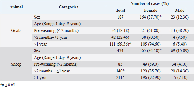
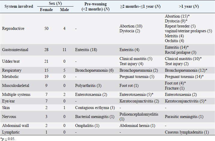
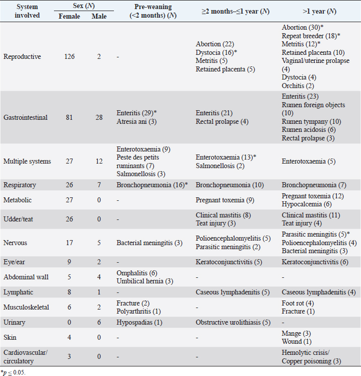
AbstractBackground: Despite major efforts that have been undertaken to improve livestock health and productivity in Jordan, infectious and non-infectious diseases continue to cause significant economic losses. Aim: The objective of this study was to report the most common diseases (infectious and non-infectious) affecting sheep (Ovis aries Linnaeus) and goats (Capra aegagrus hircus) in Jordan. Methods: Data related to sheep and goats presented for clinical evaluation to the Veterinary Health Center of the Faculty of Veterinary Medicine at Jordan University of Science and Technology between January 2015 and December 2021 extracted from the case medical records were used in this study. The data were entered into Microsoft Excel spreadsheets and descriptive analysis was performed to report the frequencies, averages, and range values. The data were categorized according to sex (female vs male), body system involved in the disease process, nature of the disease process (infectious vs non-infectious), and age [pre-weaning (less than 2 months of age), 2 months to 1 year, and older than 1 year]. Significant differences between different groups were determined using an independent t-test. Results: Medical records of 187 goats and 434 sheep were included in the analysis of this study. Females were significantly more represented in the study population for goats and sheep, 87.70% and 84.10%, respectively. The age of animals ranged between 1 day and 9 years in goats and 1 day and 7 years in sheep. In both goats and sheep, a significant number of cases (p ≤ 0.05) were presented with reproductive (28.42% and 29.49%, respectively) and gastrointestinal diseases (20.52% and 25.11%, respectively). In goats, other disease diagnoses were involving the respiratory (10.52%), udder/teat (11.05%), and metabolic systems (10.00%). In sheep, other disease diagnoses were involving multiple systems (8.98%), respiratory (7.60%), metabolic (6.22%), udder/teat (5.99%), and the nervous system (5.06%). Conclusion: Results of this study provide a list of the most likely differential diagnoses in different age groups of both sexes in goats and sheep in Jordan. This information could be used by veterinarians as well as policymakers in order to formulate and implement appropriate and effective preventive and control measures against common diseases in goats and sheep. Keywords: Small ruminants, Disease diagnosis, Animal health and production, Animal welfare. IntroductionThe number of small ruminants in Jordan is estimated to be around 3 million head of sheep and 1 million head of goats (Department of Statistics, 2020). The value of sheep and goat industries is approximately 300 million JD (428.5 million USD), which amounts to about 30% of the total livestock production in the country (Department of Statistics, 2020). The status of livestock health not only reflects the socioeconomic status of the community but also the level of animal welfare and animal husbandry standards (Rich and Perry, 2011). During the last decade, major strides have been undertaken to improve livestock productivity through the implementation of health improvement measures such as vaccination campaigns, providing subsidized good quality dietary supplements, and farm biosecurity. However, poor management, inconsistent sources of high-quality feed supplies, and diseases continue to cause dramatic losses (Food and Agriculture Organization of the United Nations, 2021). In Jordan, there are few published reports of a number of important infectious diseases in sheep and goats such as mastitis (Hawari et al., 2014), foot and mouth disease (Ababneh et al., 2020), brucellosis (Musallam et al., 2015), sheep and goat pox (Hailat et al., 1994), and peste des petits ruminants (Al-Majali et al., 2008). However, scarce information could be cited regarding the incidence and epidemiology of non-infectious and management-related diseases. Therefore, this retrospective study was conducted to report the prevalence and distribution of infectious and non-infectious diseases in sheep and goats according to age, sex, and affected body system (2015–2021). The results of this study will provide a better understanding of the most common health problems in small ruminants as well as the status of animal management and husbandry practices in Jordan. Materials and MethodsStudy area and study populationCase medical records of all sheep and goats presented for clinical evaluation to the Veterinary Health Center (VHC) of the Faculty of Veterinary Medicine at Jordan University of Science and Technology between January 2015 and December 2021 were used in this study. The VHC is a mixed animal outpatient practice located in Northern Jordan that provides specialized veterinary care to various animal species within a 50-km radius. Jordan is located in the center of the Middle East between Lat. 29° 30ʹ and 32° 31ʹ. The Northern regions are mountainous in nature with an average annual rainfall of 400–600 mm (Al-Halabi, 2019). Sheep and goats in this region of Jordan typically are raised in traditional farming conditions. Animals are allowed to graze on available grasses and fodder during the spring but are maintained on grain-based concentrate dietary supplements for the rest of the year. Case selection and data collectionTo be included in the study, the case medical record must have been completed with the basic patient identification data including animal species, breed, age, and sex, and medical data including the presenting complaint, pertinent history relevant to the farm and diseased individual animal, physical examination findings, laboratory findings, clinical diagnosis, and medical or surgical treatment performed on the animal. Case medical records with missing data were excluded from the analysis. Systemic viral and septicemic bacterial infections were classified as conditions affecting multiple systems in this study. Animals that were presented with clinical signs suggestive of a respiratory infection such as fever, coughing, nasal or ocular mucopurulent discharge, and abnormal pulmonary sounds on auscultation with no other abnormalities involving any other body system were diagnosed with bronchopneumonia regardless of the possible etiologic nature (bacterial, viral, or parasitic). Although the bacterial culture of milk from cases of clinical mastitis cases is routinely performed, a specific bacterial etiologic diagnosis will not be provided here and the term “bacterial mastitis” will be used for all cases of mastitis reported in this study. The frequencies of specific disease diagnoses were calculated by dividing the number of animals diagnosed with the disease by the total number of cases in the involved system. Data analysisDescriptive analysis was performed to report the frequencies, averages, and range values using Excel spreadsheets (Microsoft Office 10, Microsoft Co., USA). The data were then categorized based on animal species into two different groups (sheep and goats). For each animal species, the data were further categorized based on sex (female vs male), body system involved in the disease process, and nature of the disease process (infectious vs non-infectious) and age [pre-weaning (less than 2 months), 2 months to 1 year, and older than 1 year]. Significant differences between different groups were determined using an independent t-test. Statistical analysis was performed using SPSS software version 23 (IBM Corp., USA). Values were considered statistically significant at p ≤ 0.05. Ethical approvalNo ethical approvals were required to conduct this type of clinical research since no live animals were used. ResultsDemographic distribution of the study populationThe demographic data of all cases included in the study are presented in Table 1. A total number of 190 goats and 434 sheep were included in the analysis of this study. There were significantly (p ≤ 0.05) more females than males (87.70% and 84.10% females in goats and sheep, respectively). The overall age distribution was between 1 day and 9 years in goats and 1 day and 7 years in sheep. Animals older than 1 year were significantly (p ≤ 0.05) more than other age groups in both goats and sheep. In goats, the distribution of animals according to age was 59.36%, 22.46%, and 18.18% of goats aged older than 1 year, 2 months to 1 year, and less than 2 months (pre-weaning), respectively. In sheep, the distribution of animals according to age was 48.61%, 32.25%, and 19.12% for sheep older than 1 year, 2 months to 1 year, and less than 2 months (pre-weaning), respectively. Overall disease distribution according to body system in goats and sheepIn both goats and sheep, a significant number of cases (p ≤ 0.05) were presented with reproductive (28.88% and 29.49%, respectively) and gastrointestinal diseases (20.86% and 25.11%, respectively) (Tables 1 and 2). In goats, other disease diagnoses were involving the udder/teat (11.23%), respiratory (10.70%), metabolic (10.16%), musculoskeletal (4.81%), multiple systems (4.81%), eye/ear (3.74%), skin (1.60%), nervous (1.60%), abdominal wall (1.07%), and lymphatic system (0.53%) (Table 2). In sheep, other disease diagnoses were involving multiple systems (8.98%), respiratory (7.60%), metabolic (6.22%), udder/teat (5.99%), nervous system (5.06%), eye/ear (2.53%), abdominal wall (2.07%), lymphatic (2.07%), musculoskeletal (1.84%), urinary (1.38%), skin (0.92%), and cardiovascular/circulatory systems (0.69%) (Table 3). Frequencies of specific diseases in goatsThe distributions of diseases involving various body systems in goats are presented in Table 2. In the reproductive system, abortion was significantly (p ≤ 0.05) more frequently diagnosed in goats older than 2 months to 1 year (18.51%) compared to other age groups. Abortion (27.77%) and dystocia (16.66%) were significantly (p ≤ 0.05) more frequently diagnosed in goats aged older than 1 year. Other diagnoses involving the reproductive system in all age groups were repeat breeder, vaginal/uterine prolapses, and metritis. In bucks, the most frequently diagnosed condition was orchitis (4 cases) caused by Brucella spp. infection. In the gastrointestinal system, enteritis was significantly (p ≤ 0.05) more frequently diagnosed in pre-weaning age kids (46.15%) and goats aged older than 1 year (35.90%) while enteritis (4 cases) and rectal prolapse (3 cases) were diagnosed in goats older than 2 months to 1 year and goats aged older than 1 year, respectively. Table 1. Demographic data of goats and sheep cases presented for veterinary medical evaluation in Northern Jordan between 2015 and 2021.
Table 2. Distribution of diseases (absolute numbers) involving various body systems in goats (N=187).
Clinical mastitis was significantly (p ≤ 0.05) more frequently diagnosed in goats aged older than 1 year and those aged older than 2 months to 1 year (47.62% and 23.80%, respectively). Teat injuries were diagnosed in goats aged older than 2 months to 1 year and those older than 1 year in 4 and 2 cases, respectively. In the respiratory system, bronchopneumonia was significantly (p ≤ 0.05) more frequently diagnosed in goats aged older than 1 year and in pre-weaning aged kids (60% and 30%, respectively), while it was diagnosed in two cases in goats older than 2 months to 1 year. Pregnancy toxemia was significantly (p ≤ 0.05) more frequently diagnosed in goats older than 1 year (73.68%) compared to goats aged older than 2 months to 1 year (26.32%). In the musculoskeletal system, foot rot (44.44%) and polyarthritis (50%) were significantly (p ≤ 0.05) more frequently diagnosed in goats older than 1 year and pre-weaning kids, respectively. Other diseases involving the musculoskeletal system were fracture (1 case) in goats older than 1 year and foot rot (1 case) in goats aged 2 months to 1 year. Enterotoxemia was the only disease involving multiple systems in goats with significant frequency (p ≤ 0.05) in goats older than 2 months to 1 year (55.55%). Enterotoxemia was also diagnosed in 2 cases in pre-weaning aged kids and goats older than 1 year, each. In the eye/ear, keratoconjunctivitis was significantly (p ≤ 0.05) more frequently diagnosed in goats older than 1 year in goats while in goats older than 2 months to 1 year compared to other age groups. Other diseases entities diagnosed with less frequencies in various age groups in goats were contagious ecthyma (3 cases) involving the skin, bacterial meningitis (1 case), polioencephalomyelitis (1 case), and parasitic meningitis (1 case) involving the nervous system, omphalitis (1 case), and abdominal hernia (1 case) involving the abdominal wall, finally, caseous lymphadenitis (1 case) involving the lymphatic system. Table 3. Distribution of diseases (absolute numbers) involving various body systems in sheep (N=434).
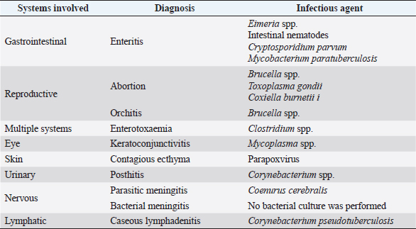
Frequencies of specific diseases in sheepThe distributions of diseases involving various body systems in sheep are presented in Table 3. In the reproductive system, a significant (p ≤ 0.05) percentage of sheep aged older than 1 year were diagnosed with abortion (23.43%), metritis (9.38%), and retained placenta (7.81%) compared to sheep aged 2 months to 1 year (17.18%, 3.90%, and 3.90%, respectively). Dystocia (12.5%) on the other hand was significantly (p ≤ 0.05) more frequently diagnosed in goats older than 2 months to 1 year compared to those older than 1 year (3.13%). Repeat breeder (14.06%) and vaginal/uterine prolapses (3.13%) were only diagnosed in sheep older than 1 year. In males, the only disease involving the reproductive system was orchitis (2 cases) diagnosed in rams aged older than 1 year. In the gastrointestinal system, enteritis was significantly (p ≤ 0.05) more frequently diagnosed in pre-weaning lambs, sheep aged older than 1 year, and sheep aged older than 2 months to 1 year (26.60%, 21.10%, and 19.27%, respectively). Rectal prolapse was diagnosed in sheep older than 2 months to 1 year (4 cases) and sheep aged older than 1 year (3 cases) while atresia ani was only diagnosed in pre-weaning lambs (3 cases). On the other hand, rumen foreign objects (10 cases), rumen tympany (10 cases), and rumen acidosis (6 cases) were only diagnosed in sheep older than 1 year. In diseases with multiple system manifestations, enterotoxaemia (33.33%) was significantly (p ≤ 0.05) more frequently diagnosed in sheep aged older than 2 months to 1 year compared to other age groups (23.08% and 12.825% in pre-weaning age lambs and sheep older than 1 year, respectively). Salmonellosis was diagnosed in pre-weaned age lambs (3 cases) and sheep older than 2 months to 1 year (2 cases) while peste des petits ruminants (7 cases) was only diagnosed in pre-weaning age lambs. In the respiratory system, bronchopneumonia was significantly (p ≤ 0.05) more frequently diagnosed in pre-weaning age lams (48.48%) compared to other age groups (30.30% and 21.21% sheep older than 2 months to 1 year and those older than 1 year, respectively). In the metabolic system, pregnancy toxemia was the most significant (p ≤ 0.05) disease condition diagnosed in sheep aged older than 2 months to 1 year and those aged older than 1 year (33.33% and 44.44%, respectively). In the udder/teat, clinical mastitis and teat injuries were diagnosed in sheep aged older than 1 year (42.30% and 15.38%, respectively) and those aged older than 2 months to 1 year (30.77% and 11.54%, respectively). In the nervous system, bacterial meningitis was diagnosed in pre-weaning age lambs (3 cases) and those older than 1 year (3 cases). Parasitic meningitis on the other hand was significantly more frequently diagnosed in sheep older than 1 year (22.73%) compared to those older than 2 months to 1 year (9.09%). On the other hand, polioencephalomyelitis was diagnosed in sheep older than 2 months to 1 year and those older than 1 year (22.73% and 18.18%, respectively). In the eye/ear, keratoconjunctivitis was the only disease condition diagnosed in sheep older than 1 year (6 cases) and those older than 2 months (5 cases). In the abdominal wall, omphalitis (6 cases) and umbilical hernia (3 cases) were diagnosed in pre-weaning age lambs. In the lymphatic system, only caseous lymphadenitis was diagnosed in sheep older than 2 months to 1 year (5 cases) and those older than 1 year (4 cases). In the musculoskeletal system, foot rot was only diagnosed in sheep older than 1 year (4 cases) while fractures were diagnosed in pre-weaning age lambs (2 cases) and sheep older than 1 year (1 case). Polyarthritis was only diagnosed in pre-weaning age lambs (1 case). In the urinary system, only males were diagnosed with conditions affecting the urinary system including obstructive urolithiasis in sheep older than 2 months to 1 year (5 cases) and hypospadias (1 case) in pre-weaning age lambs. In the skin, mange (3 cases) and traumatic skin wounds (1 case) were the only conditions diagnosed in sheep older than 1 year. In the cardiovascular/circulatory system, the only condition diagnosed was copper poisoning (3 cases) in sheep older than 1 year. Frequencies of infectious diseases in goats and sheepThe distribution of infectious diseases involving various body systems in goats is presented in Table 4. In the gastrointestinal system, the most commonly diagnosed infectious causative agents were Eimeria spp., Cryptosporidium parvum (C. parvum), and Mycobacterium paratuberculosis (M. paratuberculosis). In the reproductive system, the most common infectious causes of abortion were Brucella spp., Toxoplasma gondii (T. gondii), and Coxiella burnetii (C. burnetii). Brucella spp. was also diagnosed as the cause of orchitis in bucks. Clostridium spp. was diagnosed in goats with multiple systems manifestations. Mycoplasma spp. was the most commonly diagnosed infectious agent causing keratoconjunctivitis. In the skin, Parapoxovirus was diagnosed in goats with skin lesions suggestive of contagious ecthyma. Corynebacterium spp. was diagnosed in male goats with Posthitis. Coenurus cerebralis (C. cerebralis) was the most common cause of parasitic meningitis in goats. Corynebacterium pseudotuberculosis (C. pseudotuberculosis) was the most common infectious agent affecting the lymphatic system in goats. Table 4. Distribution of infectious diseases categorized according to the body system involved in goats.
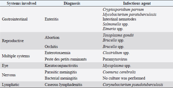
The distribution of infectious diseases involving various body systems in sheep is presented in Table 5. In the gastrointestinal system, the most commonly diagnosed infectious causative agents were C. parvum, M. paratuberculosis, Salmonella spp., Eimeria spp., and intestinal nematodes. In the reproductive system, the most common infectious causes of abortion were Brucella spp. and T. gondii. Brucella spp. was also diagnosed as the cause of orchitis in rams. Clostridium spp. and Paramyxovirus were diagnosed in sheep with multiple systems manifestations. Mycoplasma spp. was the most commonly diagnosed infectious agent causing keratoconjunctivitis in sheep. Coenurus cerebralis was the most common cause of parasitic meningitis in sheep. Corynebacterium pseudotuberculosis was the most common infectious agent affecting the lymphatic system in sheep. DiscussionIn this retrospective study, a comprehensive report of major categories of infectious and non-infectious diseases affecting small ruminants of different ages in Jordan is presented for the first time. The population of this study fairly represents the sheep and goat populations in Jordan and the diseases reported here may reflect the health situation of small ruminants at large. Abortion, dystocia, repeat breeder, vaginal or uterine prolapse, metritis in females, and infectious orchitis in breeding males were the most common diseases in goats and sheep. Globally, the most common infectious causes of abortion in small ruminants are Brucella spp., T. gondii, and C. burnetii in addition to Chlamydia spp., Leptospira spp., Campylobacter spp., and L. monocytogenes (Alemayehu et al., 2021). Furthermore, Brucella spp. has been reported as an important cause of orchitis and infertility in male small ruminants (Rossetti et al., 2022). Unfortunately, an etiological causative agent could not be identified in the majority of abortion cases in this study. This is in complete agreement with previous reports where it was suggested that less than 50% of abortion causes could have been identified using the best diagnostic capabilities (Dorsch et al., 2022). This fact could be explained by a lack of case definition, contaminated or improper sample collection, or transport. Dystocia was another major problem encountered frequently in this study. In small ruminants, dystocia has been reported most frequently in young does and ewes due to fetal oversize, malpresentation, fetal malformations, and failure of cervical dilation (Ismail, 2017). Prevention of underage pregnancy in does and ewes, proper timing of intervention, and adequate training of farm animal caretakers are required to prevent or reduce losses related to dystocia in small ruminants. Subfertility or repeat breeders were also commonly diagnosed in goats and sheep in this study. Subfertility on a herd level could significantly affect the economic status of the herd and require substantial efforts to investigate, treat and prevent. Anestrous due to anatomical, hormonal, inflammatory, or nutritional causes, failure to detect estrous, failure to conceive, early embryonic loss, along with problems related to male fertility have been cited as the most common cause of repeat breeding in goats and sheep (Ali et al., 2019). Vaginal and uterine prolapses are commonly seen in small ruminants (Makhdoomi and Gazi, 2015). Vaginal and uterine prolapse are considered emergency situations due to rapid deterioration associated with tissue necrosis and cardiovascular compromise in long-standing cases. Vaginal prolapse is commonly associated with high circulating estrogens in the prepartum period while uterine prolapse is associated with a difficult birth and forceful extraction of the fetus (Makhdoomi and Gazi, 2015). Other important risk factors to consider in cases of vaginal prolapse are obesity in late gestation, multiple fetuses, feeding bulky fibrous feedstuff, and genetics while in uterine prolapse, excessive straining following difficult birth is the most likely risk factor (Makhdoomi and Gazi, 2015). Table 5. Distribution of infectious diseases categorized according to the body system involved in sheep.
Metritis was also common in goats and sheep in this study. Metritis is characterized by infection and inflammation of the uterine wall commonly associated with dystocia with manual intervention and retained fetal membranes (Radostits et al., 2007). Bacterial infectious agents associated with metritis in small ruminants have been previously reported including Escherichia coli (E. coli), Trueperella pyogenes, Bacteriodes spp., and Fusobacterium necrophorum (Radostits et al., 2007). Clinically, acute metritis is characterized by high fever and delayed uterine involution with increased foul-odor uterine discharges (Radostits et al., 2007). The second most common disease in goats and sheep in this study involved the gastrointestinal system. Enteritis characterized by diarrhea, dehydration, and weakness was the most commonly diagnosed condition in pre-weaning age kids and lambs with Eimeria spp., and C. parvum was the most commonly diagnosed etiological agent while various species of intestinal parasites and M. paratuberculosis were the most common causes of enteritis in older goats and sheep. These results are in congruence with previous reports on neonatal and juvenile ruminants (Heller and Chigerwe, 2018). Although etiologic diagnosis is of great value in order to allow for the implementation of appropriate prevention measures on the herd level, treatment is considered vital for the survival of individual animals regardless of the cause of enteritis. Early treatment should include oral or intravenous fluid therapy supplemented with electrolytes and an alkalinizing agent, anti-inflammatory drugs, and systemic antibiotics (Heller and Chigerwe, 2018). Prevention usually requires proper nutrition of the dam in late pregnancy, prepartum dam vaccinations, hygienic kidding and lambing practices, and ingestion of colostrum as early as possible after birth (Heller and Chigerwe, 2018). Paratuberculosis or Johne’s disease, caused by M. paratuberculosis is a significant health problem facing small ruminant industries all over the world (Jiménez-Martín et al., 2022). In Jordan, previously published reports have indicated that the individual-level prevalence of paratuberculosis in sheep and goats was 22% and 50%, respectively (Al-Majali et al., 2008). Analysis of large data sets in Jordan indicated that grazing in communal areas and the addition of new animals were the most common risk factors for paratuberculosis (Al-Majali et al., 2008). Clinically, the disease causes huge economic losses due to severe weight loss in affected animals, reduced productivity, chronic diarrhea, and premature culling or death of the animal (Jiménez-Martín et al., 2022). Enteritis caused by various species of gastrointestinal parasites and protozoa was diagnosed frequently in goats and sheep in this study. Gastrointestinal parasitism caused by various helminths and protozoal species is a very common cause of poor growth and poor productivity in small ruminants (Radostits et al., 2007). Clinically, affected animals show signs of loss of appetite, loss of condition, diarrhea, anemia, and submandibular edema (Radostits et al., 2007). Further economic losses are attributed to the cost of medications used to treat internal parasites, production losses, and extra expenses associated with preventive and control measures (Radostits et al., 2007). Important risk factors for the increased prevalence of intestinal nematodes have been reported previously including the age of the animal, nutritional status, herd or flock size, housing conditions, deworming use or lack thereof, and suitability of the environmental conditions related to parasite species in an area (Dey et al., 2020). The third most common disease diagnosis in both goats and sheep in all age groups was bronchopneumonia. Bronchopneumonia is a multifactorial disease commonly seen in animals raised in poor housing and environmental conditions leading to increased susceptibility to various primary viral and secondary bacterial pathogens including Mycoplasma spp., Pasteurella multocida, Mannheimia haemolytica, and Histophilus somni (Mekibib et al., 2019). In this study, an etiological causative agent could not be reported because such an etiological diagnosis of pneumonia is often not sought unless it was a part of a herd outbreak investigation. Clinical mastitis was a common diagnosis in both goats and sheep in this study. Mastitis is an inflammation of udder tissue associated with bacterial invasion of the gland. The condition causes significant pain and substantial economic losses associated with production losses, the cost of treatment and veterinary care, and extra costs related to prevention measures. Although the bacterial culture of milk obtained from cases of clinical mastitis in goats and sheep is routinely performed at the VHC, etiological diagnosis is not reported in this study. In small ruminants, the most common udder pathogens have been reported previously including Staphylococcus aureus, Mannheimia spp., Streptococcus spp., and non-aureus staphylococci (sheep) and Staphylococcus spp., Streptococcus spp., Bacillus spp., Trueperella spp., Pseudomonas spp., Listeria spp., Mycoplasma spp., Mannheimia spp., Clostridium spp., and E. coli in goats (Kahinda, 2021). In this study, the most frequent metabolic diseases diagnosed were pregnancy toxemia in goats and sheep and hypocalcemia in sheep. Pregnancy toxemia is a potentially fatal disease affecting does and ewes in late gestation with nutritional and body condition abnormalities (Cal-Pereyra et al., 2015). Housing pregnant animals in late gestation in small and segregated groups and feeding them a high-quality energy-rich diet is sufficient enough to prevent devastating losses due to this condition (Cal-Pereyra et al., 2015). Early treatment by administering energy-boosted intravenous and oral fluids and induction of parturition or emergency cesarian section is required to save the life of the dam and potentially the newborns (Cal-Pereyra et al., 2015). In this study, other less frequently encountered disease diagnoses were involving multiple systems, musculoskeletal, ear/eye, abdominal wall, lymphatic, nervous, skin, and urinary systems in goats and the nervous, musculoskeletal, ear/eye, lymphatic, abdominal wall, cardiovascular/circulatory, skin, and urinary systems in sheep. These are considered rare conditions but are capable of causing significant losses in certain situations, therefore, the veterinary practitioner as well as farm managers or animal caretakers must take it into consideration after ruling out other disease conditions and be prepared to treat and provide the best care of affected animals to ensure favorable outcomes. ConclusionThis is the first study to provide a comprehensive view of the most commonly diagnosed diseases and their distribution according to etiology (infectious vs non-infectious, age, and body system) in sheep and goats in Jordan. The patterns of disease occurrence in goats and sheep were very similar with the top seven body systems involved in disease processes being the reproductive, gastrointestinal, multiple systems, respiratory, udder/teats, metabolic, and musculoskeletal systems for both species. These results indicated that despite major national efforts in preventative animal health, nutrition, and animal management measures, still disease – both infectious and non-infectious – presents a major hurdle in the path to achieving satisfactory status related to animal health, productivity, and welfare in goats and sheep in Jordan. More efforts at improving farm management, nutrition, and biosecurity measures such as vaccination and sanitation are required to reduce the effects of both infectious and non-infectious diseases in small ruminants. AcknowledgmentsThe authors would like to thank the students, faculty, and staff members of the Department of Veterinary Clinical Sciences and the VHC of the Faculty of Veterinary Medicine at Jordan University of Science and Technology for their support. Conflict of interestThe authors declare that there is no conflict of interest. Author contributionsMyassar Alekish conceived and designed the study, and reviewed and collected the data from the case medical records. Zuhair Bani Ismail performed data analysis, interpreted the data, and wrote the manuscript. ReferencesAbabneh, M.M., Hananeh, W., Bani Ismail, Z., Hawawsheh, M., Al-Zghoul, M., Knowles, N.J. and van Maanen, K. 2020. First detection of foot-and-mouth disease virus O/ME-SA/ Ind2001e sublineage in Jordan. Transbound. Emerg. Dis. 67(1), 455–460. Alemayehu, G., Mamo, G., Alemu, B., Desta, H., Tadesse, B., Benti, T., Bahiru, A., Yimana, M. and Wieland, B. 2021. Causes and flock level risk factors of sheep and goat abortion in three agroecology zones in Ethiopia. Front. Vet. Sci. 8, 615310. Al-Halabi, H. 2019. Government report of the Hashemite Kingdom of Jordan rainfall enhancement project: 3 year results 2016-2019. Available via https://www.weathertec-services.com/news/2019-07%20Gov%20of%20Jordan_3%20Year%20Report%20WeatherTec%20Project%20for%20Rainfall%20Enhancement.pdf. Ali, S., Zhao, Z., Zhen, Z., Kang, Z.J. and Yi, P.Z. 2019. Reproductive problems in small ruminants (sheep and goats): a substantial economic loss in the world. Large. Anim. Rev. 25, 215–223. Al-Majali, A.M., Hussain, N.O., Amarin, N.M. and Majok, A.A. 2008. Seroprevalence of, and risk factors for, peste des petits ruminants insheep and goats in Northern Jordan. Prev. Vet. Med. 85(1–2), 1–8. Al-Majali, A.M., Jawasreh, K. and Al Nsour, A. 2008. Epidemiological studies on foot and mouth disease and paratuberculosis in small ruminants in Tafelah and Ma’an, Jordan. Small. Rum. Res. 78(1–3), 197–201. Cal-Pereyra, L., González-Montaña, J.R., Benech, A., Acosta-Dibarrat, J., Martín, M., Perini, S., Abreu, M., Da Silva, S. and Rodríguez, P. 2015. Evaluation of three therapeutic alternatives for the early treatment of ovine pregnancy toxaemia. Ir. Vet. J. 68, 25. Department of Statistics, Amman, Jordan. 2020. Available via http://dosweb.dos.gov.jo/agriculture/livestock/ Dey, A.R., Begum, N., Alim, M.A., Malakar, S., Islam, M.T. and Alam, M.Z. 2020. Gastro-intestinal nematodes in goats in Bangladesh: a large-scale epidemiological study on the prevalence and risk factors. Parasite. Epidemiol. Control. 9, e00146. Dorsch, M.A., Francia, M.E., Tana, L.R., González, F.C., Cabrera, A., Calleros, L., Sanguinetti, M., Barcellos, M., Zarantonelli, L., Ciuffo, C., Maya, L., Castells, M., Mirazo, S., Silveira, S.C., Rabaza, A., Caffarena, R.D., Díaz, D.B., Aráoz, V., Matto, C., Armendano, J.I., Salada, S., Fraga, M., Fierro, S. and Giannitti, F. 2022. Diagnostic investigation of 100 cases of abortion in sheep in Uruguay: 2015–2021. Front. Vet. Sci. 9, 904786. Hailat, N., Al-Rawashdeh, O., Lafi, S. and Al-Bateineh, Z. 1994. An outbreak of sheep pox associated with unusual winter conditions in Jordan. Trop. Anim. Health. Prod. 26(2), 79–80. Hawari, A.D., Obeidat, M., Awaisheh, S., Al-Daghistani, H.I., Al-Abbadi, A.A., Omar, S.S., Qrunfleh, I.M., Al-Dmoor, H.M. and El-Qudah, J. 2014. Prevalence of mastitis pathogens and their resistance against antimicrobial agents in Awassi sheep in Al-Balqa province of Jordan. Am. J. Anim. Vet. Sci. 9(2), 116–121. Heller, M.C. and Chigerwe, M. 2018. Diagnosis and treatment of infectious enteritis in neonatal and juvenile ruminants. Vet. Clin. North. Am. Food. Anim. Pract. 34(1), 101–117. Ismail, Z.B. 2017. Dystocia in sheep and goats: outcome and fertility following surgical and non-surgical management. Mac. Vet. Rev. 40(1), 91–96. Jiménez-Martín, D., García-Bocanegra, I., Risalde, M.A., Fernández-Molera, V., Jiménez-Ruiz, S., Isla, J. and Cano-Terriza, D. 2022. Epidemiology of paratuberculosis in sheep and goats in southern Spain. Prev. Vet. Med. 202, 105637. Kahinda, C.T.M. 2021. Mastitis in small ruminants. In Mastitis in dairy cattle, sheep and goats. Eds., Dego, O.K. London, UK: IntechOpen. Available via https://www.intechopen.com/chapters/76529 doi: 10.5772/intechopen.97585. Makhdoomi, D.M. and Gazi, M.A. 2015. Uterine prolapse in a sheep and its management: a case report. Adv. Life. Sci. Technol. 28, 44–46. Mekibib, B., Mikir, T., Fekadu, A. and Abebe, R. 2019. Prevalence of pneumonia in sheep and goats slaughtered at Elfora Bishoftu export abattoir, Ethiopia: a pathological investigation. J. Vet. Med. 2019, 5169040. Musallam, I.I., Abo-Shehada, M., Omar, M. and Guitian, J. 2015. Cross-sectional study of brucellosis in Jordan: prevalence, risk factors and spatial distribution in small ruminants and cattle. Prev. Vet. Med. 118(4), 387–396. Radostits, O.M., Gay, C.C., Hinchcliff, K.W. and Constable, P.D. 2007. Veterinary medicine: a textbook of the diseases of cattle, horses, sheep, pigs and goats, 10th ed. Philadelphia, PA: WB Saunders Co. Ltd. Rich, K.M. and Perry, B.D. 2011. The economic and poverty impacts of animal diseases in developing countries: new roles, new demand for economics and epidemiology. Prev. Vet. Med. 101(3–4), 133–147. Rossetti, C.A., Maurizio, E. and Rossi, U.A. 2022. Comparative review of brucellosis in small domestic ruminants. Front. Vet. Sci. 9, 887671. The State of Food and Agriculture (SOFA). 2021. The state of food and agriculture. Livestock in the balance. Available via https://www.fao.org/publications/sofa/sofa-2021/en/ | ||
| How to Cite this Article |
| Pubmed Style Alekish MO, ZBI, . Common diseases of sheep (Ovis aries linnaeus) and goats (Capra aegagrus hircus) in Jordan: a retrospective study (2015-2021). Open Vet J. 2022; 12(6): 806-814. doi:10.5455/OVJ.2022.v12.i6.4 Web Style Alekish MO, ZBI, . Common diseases of sheep (Ovis aries linnaeus) and goats (Capra aegagrus hircus) in Jordan: a retrospective study (2015-2021). https://www.openveterinaryjournal.com/?mno=81657 [Access: July 27, 2024]. doi:10.5455/OVJ.2022.v12.i6.4 AMA (American Medical Association) Style Alekish MO, ZBI, . Common diseases of sheep (Ovis aries linnaeus) and goats (Capra aegagrus hircus) in Jordan: a retrospective study (2015-2021). Open Vet J. 2022; 12(6): 806-814. doi:10.5455/OVJ.2022.v12.i6.4 Vancouver/ICMJE Style Alekish MO, ZBI, . Common diseases of sheep (Ovis aries linnaeus) and goats (Capra aegagrus hircus) in Jordan: a retrospective study (2015-2021). Open Vet J. (2022), [cited July 27, 2024]; 12(6): 806-814. doi:10.5455/OVJ.2022.v12.i6.4 Harvard Style Alekish, M. O., , Z. B. I. & (2022) Common diseases of sheep (Ovis aries linnaeus) and goats (Capra aegagrus hircus) in Jordan: a retrospective study (2015-2021). Open Vet J, 12 (6), 806-814. doi:10.5455/OVJ.2022.v12.i6.4 Turabian Style Alekish, Myassar Omar, Zuhair Bani Ismail, and . 2022. Common diseases of sheep (Ovis aries linnaeus) and goats (Capra aegagrus hircus) in Jordan: a retrospective study (2015-2021). Open Veterinary Journal, 12 (6), 806-814. doi:10.5455/OVJ.2022.v12.i6.4 Chicago Style Alekish, Myassar Omar, Zuhair Bani Ismail, and . "Common diseases of sheep (Ovis aries linnaeus) and goats (Capra aegagrus hircus) in Jordan: a retrospective study (2015-2021)." Open Veterinary Journal 12 (2022), 806-814. doi:10.5455/OVJ.2022.v12.i6.4 MLA (The Modern Language Association) Style Alekish, Myassar Omar, Zuhair Bani Ismail, and . "Common diseases of sheep (Ovis aries linnaeus) and goats (Capra aegagrus hircus) in Jordan: a retrospective study (2015-2021)." Open Veterinary Journal 12.6 (2022), 806-814. Print. doi:10.5455/OVJ.2022.v12.i6.4 APA (American Psychological Association) Style Alekish, M. O., , Z. B. I. & (2022) Common diseases of sheep (Ovis aries linnaeus) and goats (Capra aegagrus hircus) in Jordan: a retrospective study (2015-2021). Open Veterinary Journal, 12 (6), 806-814. doi:10.5455/OVJ.2022.v12.i6.4 |


