


| Case Report | ||
Open Vet J. 2023; 13(9): 1219-1222 Open Veterinary Journal, (2023), Vol. 13(9): 1219-1222 Case Report A case of a dog with mandibular extraskeletal osteosarcoma after long-term puncture extirpation of the salivary gland cyst in the mandibleAkiko Uemura1, Io Maruyama1 and Tomohiko Yoshida2*1Division of Veterinary Research, Department of Clinical Veterinary Medicine, Laboratory of Veterinary Surgery, Obihiro University of Agriculture and Veterinary Medicine, Obihiro, Japan 2Division of Veterinary Research, Department of Clinical Veterinary Medicine, Laboratory of Veterinary Internal Medicine, Obihiro University of Agriculture and Veterinary Medicine, Obihiro, Japan *Corresponding Author: Tomohiko Yoshida. Division of Veterinary Research, Department of Clinical Veterinary Medicine, Laboratory of Veterinary Internal Medicine, Obihiro University of Agriculture and Veterinary Medicine, Obihiro, Japan. Email: tomohiko7731-yoshida [at] yahoo.co.jp Submitted: 07/06/2023 Accepted: 17/08/2023 Published: 30/09/2023 © 2023 Open Veterinary Journal
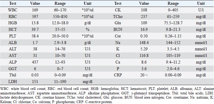
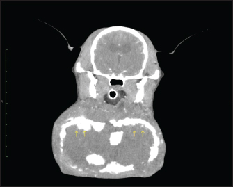
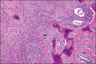
AbstractBackground: Extraskeletal osteosarcoma, unlike skeletal osteosarcoma, is a rare malignant mesenchymal tumor with a soft tissue primary that has been reported to occur in a variety of soft tissues. Case Description: The case is a 14-year-old, unneutered male Miniature Pinscher, weighing 6.7 kg, who had been treated medically for more than 5 years with a management strategy of puncture extirpation of a salivary gland cyst in the mandible; 1 month earlier, the fluid retention could not be removed, and after a computerized tomography scan showed no lesion in the mandible adjacent to the mass lesion, surgical resection was performed. Conclusion: Previous reports of extraskeletal osteosarcoma from the salivary glands in dogs have been rare. However, treatment of a salivary gland cyst in the mandible by long-term puncture extirpation may be a potential predisposing factor for the development of extraskeletal osteosarcoma around the mandible. Keywords: Dogs, Extraosseous osteosarcoma, Mandible, Salivary gland. IntroductionExtraskeletal osteosarcoma is a highly malignant mesenchymal neoplasm that arises from internal organs and soft tissues and is characterized by the formation of bone without invasion into bone or periosteal tissue (Duffy et al., 2017). Its occurrence is rare, although it has been reported in dogs and humans (Makielski et al., 2019). This case report describes a dog that developed extraskeletal osteosarcoma at the same site after long-term puncture extirpation of a salivary gland cyst in the mandible. Case DetailsCase 1 is a 14-year-old, unneutered male miniature pinscher weighing 6.7 kg. For over 5 years, the patient had repeatedly been diagnosed with salivary gland cysts at several hospitals due to the presence of mucus accumulation in the mandible. He had been routinely treated medically by aspirate removal. Three months before first visiting the university, mucus viscosity decreased and became partially indurated, leading to an increase in the frequency of puncture removal. One month earlier, the mucus in the reservoir had solidified, making it impossible to aspirate. As such, the patient was referred to our hospital for further examination and treatment. The size of the mandibular mass lesion at the time of the first visit (day 1) was 130 * 98 * 75 mm (Figs. 1 and 2). Blood tests showed hypoalbuminemia (Table 1). Fine needle aspiration of the mass lesion showed no significant cellular component. Chest and abdominal radiographs and chest ultrasound showed no specific findings, including metastatic lesions. Abdominal ultrasonography showed no noteworthy findings except a mild enlargement of the left adrenal gland (9 mm in short diameter). A computerized tomography (CT) performed on day 8 showed a hyper-absorbable area comparable to cortical bone lining the entire mass wall. In the arterial phase, the interior of the hyper-absorbable area was occupied by a slightly hypo-absorbable area compared to the margins of the mass lesion (Fig. 3). There was no vascular run inside the mass, and the mandibular glands, sublingual glands, and mandibular lymph nodes were enlarged bilaterally. There were no significant findings in the mandible. On day 14, the patient underwent surgery to resect the mass lesion. After inducing general anesthesia, the mandibular mass lesion was excised. The resected mass was a large neoplastic lesion in the subcutaneous tissue with extensive necrosis in the center. In the tumor tissue corresponding to the mass wall, bundles, sheets, and wreaths of osteoblast-like tumor cells with bone formation were observed (Fig. 4). These tumor cells were moderately atypical, and numerous mitotic figures were observed. No vascular invasion was observed, and the resection margin was good. Together with the CT scan results, the diagnosis of extraskeletal osteosarcoma was made.
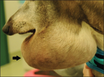
Fig. 1. External appearance of case 1 at the initial examination. A large mass (130 × 98 × 75 mm; arrow) was found in the mandible.
Fig. 2. X-ray examination of the skull. Calcification was seen inside the mass lesion that increased from the mandible to the neck (arrow). (a) AP, anterior-posterior view and (b) LR, right lateral view. Postoperatively, the patient was in good general condition, including appetite and activity, and was treated with antibiotics until day 25. Since the owner did not wish to receive concurrent chemotherapy, patient follow-ups were carried out by her family doctor from day 29 onward. DiscussionThe exact incidence of extraskeletal osteosarcoma in dogs is not known (Makielski et al., 2019), but it has been reported that there were 11 cases of extraskeletal osteosarcoma out of approximately 1,000 cases of skeletal osteosarcoma (Patnaik, 1990). The spleen (Kuntz et al., 1998; Duffy et al., 2017), gastrointestinal tract, and mammary gland (Langenbach et al., 1998) have also been reported as the common sites of extraskeletal osteosarcoma in dogs. Although four cases of extraskeletal osteosarcoma in the cervical salivary glands of dogs have been reported in the past (Langenbach et al., 1998; Thomsen and Myers, 1999; Umeda et al., 2023), with no report of a common site of occurrence. In addition to the reported hereditary occurrence of osteosarcoma in dogs (Phillips et al., 2007), physical irritation at the site of bone plate insertion has been reported as a suspected predisposing factor for tumor development (Boudrieau et al., 2005). The factors leading to extraskeletal osteosarcoma development are still largely unknown. However, since there have been reports of dogs with extraskeletal osteosarcoma caused by residual surgical sponges (Miller et al., 2006; Slovak et al., 2015) and cotton swabs (Goto et al., 2022), long-term physical irritation by foreign objects may be associated with the development of the disease. The occurrence of fibrosarcoma due to trauma or microchip insertion sites is well-known in cats (Daly et al., 2008). In dogs also, there is a risk of postinjection sarcoma, and the known occurrence of extraskeletal osteosarcoma at injection sites is suggested to result from severe local inflammation caused by the adjuvant contained in the vaccine (Selmic et al., 2016). In this case, the accumulation of mucus was treated internally as a salivary gland cyst by long-term puncture extirpation. Drainage of salivary gland cysts by puncture extirpation is sometimes performed as an emergency evacuation. However, healing salivary gland cysts without surgery is rare, and long-term continuous use of internal puncture extractions is not recommended as a treatment (Peeters, 1991; Waldron and Smith, 1991). On imaging examination, the typical case of extraskeletal osteosarcoma is a soft-tissue mass with localized sclerosis and no lesion in the adjacent bone (Schena et al., 1989). In the present report, the mass lesion and the adjacent mandible in this case were consistent with this finding on both X-ray and CT. Together with the histopathological examination, these were some of the determining factors in diagnosing extraskeletal osteosarcoma in this case. Extraskeletal osteosarcoma may be considered a differential diagnosis in soft tissue mass lesions with sclerosis in the submandibular region in the absence of an adjacent mandible or other bone lesions. Unfortunately, there were no clear histologic findings that the extra salivary osteosarcoma in this case is of salivary gland origin. Table 1. Blood test results, day 1.
Fig. 3. CT scan on day 8. A hyper-absorptive area as large as the cortical bone was observed lining the entire mass wall (arrow). In the arterial phase, the interior of the highly absorbable area was occupied by a slightly less absorbable area compared to the margins of the mass lesion.
Fig. 4. Histopathological examination results of the resected mass lesion. In the tumor tissue corresponding to the mass wall, bundles, sheets, and wreaths of osteoblast-like tumor cells (arrow) with bone formation were observed. The tumor cells were moderately atypical, and numerous mitotic figures were observed; bar, 100 μm. In this case, the physical irritation from the puncture needle and the iodine injected by the primary veterinarian caused regression of the salivary gland cyst. The long-term presence of chronic inflammation due to prolonged saliva accumulation in the soft tissue may have predisposed the patient to develop extraskeletal osteosarcoma. Salivary gland cysts in the submandibular region are commonly encountered clinically and often heal without major complications with appropriate surgical procedures. On the other hand, extraskeletal osteosarcoma has a poor prognosis, with a reported median survival time of 26 days (Langenbach et al., 1998), and a high rate of metastases (Kuntz et al., 1998; Duffy et al., 2017). Given the history of this case, surgical treatment should be promptly considered for salivary gland cysts around the mandible rather than continuing uncontrolled medical treatment. This report provides an overview of a dog with a salivary gland cyst in the mandible that had been under medical management for over 5 years that developed extraskeletal osteosarcoma in the same region. This case also suggests that surgical resection of salivary gland cysts should be performed at the appropriate time. AcknowledgmentThe authors thank Prof. Yasuyoshi Kobayashi, Dr. Kenichi Watanabe, and Dr. Mineshige Takayuki for conducting the histopathological examinations. Conflict of interestThe authors declare that there is no conflict of interest. Author contributionsAU: conceptualization, data curation, formal analysis, investigation, methodology, project administration, resources, software, supervision, validation, visualization, writing original draft, and writing review and editing; IM: writing original draft; TY: methodology; and data curation, formal analysis, investigation, methodology, software, and writing review and editing. FundingNo funding was received to assist with the preparation of this manuscript. Data availabilityThe datasets generated and/or analyzed during the current study are available from the corresponding author upon reasonable request. ReferencesBoudrieau, R.J., Mccarthy, R.J. and Sisson, R.D., Jr. 2005. Sarcoma of the proximal portion of the tibia in a dog 5.5 years after tibial plateau leveling osteotomy. J. Am. Vet. Med. Assoc. 227(10), 1613–1617. Daly, M.K., Saba, C.F., Crochik, S.S., Howerth, E.W., Kosarek, C.E., Cornell, K.K., Roberts, R.E. and Northrup, N.C. 2008. Fibrosarcoma adjacent to the site of microchip implantation in a cat. J. Feline Med. Surg. 10(2), 202–205. Duffy, D., Selmic, L.E., Kendall, A.R. and Powers, B.E. 2017. Outcome following treatment of soft tissue and visceral extraskeletal osteosarcoma in 33 dogs: 2008-2013. Vet. Comp. Oncol. 15(1), 46–54. Goto, M., Owaki, K., Hirata, A., Murakami, M. and Sakai, H. 2022. Extraskeletal osteosarcoma associated with two different types of synthetic fibers derived from a surgical swab in a dog. J. Vet. Med. Sci. 84(8), 1056–1060. Kuntz, C.A., Dernell, W.S., Powers, B.E. and Withrow, S. 1998. Extraskeletal osteosarcomas in dogs: 14 cases. J. Am. Anim. Hosp. Assoc. 34(1), 26–30. Langenbach, A., Anderson, M.A., Dambach, D.M., Sorenmo, K.U. and Shofer, F.D. 1998. Extraskeletal osteosarcomas in dogs: a retrospective study of 169 cases (1986-1996). J. Am. Anim. Hosp. Assoc. 34(2), 113–120. Makielski, K.M., Mills, L.J., Sarver, A.L., Henson, M.S., Spector, L.G., Naik, S. and Modiano, J.F. 2019. Risk factors for development of canine and human osteosarcoma: a comparative review. Vet. Sci. 6(2), 1–19. Miller, M.A., Aper, R.L., Fauber, A., Blevins, W.E. and Ramos-Vara, J.A. 2006. Extraskeletal osteosarcoma associated with retained surgical sponge in a dog. J. Vet. Diagn. Invest. 18(2), 224–228. Patnaik, A.K. 1990. Canine extraskeletal osteosarcoma and chondrosarcoma: a clinicopathologic study of 14 cases. Vet. Pathol. 27(1), 46–55. Peeters, M.E. 1991. The treatment of salivary cysts in dogs and cats. Tijdschr. Diergeneeskd. 116(4), 169–172. Phillips, J.C., Stephenson, B., Hauck, M. and Dillberger, J. 2007. Heritability and segregation analysis of osteosarcoma in the Scottish deerhound. Genomics 90(3), 354–363. Schena, C.J., Stickle, R.L., Dunstan, R.W., Trapp, A.L., Reimann, K.A., White, J.V., Killingsworth, C.R. and Hauptman, J.G. 1989. Extraskeletal osteosarcoma in two dogs. J. Am. Vet. Med. Assoc. 194(10), 1452–1456. Selmic, L.E., Griffin, L.R., Rector, M.H., Lafferty, M., Pool, R. and Ehrhart, N.P. 2016. Treatment of extraskeletal osteosarcoma at a previous injection site resulting in prolonged survival in 1 dog. Can. Vet. J. 57(9), 950–954. Slovak, J.E., Kieves, N.R. and Haynes, J. 2015. Extraskeletal osteosarcoma induced by a foreign body granuloma. J. Am. Anim. Hosp. Assoc. 51(5), 315–319. Thomsen, B.V. and Myers, R.K. 1999. Extraskeletal osteosarcoma of the mandibular salivary gland in a dog. Vet. Pathol. 36(1), 71–73. Umeda, N., Yamazoe, H., Wada, A. and Nagata, K. 2023. A dog with extraskeletal osteosarcoma of the salivary glands survived long-term, following surgical resection and adjuvant therapy. J. Vet. Med. Sci. 85(3), 358–362. Waldron, D.R. and Smith, M.M. 1991. Salivary mucoceles. Probl. Vet. Med. 3(2), 270–206. | ||

| How to Cite this Article |
| Pubmed Style Uemura A, Maruyama I, Yoshida T. A case of a dog with mandibular extraskeletal osteosarcoma after long-term puncture extirpation of the salivary gland cyst in the mandible. Open Vet J. 2023; 13(9): 1219-1222. doi:10.5455/OVJ.2023.v13.i9.18 Web Style Uemura A, Maruyama I, Yoshida T. A case of a dog with mandibular extraskeletal osteosarcoma after long-term puncture extirpation of the salivary gland cyst in the mandible. https://www.openveterinaryjournal.com/?mno=156425 [Access: May 19, 2024]. doi:10.5455/OVJ.2023.v13.i9.18 AMA (American Medical Association) Style Uemura A, Maruyama I, Yoshida T. A case of a dog with mandibular extraskeletal osteosarcoma after long-term puncture extirpation of the salivary gland cyst in the mandible. Open Vet J. 2023; 13(9): 1219-1222. doi:10.5455/OVJ.2023.v13.i9.18 Vancouver/ICMJE Style Uemura A, Maruyama I, Yoshida T. A case of a dog with mandibular extraskeletal osteosarcoma after long-term puncture extirpation of the salivary gland cyst in the mandible. Open Vet J. (2023), [cited May 19, 2024]; 13(9): 1219-1222. doi:10.5455/OVJ.2023.v13.i9.18 Harvard Style Uemura, A., Maruyama, . I. & Yoshida, . T. (2023) A case of a dog with mandibular extraskeletal osteosarcoma after long-term puncture extirpation of the salivary gland cyst in the mandible. Open Vet J, 13 (9), 1219-1222. doi:10.5455/OVJ.2023.v13.i9.18 Turabian Style Uemura, Akiko, Io Maruyama, and Tomohiko Yoshida. 2023. A case of a dog with mandibular extraskeletal osteosarcoma after long-term puncture extirpation of the salivary gland cyst in the mandible. Open Veterinary Journal, 13 (9), 1219-1222. doi:10.5455/OVJ.2023.v13.i9.18 Chicago Style Uemura, Akiko, Io Maruyama, and Tomohiko Yoshida. "A case of a dog with mandibular extraskeletal osteosarcoma after long-term puncture extirpation of the salivary gland cyst in the mandible." Open Veterinary Journal 13 (2023), 1219-1222. doi:10.5455/OVJ.2023.v13.i9.18 MLA (The Modern Language Association) Style Uemura, Akiko, Io Maruyama, and Tomohiko Yoshida. "A case of a dog with mandibular extraskeletal osteosarcoma after long-term puncture extirpation of the salivary gland cyst in the mandible." Open Veterinary Journal 13.9 (2023), 1219-1222. Print. doi:10.5455/OVJ.2023.v13.i9.18 APA (American Psychological Association) Style Uemura, A., Maruyama, . I. & Yoshida, . T. (2023) A case of a dog with mandibular extraskeletal osteosarcoma after long-term puncture extirpation of the salivary gland cyst in the mandible. Open Veterinary Journal, 13 (9), 1219-1222. doi:10.5455/OVJ.2023.v13.i9.18 |

