


| Research Article | ||
Open Vet J. 2024; 14(2): 652-663 Open Veterinary Journal, (2024), Vol. 14(2): 652-663 Original Research Depraved appetite in dromedary camels: Clinical, ultrasonographic, and postmortem findingsMohamed Tharwat1,2, Waleed R. El-Ghareeb3 and Tariq I. Almundarij4*1Department of Clinical Sciences, College of Veterinary Medicine, Qassim University, Buraidah, Saudi Arabia 2Department of Animal Medicine, Faculty of Veterinary Medicine, Zagazig University, Zagazig, Egypt 3Department of Public Health, College of Veterinary Medicine, King Faisal University, Hofuf, Saudi Arabia 4Department of Biomedical Sciences, College of Veterinary Medicine, Qassim University, Buraidah, Saudi Arabia *Corresponding Author: Tariq I. Almundarij. Department of Biomedical Sciences, College of Veterinary Medicine, Qassim University, Buraidah, Saudi Arabia. Email: tmndrj [at] qu.edu.sa Submitted: 21/10/2023 Accepted: 16/01/2024 Published: 29/02/2024 © 2024 Open Veterinary Journal
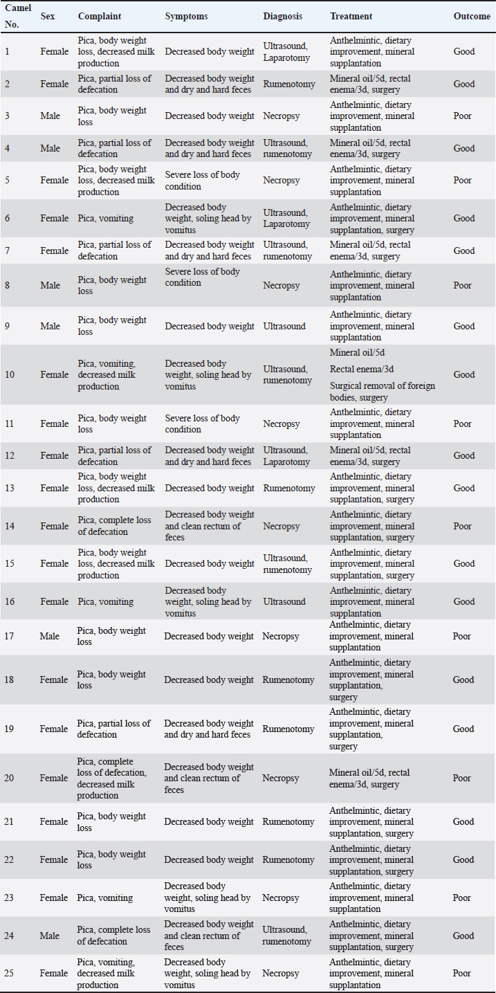
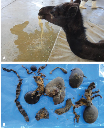
AbstractBackground: Camels are subjected to a wide variety of nutritional deficiencies as they are largely dependent upon grazing desert plants. As a consequence, the syndrome of pica or depraved appetite is occasionally seen in dromedary camels. The condition is manifested as chewing or eating abnormal things such as wood, dirt, bones, stones, clothes, plastics, mud, sand, or other inanimate objects. Aim: This study was designed to investigate the clinical, ultrasonographic, and postmortem findings in dromedary camels with pica or depraved appetite. Methods: Twenty-five camels of 5 days to 15 years were examined. Owner complaints included depraved appetite, loss of body condition, regurgitation of stomach content, and partial or complete absence of feces. Symptoms described were present for a period varying between 3 days, up to 12 months. The stomach compartments and small and large intestines were scanned using ultrasonography from the right and left sides of the abdomen. Necropsy was carried out on six female and three male camels where the thoracic and abdominal organs were examined with special attention to the digestive system. Results: The affected animals had a history of gradual loss of body conditions, eating foreign objects, decreased or total absence of feces, and regurgitation of stomach content. Using ultrasound, the foreign body was imaged occluding completely or partially the intestines. Foreign bodies within the rumen could not be visualized with ultrasound. In cases where the rumen is impacted by sand, small pin-points revealing acoustic enhancement were imaged. Foreign bodies were removed from the rumen at exploratory rumenotomy (n =11), laparotomy (n=3), or at necropsy (n =8) in the form of plastics, cloths, sand, mud, wool balls, robes, glasses, or even metallic objects which may be blunt or sharp. Sixteen (64%) of the camels were recovered while the remaining 9 (36%) did not survive. Conclusion: The syndrome of pica or depraved appetite is an important condition in dromedary resulting in the ingestion of objects other than normal feed. Substantial economic losses are expected as a result of this syndrome. Ultrasonography of the digestive system may help the clinician in some cases to localize of occluding foreign bodies in the intestines, while in the transabdominal scanning of the stomach is valuable only in cases of sand impaction. Keywords: Animals, Animal behavior, Nutrition, Feeding behavior, Ultrasound. IntroductionIn human life, the camel holds a crucial value especially in desert and rural regions, due to its multi-purpose function and its ability to adapt to harsh environmental conditions. In addition, it offers enormous economic, cultural, and biological benefits. Camels have a considerable role in the lifestyle of various societies, particularly those in dry zones in the Middle East and the Arabian territory (Kaskous, 2016). Camels exhibit unique morphological, biochemical, and behavioral traits, which arose from natural and artificial selection (Alhaddad and Alhajeri, 2019; AlAskar et al., 2020; Alaqeely et al., 2021). Approximately, 29 million camels are distributed all over the world, 95% of them are dromedaries (Sikkema et al., 2019). However, based on an official report issued by the Food and Agriculture Organization (FAO), there are about 35 million dromedaries over the world (FAO, 2018). The dromedary camel (Camelus dromedarius) is singular among other livestock in both its high adaptation to deserts and its multiple uses to humans (Alaskar et al., 2021). Camels are used for the transportation of humans and goods across deserts with comparative ease. More importantly, due to the composition and nutritional values, dromedary milk is similar to human milk, and therefore perhaps more superior to bovine milk for human consumption (Ho et al., 2022). It contains high concentrations of several bioactive compounds besides its antioxidant, anti-inflammatory, immunomodulatory, and medical effects (Al Nohair, 2021; Behrouz et al., 2022; Ho et al., 2022). Likewise, camel meat is of high nutritional value, with 78% water, 19%–22% protein, and 3% fat content (Kadim et al., 2022). Camels are capable of traveling long distances (up to 50 km per day) and they primarily graze on desert plants such as trees, shrubs, and thorny or salty herbaceous plants that are usually not consumed by other livestock species. It is generally agreed that camel is predominantly a browser, although it also grazes on tall succulent young grass. At large, dromedaries graze a broad spectrum of fodder plants, including thorny bushes, halophytes, and aromatic species generally avoided by other domestic herbivores (Iqbal and Khan, 2001). In some countries, the production of camel meat and milk has intensified; therefore, camels are now reared in intensive and semi-intensive systems (Padalino and Menchetti, 2021). In addition, some modernizations in farming techniques, such as machine milking, have been introduced but room for improvement still exists (Faye, 2014; Nagy and Juhasz, 2016). Since the grazing of dromedaries mostly happens in desert-like areas, camels are subjected to a wide variety of nutritional deficiencies, since these sand soils are often deficient in crucial elements (Firyal, 2007; Smith, 2015; Nikvand et al., 2018). As a consequence, the syndrome of pica (allotrophagia, geophagia, or depraved appetite) is occasionally seen in dromedary camels. Pica is therefore associated in most cases with dietary abnormalities, either of bulk or, in some cases, more specifically fiber, or because of individual nutrients, particularly mineral deficiency (Constable et al., 2017). The condition starts when the animal refuses to eat its normal feed and eats or chews abnormal things such as wood, dirt, bones, stones, cloths, plastics, mud, sand, or other inanimate objects (Firyal, 2007; Smith, 2015; Nikvand et al., 2018). According to Constable et al. (2017), pica can be classified into three main categories; (1) osteophagia, the chewing of bones, (2) infantophagia, the eating of young, and (3) coprophagia, the eating of feces. However, several other classes are reported and include bark eating, wood eating in sheep, cannibalism, and the eating of carrion. Salt deficiency can result in coat licking, leather chewing, earth-eating, and the drinking of urine (Constable et al., 2017). This article was designed to shed light on the syndrome of depraved appetite in the dromedary camel. It focuses on predisposing and existing causes, clinical, laboratory, and postmortem findings, and control measures. Materials and MethodsCamels, history, clinical examination and treatmentTwenty-five camels (19 females and 6 males) of 5 days to 15 years were examined at the Veterinary Hospital of the University of Qassim, and neighboring camel farms in Saudi Arabia between 2014 and 2023. These animals were referred with a history of depraved appetite, progressive weight loss, decreased milk production, regurgitation of stomach content, and partial or complete loss of defecation. The duration of these symptoms varied from 3 days to 12 months. Upon admission, each animal gained a full clinical examination including rectal temperature, pulse and respiratory rates, mucus membranes, and examination of the cardiopulmonary and digestive systems. Treatment options included oral and parenteral anthelmintic, dietary improvement, and oral and parenteral mineral supplantation. In camels with foreign bodies in the rumen or intestines, rumenotomy or laparotomy was performed (Table 1). Animals were maintained and treated according to the Laboratory Animal Control Guidelines of Qassim University, which basically conforms to the Guide for the Care and Use of Laboratory Animals of the National Institutes of Health in the USA (NIH publications No. 86 to 23, revised 1996). Ultrasonography, rumenotomy, laparotomy, and postmortem examinationsThe stomach and intestines were imaged with ultrasound using 3.5 and 7.5 MHz convex transducers (SonoScape, Sonoscape Medical Corporation, China). The stomach compartments and small and large intestines were scanned transabdominally from the right and left sides of the abdomen as previously reported (Tharwat et al., 2012a; Tharwat, 2020a). When necessary, the digestive tract, cardiovascular, respiratory, and urinary systems, and the liver were scanned as reported (Tharwat et al., 2012a,b,c,d,e; Tharwat, 2020a,b,c; Tharwat, 2021; Tharwat, 2023a; Sadan et al., 2024). Exploratory rumenotomy was performed in 11 camels (nine females and two males) where abdominal ultrasound did not reveal abnormalities. Intestinal surgery was also performed in three female camels. Nine camels that did not respond to treatment (six females and three males), were euthanized and a postmortem examination was performed. The thoracic and abdominal organs were examined thoroughly at necropsy and special attention was given to the digestive system. Ethical approvalAnimal Care and Welfare Committee of the Deanship of Scientific Research at Qassim University, Kingdom of Saudi Arabia approved the study design. ResultsFull details including presenting complaints, symptoms, diagnosis, treatment, and outcome of the 25 dromedary camels with depraved appetites are listed in Table 1. The dromedary camels with depraved appetites had different presentations, but all of the examined animals had a history of eating foreign or inanimate objects. Pica was evident in some cases upon initial clinical examination (Fig. 1). Twelve of the camels (48%) were admitted with a history of loss of body weight and 8 (32%) with partial or complete loss of defection. Seven out of the nine (77.8%) lactating camels in this study suffered from decreased milk production. An example of such a case is shown in Figure 2. In animals undergoing laparotomy, the foreign bodies were surgically removed as shown in Figure 3. Bouts of repeated vomiting were also found in 5 (20%) of the camels. In animals that underwent rumenotomy, the foreign bodies were removed as in Figure 4. Eating feces (coprophagia) was also found in six camels as shown in Figure 5. Sixteen (64%) of the camels were recovered while the remaining 9 (36%) did not survive. Table 1. Owner complaints, symptoms, diagnosis, treatment, and outcome in 25 dromedary camels with depraved appetite.
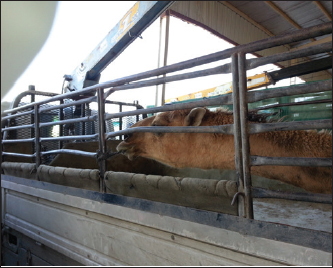
Fig. 1. Pica in a female camel. The animal tries licking the iron bars and eating a robe. Sonographic and postmortem findings are summarized in Table 2. In cases of camels presented with a history of pica and with decreased or total absence of feces, the obstructing foreign body was imaged in 10 of the 25 camels (40%) occluding completely or partially the intestines that may be confirmed at abdominal surgery (Figs. 2, 3, and 6). Foreign bodies within the rumen could not be visualized with ultrasound owing to the gas content. In cases where the rumen is impacted by sand, small pin-points revealing acoustic enhancement were imaged in 5 (20%) of the camels. An example of these cases is shown in Figure 7. Foreign bodies were removed from the rumen or intestines at exploratory rumenotomy and laparotomy, which included plastics, cloths, sand, mud, robes, wool balls, glasses, or even metallic objects that were blunt or sharp (Figs. 8 and 9). DiscussionIn camels, pica was reported to have a variety of contributing causes. Dietary deficiency and an imbalanced diet especially the shortage of major and trace elements such as phosphorus, calcium, iron, copper, cobalt, selenium, manganese sodium, and chloride are a common factor (Firyal, 2007; Smith, 2015; Nikvand et al., 2018). Ingest foreign objects such as plastics, cloth, and robes and the low hygienic standards of the surrounding environment are other causes (Singh et al., 2011; Sadan et al., 2020; Sadan et al., 2023).
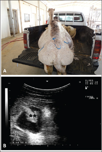
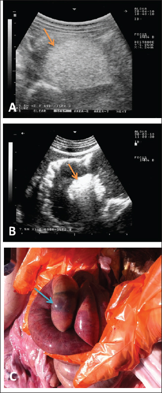
Fig. 2. Ultrasonographic findings in an adult female camel with a history of pica. The animal was admitted with a 7-day history of decreased fecal output and abdominal distension (A). Image (B) shows an obstructing foreign body occluding the small intestines partially (stars) using a 3.5 MHz sector transducer.
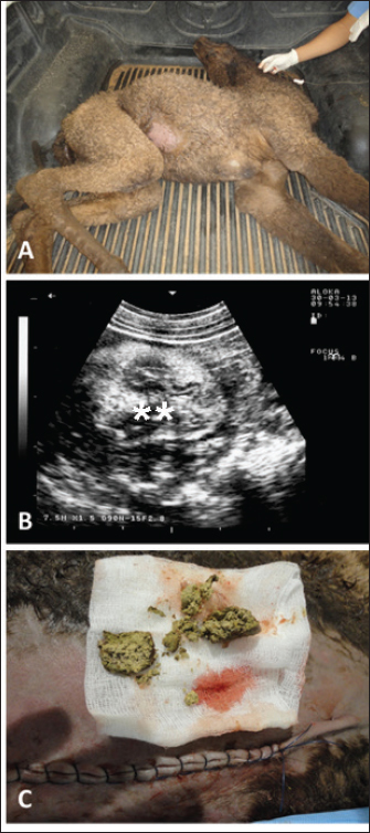
Fig. 3. Ultrasonographic findings in a 30-day camel calf with a history of pica. The calf was admitted with a 5-day history of no defection and abdominal distension (A). Image (B) shows an obstructing enterolith occluding the small intestines (stars) using a 7.5 MHz sector transducer. Image (C) shows the obstructing enterolith detected at right-sided surgical laparotomy.
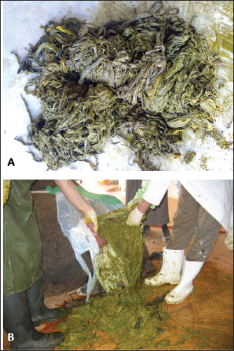
Fig. 4. Pica in a female camel referred with a history of depraved appetite and repeated volition (A). Exploratory rumenotomy revealed wool balls, plastics, and robes within the rumen (B). Pica in farm animals has been reported to have various contributing causes. For instance, in cattle, low levels of calcium, phosphorus, iron, zinc, chloride, total proteins, sodium, copper, and selenium were detected, along with reduced antioxidant activity. (Ocal et al., 2008; Mohamed, 2018; Nikvand et al., 2018; Onmaz et al., 2019). Calcium phosphorus ratio should be 2:1. Ketosis and trace element deficiency especially for cobalt, copper, iron, zinc, and manganese should also be treated, and therefore, mineral blocks should be provided all over the year and not on special occasions (Constable et al., 2017; Asín et al., 2021; Helmer et al., 2021). Phosphorus is essential for the production of milk and the building of bones (Begum et al., 2010). Its dietary deficiency may originate as a primary condition due to a shortage in feed or secondary due to increases in dietary calcium (Haskell and Anttilla, 2001). Both trace element deficiency and toxicity were reported to affect the camels’ reproductive and productive efficiency, as well as different aspects of their metabolism and growth. Therefore, assessing the status of trace elements in camels is an important step toward improving the health and productivity of camels (Abdelrahman et al., 2022). Among these elements, cobalt plays an important role as it is utilized by rumen microflora for synthesizing vitamin B12 resulting in chronic anemia. Deficiency of iron, copper, zinc, manganese, and selenium may be either primary, resulting from low availability in the diet, or secondary, due to decreased absorption (Cantile and Youssef, 2016; Hill and Shannon, 2019).
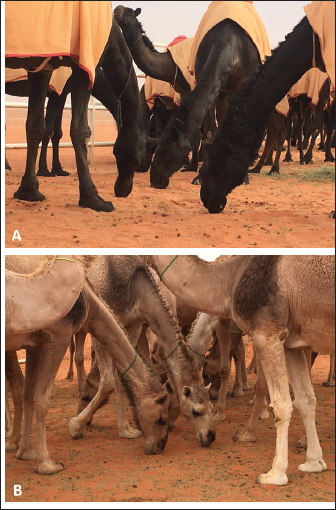
Fig. 5. Pica in two camel groups. Although receive a full balanced diet, some camels in (A) and (B) eat feces (coprophagia) due to deed deprivation. The consequences of pica in farm animals are numerous. In dairy cattle, the condition is also a predisposing cause for traumatic reticuloperitonitis (Ocal et al., 2008). Toxicity caused by lead, botulism, traumatic reticuloperitonitis, intestinal obstruction, and sand impaction are examples of these consequences (Melendez et al., 2007; Tharwat and Al-Hawas, 2024). The mechanisms of pica are not yet fully understood. The condition has also been found with other conditions such as ketosis and chronic parasitism (Firyal, 2007; Smith, 2015). Therefore, pica in farm animals should be differentiated from abnormal behavior associated with central nervous system diseases, bovine ketosis, and equine behavioral abnormalities associated with boredom (Tharwat and Al-Hawas, 2024). In camels, feeding foreign objects has several complications. One of these complications is the obstruction of the esophagus which is frequently reported in adult and young camels (Hass, 2004; Ahmed, 2011; Singh, 2011; Ramadan, 2017; Sadan, 2023). In addition, pica leads also to different undesirable effects on camel health, the economy, and a serious impact on productivity (Sadan, 2020). Obstruction of the intestines, whether partial or complete, caused by ingested foreign bodies, is the most fatal outcome of this syndrome in dromedary camels (Tharwat et al., 2012a). In this study, economic losses were observed following pica in the form of progressive weight losses. Consequently, there were decreases in meat and milk production, as well as instances of partial or complete intestinal obstructions. Table 2. Ultrasonographic and postmortem findings in dromedary camels with depraved appetite.
Predisposing causes of depraved appetite should be corrected. Proper disposal of foreign materials should be carried out from livestock farms. However, this is not possible on a large scale. Internal and external parasites should be periodically controlled. In addition, addressing any nutrient deficiencies in the diet must be corrected to maintain the health of the animals; therefore, dietary improvement is also deemed crucial (Tharwat and Al-Hawas, 2024). However, it should be stated that in this study, coprophagia was found in 6 animals that were reared on a well-balanced diet and did not have any other, non-related medical problems. The latter case is usually observed in farms where camels are deprived of feed for long periods or when the time between meals is prolonged.
Fig. 6. Ultrasonographic findings in camels with depraved appetite. In image (A), a wool ball (5.4 × 4.2 cm) was detected obstructed the small intestines in an adult male camel (arrow). Image (B) shows a foreign body detected within the small intestines in a 5-day female camel calf (arrow). Images were taken from the right lower abdomen using 3.5 and 7.5 MHz sector transducers, respectively. Image (C) shows the obstructing wool ball detected at necropsy of the camel shown in Fig. 6A.
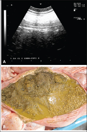
Fig. 7. Ultrasonographic and postmortem findings in an adult female camel with a history of pica and decreased fecal output and abdominal distension. Image (A) was taken using a 3.5 MHz sector transducer and shows sonography of the rumen from the left lower abdomen where several pin-points acoustic shadowing were imaged. Image (B) shows large amounts of sand within the rumen detected at postmortem examination. During the past 13 years, our research group has focused on the effectiveness of diagnostic imaging techniques in dromedary camels either in the healthy or diseased conditions (El-Tookhy and Tharwat, 2012; Ali et al., 2018; Al-Sobayil et al., 2018; Tharwat et al., 2012a,b,c,d; Tharwat, 2013a; Tharwat et al., 2013; Tharwat and Al-Sobayil, 2016a; Tharwat et al., 2018a; Tharwat, 2019; Tharwat, 2020a,b,c; Tharwat, 2021a,b; Tharwat and Al-Hawas, 2021; Tharwat and El-Tookhy, 2021; Tharwat et al., 2021; Tharwat and Al-Hawas, 2023; Tharwat et al., 2023; Tharwat,$ 2024; Tharwat and Al-Hawas, 2024). In these studies, the occluding foreign body was imaged obstructing the intestines either partially or completely. Because of the gaseous contents, the foreign bodies could not be localized within the rumen. However, in camels with rumen sand impaction, shining pin-points with acoustic enhancement are clearly visualized.
Fig. 8. Pica in two female camels with a history of reduced feed intake and weight loss. Exploratory rumenotomy revealed large amounts of plastics, cloths, and robes in the image (A), and plastics and robes in image (B). ConclusionThe syndrome of pica or depraved appetite is an important condition in dromedary camels. It was found in camels fed even a well-balanced diet resulting in ingestion of any objects other than normal feed. Substantial economic losses can be expected as a result of this syndrome as 36% of camels under investigation did not respond to treatment and collapsed. Death might be attributed to intestinal obstruction or some other critical conditions. Dietary abnormalities such as mineral or protein deficiency, parasitism, malnutrition, and ketosis are predisposing factors. Major complications may include progressive weight loss, choking, poisoning, and even death. Imaging of the digestive system enabled the clinician in 10 out of 25 cases (40%) for the localization of occluding foreign bodies in the intestines, while in the rumen, ultrasonography was only valuable in cases of sand impaction.
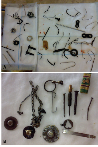
Fig. 9. Pica in two female camels with a history of reduced feed intake and weight loss. Exploratory rumenotomy in the first animal (A) revealed numerous sharp and blunt metals and glasses. In the second female (B), sharp and blunt metals, chains, and sharp nails were found. AcknowledgmentsResearchers would like to thank the Deanship of Scientific Research, Qassim University for funding the publication of this project. Conflict of interestThe authors declare that there is no conflict of interest. Authors’ ContributionConceptualization and design: MT; acquisition of data: MT, WRE, and TIA; formal analysis and interpretation of data: MT, WRE, and TIA; writing-original draft preparation: MT; writing-review and editing: MT, WRE, and TIA. All authors revised and approved the final manuscript for publication. FundingPublication of this research was funded by the Deanship of Scientific Research, Qassim University, Saudi Arabia. Data availabilityAll data supporting the findings of this study are available within the manuscript and no additional data sources are required. ReferencesAbdelrahman, M.M., Alhidary, I.A., Aljumaah, R.S. and Faye, B. 2022. Blood trace element status in camels: a review. Animals 12, 2116. Ahmed, A.F. 2011. Esophageal obstruction in young camel calves (Camelus dromedarius). Res. J. Vet. Sci. 4, 20–26. Al Nohair, S.F. 2021. Medical benefits of camel’s milk: a comprehensive review. J. Pak. Med. Assoc. 71, 933–937. Alaqeely, R., Alhajeri, B.H., Almathen, F. and Alhaddad, H. 2021. Mitochondrial sequence variation, haplotype diversity, and relationships among dromedary camel-types. Front. Genet. 12, 723964. Alaskar, H., Alaqeely, R., Alhajeri, B.H. and Alhaddad, H. 2021. The enigma of camel-types: localities, utilities, names, and breed statuses. J. Camelid Sci. 14, 22–34. AlAskar, H., Alhajeri, B.H., Almathen, F. and Alhaddad, H. 2020. Genetic diversity and population structure of dromedary camel-types. J. Hered. 111, 405–413. Alhaddad, H. and Alhajeri, B.H. 2019. Cdrom archive: a gateway to study camel phenotypes. Front. Genet. 10, 48. Ali, A., Derar, D., Al-Sobayil, F., Tharwat, M., Ahmed, F. and Khodeir, M. 2018. Adenocarcinoma in the genital tract of female dromedary camels. J. Camel Pract. Res. 25, 181–187. Al-Sobayil, F., Ali, A., Derar, D., Tharwat, M., Ahmed, F. and Khodeir, M. 2018. Tumors in dromedary camels: prevalence, types and locations. J. Camel Pract. Res. 25, 189–197. Asín, J., Ramírez, G.A., Navarro, M.A., Nyaoke, A.C., Henderson, E.E., Mendonça, F.S., Molín, J. and Uzal, F.A. 2021. Nutritional wasting disorders in sheep. Animals 11, 501. Begum, I., Azim, A., Akhter, S., Anjum, M. and Afzal, M. 2010. Mineral dynamics of blood and milk buffaloes fed on calcium and phosphorus supplementation. Pak. Vet. J. 30, 105–109. Behrouz, S., Saadat, S., Memarzia, A., Sarir, H., Folkerts, G. and Boskabady, M.H. 2022. The antioxidant, anti-inflammatory and immunomodulatory effects of camel milk. Front. Immunol. 13, 855342. Cantile, C.Y. and Youssef, S. 2016. Nervous system. In Jubb, Kennedy & Palmer’s Pathology of Domestic Animals: Volumen 1, 6th ed.; Maxie, M.G., Ed.;W.B. Saunders: St. Louis, MO, USA, pp: 250–406. Constable, P.D., Hinchcliff, K.W., Done, S.H. and Gruenberg, W. 2017. Disturbances of appetite, food intake, and nutritional status. In Veterinary medicine: a textbook of the diseases of cattle, Horses, Sheep, Pigs and Goats. Saunders Ltd.; 11th edition, pp: 87-90. El-Tookhy, O. and Tharwat, M. 2012. Clinical and ultrasonographic findings of some ocular affections in dromedary camels. J. Camel Pract. Res. 19, 183–191. FAO. 2018. ‘Food and Agricultural Organization, United Nations. http://www.fao.org/faostat/en/#data Faye, B. 2014. The camel today: assets and potentials. Anthropozoologica 49, 167–76. Firyal, S. 2007. Pica (depraved appetite; allotrophagia) in domestic Animals and man. Pakistan Vet. J. 27, 208–210. Haskell, S. and Anttilla, T. 2001. Small ruminant clinical diagnosis and therapy. 1st ed., University of Minnesota. Hass, J. 2004. Esophageal foreign body in a 2-day-old calf. Can. Vet. J. 51, 406–408. Helmer, C., Hannemann, R., Humann-Ziehank, E., Kleinschmidt, S., Koelln, M., Kamphues, J. and Ganter, M. 2021. A case of concurrent molybdenosis, secondary copper, cobalt and selenium deficiency in a small sheep herd in northern Germany. Animals 11, 1864. Hill, G. M. and Shannon, M.C. 2019. Copper and zinc nutritional issues for agricultural animal production. Biol. Trace Elem. Res. 188, 148–159. Ho, T. M., Zou, Z. and Bansal N. 2022. Camel milk: a review of its nutritional value, heat stability, and potential food products. Food Res. Int. 153, 110870. Iqbal, A. and Khan, B.B. 2001. Feeding behavior of camel: a review. Pak. J. Agri. Sci. 38, 58–63. Kadim, I.T., Al-Amri, I.S., Alkindi, A.Y. and Haq, Q.M.I. 2022. Nutritional values and health benefits of dromedary camel meat. Anim. Front. 12, 61–70. Kaskous, S. 2016. Importance of camel’s milk for human health. Emir J. Food Agri. 28, 158–163. Melendez, P., Krueger, T., Benzaquen, M. and Risco, C. 2007. An outbreak of sand impaction in postpartum dairy cows. Can. Vet. J. 48, 1067–1070. Mohamed, M.M.S. 2018. Studies of depraved appetite in Egyptian cattle. Int. J. Res. 11, 1–6. Nagy, P. and Juhasz, J. 2016. Review of present knowledge on machine milking and intensive milk production in dromedary camels and future challenges. Trop. Anim. Health Prod. 48, 915–26. Nikvand, A.B., Rashnavadi, M. and Tabandeh, M.R. 2018. A study of pica in cattle in Iran. J. Vet. Behav. 23, 15–18. Ocal, N., Gokce, G., Gucu, A.I., Uzlu, E., Yagci, B.B. and Ural, K. 2008. Pica as a predisposing factor for traumatic reticuloperitonitis in dairy cattle: serum mineral concentrations and hematological findings. J. Anim. Vet. Adv. 7, 651–656. Onmaz, A.C., Güneş, V., Çınar, M., Çitil, M. and Keleş, I. 2019. Hematobiochemical profiles, mineral concentrations and oxidative stress indicators in beef cattle with pica. Ital. J. Anim. Sci. 18, 162–167. Padalino, B. and Menchetti, L. 2021. The first protocol for assessing welfare of camels. Front. Vet. Sci. 7, 631876. Ramadan, R.O. 2017. Esophageal obstruction in camels (Camillus dromedaries). J. Vet. Sci. Technol. 8(Suppl. 4), 57. Sadan, M., El-Khodery, S., Almatroodi, S., Alsobayil, F. and El-Shafaey, E. 2023. Diagnosing and treating esophageal obstruction in camels (Camelus dromedarius). Vet. World 16, 735–742. Sadan, M., El-Shafaey, E. and Al-Sobayil, F. 2020. Diagnosis and treatment of foreign bodies swallowing syndrome in camels (Camelus dromedarius) with special reference to the role of mineral deficiency. J. Vet. Med. Sci. 82, 1097–1103. Sadan, M., Tharwat, M., Alkhedhairi, S., Refaai, W., Moghazy, HMEL., Khodier, MM., Alkhamiss, AS. and Ghallab, A. 2024. Abdominal pedunculated leiomyoma in a male dromedary camel: clinical, hematobiochemical, ultrasonographic and pathologic findings. Int. J. Vet. Sci. 13, 458–462. Sikkema, R.S., Farag, E.A.B.A., Islam, M., Atta, M., Reusken, C.B.E.M., Al-Hajri, M.M. and Koopmans, M.P.G. 2019. Global status of Middle East respiratory syndrome coronavirus in dromedary camels: a systematic review. Epidemiol. Infect. 147, e84–e84. Singh, P., Chandolia, R.K., Behl, S.M., Tayal, R., Chawla, S.K. and Bugalia, N.S. 2011. Surgical management of esophageal obstruction in camels-report of 5 clinical cases. J. Camel Pract. Res. 18, 347–349. Smith, B.P. 2015. Pica. In Large animal internal medicine. 5th edition. Mosby: Elsevier, p. 152. Tharwat, M. and Al-Hawas, A. 2023. Infrared thermography in healthy Arabian camels (Camelus dromedarius). Int. J. Vet. Sci. 12, 762–767. Tharwat, M. 2013a. Ultrasonographic findings in camels (Camelus dromedarius) with trypanosomiasis. J. Camel Pract. Res. 20, 283–287. Tharwat, M. 2013b. Ultrasonography of the lungs and pleura in healthy camels (Camelus dromedarius). Acta Vet. Hung. 61, 309–318. Tharwat, M. 2019. Chronic peritonitis in dromedary camels: clinical, hematobiochemical, ultrasonographic and pathologic findings. J. Camel Pract. Res. 26, 169–172. Tharwat, M. 2020a. Ultrasonography of the abdomen in healthy and diseased camels (Camelus dromedaries). J. Appl. Anim. Res. 48, 300–312. doi: 10.1080/09712119.2020.1788035. Tharwat, M. 2020b. Ultrasonography of the liver in healthy and diseased camels (Camelus dromedaries). J. Vet. Med. Sci. 82, 399–407. Tharwat, M. 2020c. Ultrasonography of the kidneys in healthy and diseased camels (Camelus dromedaries). Vet. Med. Intern. 2020, 7814927. Tharwat, M. 2021a. Obstructive urolithiasis in dromedary camels: clinical, ultrasonographic and postmortem findings. J. Camel Pract. Res. 28, 85–93. Tharwat, M. 2021b. Ultrasonography of the thorax in healthy and diseased camels (Camelus dromedarius)—an overview. J. Camel Pract. Res. 28, 313–318. Tharwat, M. 2024. Fundamentals of diagnostic ultrasound in dromedary camel medicine. Int. J. Vet. Sci. 13, 1–6. Tharwat, M. and Al-Hawas, A. 2021. Ultrasound detection of cosmic fillers injection of lips in camel beauty pageants: first report in veterinary medicine. Trop. Anim. Health Prod. 53, 53. Tharwat, M. and Al-Hawas, A. 2024. The emerging topic of cosmetic medicine in dromedary camels. Int. J. Vet. Sci. 13, 139–146. Tharwat, M. and Al-Hawas, A. 2024. The syndrome of pica or depraved appetite in small ruminants: A mini-review. Int. J. Vet. Sci. 13, 341–348. Tharwat, M. and Al-Sobayil, F. 2016a. Ultrasonographic findings in camels (Camelus dromedarius) with abdominal disorders. J. Camel Pract. Res. 23, 291–299. Tharwat, M. and El-Tookhy O. 2021. Ocular ultrasonography in camels (Camelus dromedarius): a review. J. Camel Pract. Res. 28, 185–190. Tharwat, M., Al-Hawas, A. and Albotti, Y. 2021. Infrared thermography in healthy dromedary camels and its feasibility in injected and stretched lips in camel beauty pageants. J. Camel Pract. Res. 28, 355–359. Tharwat, M., Ali, A., Al-Sobayil, F. and Buczinski, S. 2013. Ultrasound-guided collection of peritoneal fluid in healthy camels (Camelus dromedarius) and its biochemical analysis. Small Rum. Res. 113, 307–311. Tharwat, M., Al-Sobayil, F., Ali, A. and Buczinski, S. 2012a. Transabdominal ultrasonographic appearance of the gastrointestinal viscera of healthy camels (Camelus dromedarius). Res. Vet. Sci. 93, 1015–1020. Tharwat, M., Al-Sobayil, F., Ali, A. and Buczinski, S. 2012b. Ultrasonographic evaluation of abdominal distension in 52 camels (Camelus dromedarius). Res. Vet. Sci. 93, 448–456. Tharwat, M., Al-Sobayil, F., Ali, A. and Buczinski, S. 2012e. Ultrasonography of the liver and kidneys of healthy camels (Camelus dromedarius). Can. Vet. J. 53, 1273–1278. Tharwat, M., Al-Sobayil, F., Ali, A. and Buczinski, S. 2012f. Echocardiography of the normal camel (Camelus dromedarius) heart: technique and cardiac dimensions. BMC Vet. Res. 8, 130. Tharwat, M., Al-Sobayil, F., Ali, A., Derar, D. and Khodeir, M. 2017. Renal cell carcinoma in a female Arabian camel: clinical, hematobiochemical, ultrasonographic and pathologic findings. J. Camel Pract. Res. 24, 61–66. Tharwat, M., Al-Sobayil, F., Ali, A., Hashad, M. and Buczinski, S. 2012d. Clinical, ultrasonographic, and pathologic findings in 70 camels (Camelus dromedarius) with Johne’s disease. Can. Vet. J. 53, 543–548. Tharwat, M., Al-Sobayil, F., Ali, A., Wittek, T. and Floeck, M. 2012c. Percutaneous ultrasound-guided portocentesis in camels (Camelus dromedaries). J. Camel Pract. Res. 19: 193–196. Tharwat, M., and Al-Sobayil, F. 2016b. Ultrasonographic findings in camel calves (Camelus dromedarius) with thoracic affections. J. Camel Pract. Res. 23, 287–290. Tharwat, M., El Moghazy, H.M. and Oikawa, S. 2023. Ultrasonographic verification of hepatic hydatidosis in a female dromedary camel: a case report. J. Vet. Med. Sci. 85, 1286–1290. Tharwat, M., El-Shafaey, E., Sadan, M., Ali, A., Al-Sobayil, F. and Al-Hawas, A, 2018a. Omaso-abomasal adenocarcinoma in a female Arabian camel (Camelus dromedarius). J. Appl. Anim. Res. 46, 1268–1271. Tharwat, M., Sadan, M., El-Shafaey, E., Al-Hawas, A. and Saeed, E.M.A. 2018b. Unilateral nephrectomy in a female dromedary camel with pyelonephritis caused by Staphylococcus lugdunensis. Pakistan Vet. J. 38, 116–118. Tharwat, M., Sadan, M., El-shafaey, E., Saeed, E. and Al-hawas, A. 2018c. Bilateral renal abscessation and chronic active pyelonephritis in a male camel (Camelus dromedarius) caused by Escherichia coli. J. Vet. Med. Sci. 80, 778–783. | ||

| How to Cite this Article |
| Pubmed Style Tharwat M, El-Ghareeb WR, Almundarij T. Depraved appetite in dromedary camels: Clinical, ultrasonographic and postmortem findings. Open Vet J. 2024; 14(2): 652-663. doi:10.5455/OVJ.2024.v14.i2.5 Web Style Tharwat M, El-Ghareeb WR, Almundarij T. Depraved appetite in dromedary camels: Clinical, ultrasonographic and postmortem findings. https://www.openveterinaryjournal.com/?mno=173288 [Access: April 27, 2024]. doi:10.5455/OVJ.2024.v14.i2.5 AMA (American Medical Association) Style Tharwat M, El-Ghareeb WR, Almundarij T. Depraved appetite in dromedary camels: Clinical, ultrasonographic and postmortem findings. Open Vet J. 2024; 14(2): 652-663. doi:10.5455/OVJ.2024.v14.i2.5 Vancouver/ICMJE Style Tharwat M, El-Ghareeb WR, Almundarij T. Depraved appetite in dromedary camels: Clinical, ultrasonographic and postmortem findings. Open Vet J. (2024), [cited April 27, 2024]; 14(2): 652-663. doi:10.5455/OVJ.2024.v14.i2.5 Harvard Style Tharwat, M., El-Ghareeb, . W. R. & Almundarij, . T. (2024) Depraved appetite in dromedary camels: Clinical, ultrasonographic and postmortem findings. Open Vet J, 14 (2), 652-663. doi:10.5455/OVJ.2024.v14.i2.5 Turabian Style Tharwat, Mohamed, Waleed Rizk El-Ghareeb, and Tariq Almundarij. 2024. Depraved appetite in dromedary camels: Clinical, ultrasonographic and postmortem findings. Open Veterinary Journal, 14 (2), 652-663. doi:10.5455/OVJ.2024.v14.i2.5 Chicago Style Tharwat, Mohamed, Waleed Rizk El-Ghareeb, and Tariq Almundarij. "Depraved appetite in dromedary camels: Clinical, ultrasonographic and postmortem findings." Open Veterinary Journal 14 (2024), 652-663. doi:10.5455/OVJ.2024.v14.i2.5 MLA (The Modern Language Association) Style Tharwat, Mohamed, Waleed Rizk El-Ghareeb, and Tariq Almundarij. "Depraved appetite in dromedary camels: Clinical, ultrasonographic and postmortem findings." Open Veterinary Journal 14.2 (2024), 652-663. Print. doi:10.5455/OVJ.2024.v14.i2.5 APA (American Psychological Association) Style Tharwat, M., El-Ghareeb, . W. R. & Almundarij, . T. (2024) Depraved appetite in dromedary camels: Clinical, ultrasonographic and postmortem findings. Open Veterinary Journal, 14 (2), 652-663. doi:10.5455/OVJ.2024.v14.i2.5 |

