


| Research Article | ||
Open Veterinary Journal, (2024), Vol. 14(1): 154-163 Original Research Assessment of some toxic elements (Co, Cr, Mn, Se, and As) in muscle, offal, hair, and blood of camels (Camelus dromedaries) and their risk assessmentAhmed M. A. Meligy1,2, Waleed R. El-Ghareeb3,4*, Sherief M. Abdel-Raheem3,5, Hesham A. A. Ismail3,6, Wageh S. Darwish4, Mahmoud Kandeel7,8, Ahmed E. Alfifi3, Saad S. Al-Shokair1 and Mohamed A. Hussein41Department of Clinical Sciences, College of Veterinary Medicine, King Faisal University, P.O. Box 400, Al-Hofuf 31982, Al-Ahsa, Saudi Arabia 2Department of Physiology, PPRI Institute, Agriculture Research Center, Giza, Egypt 3Department of Public Health, College of Veterinary Medicine, King Faisal University, P.O. Box 400, Al-Hofuf 31982, Al-Ahsa, Saudi Arabia 4Food Control Department, Faculty of Veterinary Medicine, Zagazig University, Zagazig, Egypt 5Department of Animal Nutrition and Clinical Nutrition, Faculty of Veterinary Medicine, Assiut University, Assiut, Egypt 6Department of Food Hygiene, Faculty of Veterinary Medicine, Assiut University, Assiut, Egypt 7Department of Biomedical Sciences, College of Veterinary Medicine, King Faisal University, Al Hofuf, Saudi Arabia 8Department of Pharmacology, Faculty of Veterinary Medicine, Kafrelsheikh University, Kafrelsheikh, Egypt *Corresponding Author: Waleed R. El-Ghareeb. Department of Public Health, College of Veterinary Medicine, King Faisal University, Hofuf, Saudi Arabia. Email: welsaid [at] kfu.edu.sa Submitted: 01/10/2023 Accepted: 15/12/2023 Published: 31/01/2024 © 2024 Open Veterinary Journal
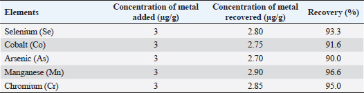
AbstractBackground: Camel meat tainted with heavy metals or trace elements may pose a health risk to consumers. Heavy metal contamination poses a severe danger due to both their toxicity and bioaccumulation in the food chain. Aim: To estimate the residual levels of heavy metals (Co, Cr, Mn, Se, and As) in muscle, liver, kidney, hair, and serum of three camel breeds (Magaheem, Maghateer, and Wadha) collected from Al-Omran abattoir, Al-Ahsa, Saudi Arabia. Methods: A total of 225 tissue samples (muscles, liver, kidney, serum, and hair) were taken and analyzed using an Atomic Absorption Spectrophotometer. Health risk assessment was assessed using the guidelines set by the US Environmental Protection Agency. Results: Camel breed significantly (p < 0.05) influences Co, Cr, Mn, and Se accumulation and distribution in organs and muscle; however, arsenic accumulation was not significantly affected (p < 0.05) by camel breeds. The highest values of Co, Cr, Se, and Mn in all examined samples were detected in the liver samples of Maghateer and Magaheem breeds. Furthermore, significant strong positive correlation between serum and liver cobalt, chromium, manganese, and arsenic. The estimated daily intake owing to camel meat consumption was less than the tolerated daily intake. Conclusion: Heavy metals were distributed among different breeds of camel. Trace elements (Pb and Cd) in meat and offal were below the international maximum permissible limit. The correlation between samples reflects the role of hair as a good tool for the identification of heavy metal pollution. Keywords: Camel carcass, Toxic metals, Risk assessment. IntroductionSaudi Arabia is the Middle East’s largest producer of camel meat, accounting for 62% of the total volume with annual per capita consumption of 3.10 kg (Index box, 2022). Camel meat is becoming increasingly popular due to its low fat, low cholesterol, and high polyunsaturated fatty acids (Kadim et al., 2008). Because of its comparable nutritional value, camel meat is a good alternative to other red meat (Kadim et al., 2022). The hazard of heavy metals in meat is of big concern for food safety and public health as well due to their toxic effect at very low concentrations (Santhi et al., 2008). When camels graze freely and drink water from contaminated sources, heavy metals may bio-accumulate and bio-magnify in their tissues and organs (Bala et al., 2018). Some heavy metals were proven to have carcinogenic, mutagenic, or teratogenic effects (Pitot and Dragon, 1995). In addition, toxic metals may compromise the metabolism and bioavailability of essential metals and decrease their body concentration (Lazarus, 2010; Matović et al., 2011). Trace elements, such as selenium, manganese, and cobalt, are essential metals where they play a significant role in biological systems. However, they can be very harmful and produce toxic effects if taken in excessive amounts (Mohammed et al., 2011). Chronic arsenic toxicity increases the risk of developing various cancers, such as skin, liver, lung, bladder, kidney, and colon cancer (Hu, 2002). Manganese toxicity can cause many pathological alterations in the CNS, reproductive and immune system dysfunction, damage to the testicles and pancreas, and hepatitis (Keen and Leach, 1987). Selenium plays a significant role in the health of both animals and humans. It is a crucial component of several enzymes that are required for the immune system as well as anti-carcinogenic activity (Navarro and Lopez, 2000). Monitoring the levels of trace elements and heavy metals in camel meat, as a human food, is of significant importance for both food safety and human health. Therefore, the goal of this research is to assess the levels of trace elements (selenium, manganese, and cobalt) and heavy metals (arsenic and chromium) in the blood, hair, and offal of three different camel breeds (Magaheem, Maghateer, and Wadha) in Al-Ahsa province, Saudi Arabia. In addition, human dietary intake of these metals and risk assessment associated with the consumption of such camel meat were estimated. Materials and MethodsSamples collectionA total of 225 tissue samples of muscles, liver, kidney, serum, and hair (n=45 for each) were taken from three local camel breeds directly after slaughter. Samples were obtained from Al-Omran central slaughterhouse, Al-Ahsa, Saudi Arabia. Al-Ahsa is classified as rural since it relies primarily on livestock production, but industrial activities are restricted. From October 2021 until January 2022, samples were gathered. The ages of the animals ranged from less than 5 to more than 10 years. All animals appeared to be in good health, active and disease-free. The samples were kept at a temperature of −20°C in falcon tubes until analysis. Sample preparation and extractionThe Shimadzu AA-7000 Atomic Absorption Spectrophotometer was used in conjunction with a graphite furnace atomic absorption spectrometry system (GFAAS) to assess the amounts of the trace elements (Se, Mn, Cr, Co, and As) (Table 1). In addition, hollow cathode lamps were used for the analysis. For Se, Mn, Cr, Co, and As analysis, were measured using the GFAAS system. The Shimadzu ASO6100 Automatic Sampler was used to inject the samples into the GFAAS (Waheed et al., 2022). After dilution and filtration with Whatman filter paper 1, the digested samples were analyzed using atomic absorption spectrophotometry according to the method mentioned by Meligy et al. (2019) and Hussein et al. (2022). Metal concentrations were calculated using standard curves for all metals studied. Quality assurance and controlMeasurement of IAEA142/TM from IAEA-certified reference materials (muscle homogenate) was used to ensure the accuracy of the assay (Vienna, Austria). The certified samples’ recovered concentrations were 5% of the certified values. Triplicates of each sample were evaluated. Estimated daily intake (EDI)As described by the Human Health Evaluation Manual (United States Environmental Protection Agency, 2002), the equation was used to calculate the EDI of the metals studied: EDI=Cm × FIR/BW. Where EDI is expressed in µg/kg/day; Cm is the metal concentration in the sample (measured in mg/kg wet weight); FIR is for Saudi Arabia’s meat intake rate, which was assessed to be 146 g per day; BW stands for Saudi adults and children body weight, which was assessed to be 70 kg for adults and 30 kg for children (Adam et al., 2014, Hussein et al., 2022). Health risk assessmentThe noncancer risk caused by the consumption of metal-contaminated edible tissues by the Saudi population (adults and children) was assessed using the guidelines set by the (United States Environmental Protection Agency, 2002). Statistical analysisSPSS (2010) was utilized to conduct statistical analysis. To see if variables were normally distributed, the Kolmogorov-Smirnov normality test was used. One-way analysis of variance was applied to compare the means of the groups when breed was used as a factor. To examine the impact of breed on the analyzed parameters, the Duncan multiple range test (Steel and Torrie, 1980) was used. The differences between genders were tested by independent t-test. Table 1. Recovery of trace elements and heavy metals from homogeneous muscle samples.
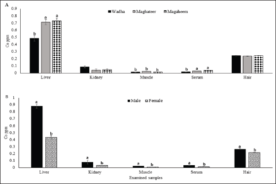
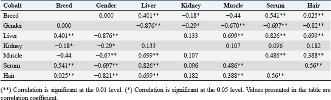
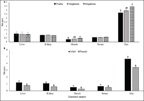
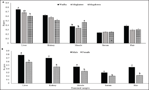
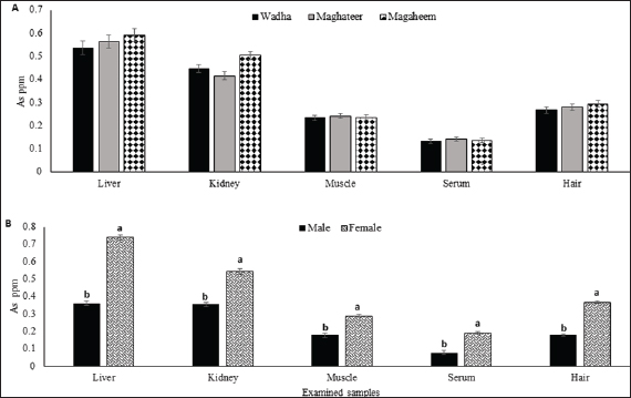
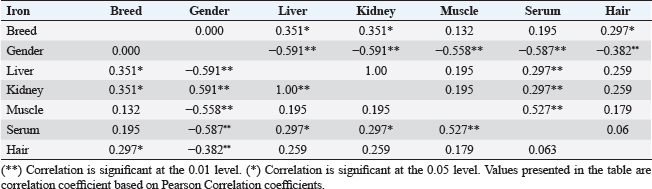
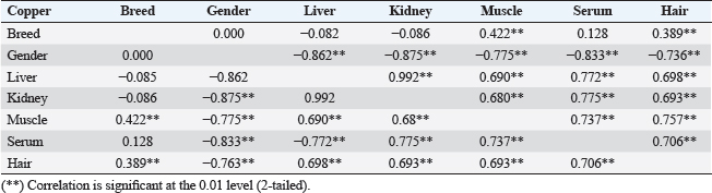
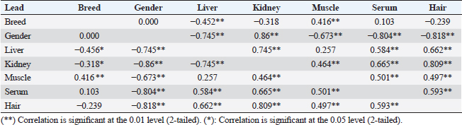
Ethical approvalAll research procedures were carried out by King Faisal University’s regulations and requirements. ResultsCobaltThe recorded data in Figure 1A declared that mean values of Cobalt ranged from 0.246 to 1.03, 0.024 to 0.652, 0.004 to 0.043, 0.008 to 0.066, and 0.205 to 0.326 mg.kg−1 in liver, kidney, muscle, serum, and hair, respectively. The cobalt values significantly varied between breeds (p < 0.05). Furthermore, values are arranged in a descending manner liver > kidney> hair > serum > muscle. Regarding the effect of gender as shown in Figure 1B, all male collected samples had substantially greater (p < 0.05) Co levels than female samples. The correlation coefficient between Co concentrations in organs and serum with breed and gender in Table 2 revealed a strong positive correlation between muscle and liver cobalt levels. ChromiumThe recorded data in Figure 2A declared that mean values of chromium ranged from 0.41 to 0.75, 0.23 to 0.57, 0.02 to 0.20, 0.12 to 0.41, and 0.05 to 0.95 mg.kg−1 in liver, kidney, muscle, serum, and hair, respectively. Furthermore, values are arranged in a descending manner hair > liver > kidney > serum > muscle. The chromium content in all male samples studied was considerably greater (p < 0.001) than in female samples (Fig. 2B). Manganese (Mn)The recorded data in Figure 3A declared that mean values of Mn ranged from 0.460 to 1.6, 0.310 to 1.5, 0.000 to 0.690, 0.41 to 0.77, and 3.06 to 5.761 mg.kg−1 in liver, kidney, muscle, serum, and hair, respectively. The Mn content in all male camel samples studied was considerably greater (p < 0.001) than in female camel samples (Fig. 3B). SeleniumThe recorded data in Figure 4A declared that mean values of selenium ranged from 0.424 to 0.424, 0.28 to 0.76, 0.211 to 0.587, 0.156 to 0.367, and 0.11 to 0.589 mg.kg−1 in liver, kidney, muscle, serum, and hair, respectively. The selenium content in all male camel samples studied was considerably greater (p < 0.05) than in female samples (Fig. 4B). The correlation coefficient between Se concentrations in organs and serum with breed and gender in Table 5 revealed a strong positive correlation between muscle and kidney cobalt level. Moreover, a positive correlation was detected between serum, liver, kidney, and hair. ArsenicThe obtained data in Figure 5A declared that mean values of As ranged from 0.304 to 0.887, 0.224 to 0.723, 0.15 to 0.44, 0.12 to 0.37, and 0.02 to 0.27 mg.kg−1 in liver, kidney, hair, muscle, and serum, respectively. The correlation coefficient between As concentrations in organs and serum with breed and gender in Table 6 revealed a strong positive correlation between gender and all examined samples (Fig. 5B). Meanwhile, breeds negatively correlated with liver, kidney, and hair.
Fig. 1. Cobalt mean values (ppm) in liver, kidney, muscle, serum, and hair A. Effect of breed on cobalt concentrations (ppm). B. Effect of Gender on cobalt concentration (ppm). a,b Means with different superscript letters in the same sample type are significantly different (p ≤ 0.05). Table 2. Correlations between cobalt concentrations in organs and serum with breed and gender.
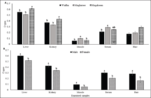
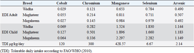
Fig. 2. Chromium mean values (ppm) in liver, kidney, muscle, serum, and hair A. Effect of breed on chromium concentrations (ppm). B. Effect of Gender on chromium concentration (ppm). a,b Means with different superscript letters in the same sample type are significantly different (p ≤ 0.05). Health risk assessmentThe presented results in Table 7 indicated that the EDI of all investigated metals from all breeds were below the tolerated daily intake (TDI) approved by FAO/WHO (2010). The noncarcinogenic hazard ratios (HRs) and hazard indices (HIs) were assessed in Table 8. DiscussionCobaltThe maximum Co concentration found in liver samples from different camel breeds was equivalent to the finding of Mahmud et al. (2011) and Asli et al. (2020) who examined muscle and liver in Iranian camels and found low concentrations in the liver and muscle of camels. Relatively higher levels of Co were reported in the liver compared with meat, liver, lung, heart, and kidney in previous studies as 1.10–14.22 mg kg−1 (Chafik et al., 2014) and 1.913–8.194 mg kg−1 (Al-Perkhdri, 2021). Camel breed influences Co accumulation and distribution in camel organs and muscle. Significant variations (p < 0.05) were found across breeds, which can be explained by each breed’s environment being in a distinct geographical location with varying concentrations of metals in soil, forages, and water. Faye et al. (2008) detected significantly higher cobalt levels in male than female camels collected from Emirates. The positive correlation attributed to Co can enter the body through the digestive tract from food or drink water or lungs after inhalation of Co dust.
Fig. 3. Manganese mean values (ppm) in liver, kidney, muscle, serum, and hair A. Effect of breed on manganese concentrations (ppm). B. Effect of Gender on manganese concentration (ppm). a,b Means with different superscript letters in the same sample type are significantly different (p ≤ .0.05).
Fig. 4. Selenium mean values (ppm) in liver, kidney, muscle, serum, and hair A. Effect of breed on selenium concentrations (ppm). B. Effect of Gender on selenium concentration (ppm). a,b Means with different superscript letters in the same sample type are significantly different (p ≤ 0.05). ChromiumThe chromium concentration in the current study ranged from 0.032 to 96.62 mg/kg. There are significant differences (p < 0.05) between breeds according to their contents of Cr between liver kidney, muscle, and hair (Fig. 2A). The Cr variations in our findings are attributable to variances in the availability of Cr in forages that are grown in the grazing areas. The highest values of Cr in all examined samples were detected in the liver samples of maghateer magaheem breeds.The correlations between chromium concentrations in organs and serum with breed and gender was displayed in Table (3). The average of Cr in this study was (0.4) mg/kg in meat and organs which coincided with USDA (2006) and China standards (2006) which stated the maximum level of chromium in meat as 1.0 mg/kg. Higher Cr values obtained were 2.333–4.92 mg kg−1 of muscle and 4.256–9.878 mg kg−1 of liver samples in Iran (Asli et al., 2020).
Fig. 5. Arsenic mean values (ppm) in liver, kidney, muscle, serum, and hair A. Effect of breed on arsenic concentrations (ppm). B. Effect of gender on arsenic concentration (ppm). a,b Means with different superscript letters in the same sample type are significantly different (p ≤ 0.05). Table 3. Correlations between chromium concentrations in organs and serum with breed and gender.
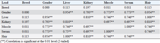
Table 4. Correlations between manganese concentrations in organs and serum with breed and gender.
Table 5. Correlations between selenium concentrations in organs and serum with breed and gender.
Table 6. Correlations between arsenic concentrations in organs and serum with breed and gender.
Manganese (Mn)Manganese values significantly varied between breeds (p < 0.05). Furthermore, values are arranged in a descending manner hair > liver > kidney > serum > muscle. The Mn content in all male camel samples studied was considerably greater (p < 0.001) than in female camel samples (Fig. 3B). Similar to our finding male and female camels had Mn in hair (Faraz et al., 2020). In addition, adult male and female camel from Pakistan dromedary calves contained 45.8 ± 1.8 and 32.9 ± 4.4 ppm in male and female hair, respectively (Faraz et al., 2020). On the other hand, there were significant variations (p < 0.05) between Mn content in collected meat samples from middle and southern districts and other districts in the Saudi Kingdom (Faraz et al., 2020). The correlation coefficient between Mn concentrations in organs and serum with breed and gender in Table 4 revealed a strong positive correlation between muscle and hair Mn levels. Moreover, a positive correlation was detected between serum and liver. The negative correlation obtained for both breed and gender may attributed to the accumulation of heavy metals in the animal body related to environmental factors such as air pollution, feeding, and available water sources. SeleniumThe Se values significantly varied between breeds (p < 0.05). Furthermore, values are arranged in a descending manner kidney > hair > muscle > liver > serum. The current findings of higher values of Se in kidney samples come in accordance with Seboussi et al. (2010) reported the selenium concentrations in liver (216.1 µg/kg), kidney (1,006 µg/kg), for limb muscle (368.7 µg/kg), and hair (80.7 µg/kg). It is proved that the kidney was the richest organ in Se. In addition, the hair seemed to be the best indicator of selenium intake in camel (Seboussi et al., 2010). In Saudi Arabia, serum Se values reported in young camels at the slaughterhouse varied between 5.3 and 131 ng/ml. Serum concentrations in camels were 0.281 ppm on average in sera coming from the Sultanate of Oman (Faye et al., 2008). The threshold for serum selenium to be considered a deficiency is below 35 ng/ml as indicated by El-Khouly et al. (2001). However, in young adults, Al-Qarawi et al. (2001) reported the appearance of clinical signs of Se deficiency only in animals with selenium values below 5 ng/ml. The selenium content in all male camel samples studied was considerably greater (p < 0.05) than in female samples (Fig. 4B). Meanwhile, Seboussi et al. (2004) recorded that adult males presented lower values than females 139 ± 5 versus 229 ± 7 ng/ml, respectively. The correlation coefficient between Se concentrations in organs and serum with breed and gender in Table 5 revealed a strong positive correlation between muscle and kidney cobalt levels. Moreover, a positive correlation was detected between serum, liver, kidney, and hair. The negative correlation obtained for both breed and gender may attributed to the accumulation of trace elements in the animal body related to environmental factors. The highest values for Se were detected in the Wadha breed than the other two breeds. There was a contradiction in the breed effect when compared to the work done by Abdelrahman et al. (2013), who reported a two-fold higher concentration in Majaheem camels (147.1 ng/ml) than in Waddha camels (73.3 ng/ml), while Seboussi et al. (2004) indicated no genetic difference. Table 7. EDI in comparison with TDI (μg/kg/day) due to ingestion of camel meat from different breeds.
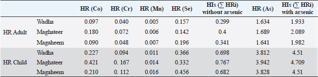
Table 8. The HR and HIs are due to ingestion of camel meat from different breeds.
ArsenicThe As values were not significantly varied between breeds but were highly significant with the effect of Gender. The obtained results were in comparable with the findings of El-Ghareeb et al. (2019). One probable explanation for the high As level in the studied samples is the use of feed additives rich in arsenic during intensive production, which is common in several regions worldwide (Hu et al., 2017). Inorganic arsenic is naturally present at high levels in the groundwater of a number of countries. Sources of arsenic exposure could include drinking water, crops irrigated with such water, and food produced with such water as well (Flanagan et al., 2012). The correlation coefficient between As concentrations in organs and serum with breed and gender in Table 5 revealed a strong positive correlation between gender and all examined samples. Meanwhile, breeds negatively correlated with liver, kidney, and hair. Health risk assessmentThe presented results in Table 7 indicated that the EDI of all investigated metals from all breeds were below the TDI approved by FAO/WHO. The EDI of Pb and Cd were lower than those reported from camel meat in Saudi Arabia (El-Ghareeb et al., 2019). The noncarcinogenic HRs and HIs were assessed in this study in Table 8. The HR from all examined metals below 1 meanwhile, HR(As) exceeding 1 for adults and children as 1.634 and 3.812, 1.689 and 3.942, and 1.641 and 3.828 from meat consumption of Wadha, Maghateer, Magaheem breeds, respectively. Since the toxicological profile primarily takes into account the inorganic chemical forms of arsenic, HR values for As bigger than one are not relevant for human wellness. This is because organic forms of arsenic are relatively nontoxic to humans (ATSDR, 2005). The HIs due to the consumption of the Maghateer breed are slightly higher than Wadha or Magaheem. On neglecting HR (As), HIs values were below 1, which proved no potential exposure to risk due to the consumption of camel meat from different breeds. The obtained values for HR and HIs were lower than those in Ghana (Bortey et al., 2015) and Saudi Arabia (El-Ghareeb et al., 2019). ConclusionIt can be concluded that heavy metals are distributed among camel samples of different breeds. Trace elements (Pb and Cd) in meat and offal were below the international maximum permissible limit. The correlation between samples reflects the role of hair as a good tool for the identification of heavy metal pollution. In addition, no potential health hazards among camel meat consumers in Saudi Arabia especially, adults. We need some future studies to differentiate between the percentages of organic and inorganic arsenic in camel meat and offal. AcknowledgmentThis work was supported through the Annual Funding track by the Deanship of Scientific Research, Vice Presidency for Graduate Studies and Scientific Research, King Faisal University, Saudi Arabia [Project No 4002]. Conflict of interestThe authors declare that there is no conflict of interest. FundingThe Annual Funding track by the Deanship of Scientific Research, Vice Presidency for Graduate Studies and Scientific Research, King Faisal University, Saudi Arabia [Project No. 4002]. Data availabilityAll data are provided in the manuscript. Any extra data needed can be provided by the corresponding author upon reasonable request. ReferencesAbdelrahman, M.M., Aljumaah, R.S. and Ayadi, M. 2013. Selenium and iodine status of two camel breeds (Camelus dromedaries) raised under semi intensive system in Saudi Arabia. Ital. J. Anim. Sci. 12(1), e14. Adam, A., Osama, S. and Muhammad, K.I. 2014. Nutrition and food consumption patterns in the Kingdom of Saudi Arabia. Pak. J. Nut. 134, 181–190. Al-Perkhdri, A.S.A. 2021 April. Study of the some heavy metals residues in the camel meat in different regions of Kirkuk governorate during the spring and summer seasons. IOP. Conf. Ser. Earth. Environ. Sci. 735(1), 012080. Al-Qarawi, A.A., Abbas, B., Haroun, E.M., Mahmoud, O.M. and Al-Hawas, A. 2001. Clinicopathological investigation of selenium responsive myopathy in young adult camels. J. Camel. Pract. Res. 8, 23–27. Asli, M., Azizzadeh, M., Moghaddamjafari, A. and Mohsenzadeh, M. 2020. Copper, iron, manganese, zinc, cobalt, arsenic, cadmium, chrome, and lead concentrations in liver and muscle in Iranian camel (Camelus dromedarius). Biol. Trace. Elem. Res. 194, 390–400. ATSDR. 2005. Agency for toxic substances and disease registry. Public health assessment guidance manual. Atlanta, Georgia: US Department of Health and Human Services. Available via https://www.atsdr.cdc.gov/pha-guidance/index.html (Accessed 07 October 2023). Bala, A., Junaidu, A.U., Salihu, M.D., Agaie, B.M., Saulawa, M.A., Musawa, A.I. and Ahmad, K.H. 2018. Determination of heavy metal residues in slaughtered camels at Sokoto and Gusau modern abattoirs, Nigeria. J. Health. Poll. 8(20), 181204. Bortey-Sam, N., Nakayama, S.M., Ikenaka, Y., Akoto, O., Baidoo, E., Yohannes, Y.B., Mizukawa, H. and Ishizuka, M. 2015. Human health risks from metals and metalloid via consumption of food animals near gold mines in Tarkwa, Ghana: estimation of the daily intakes and target hazard quotients (THQs). Ecotoxicol. Environ. Saf. 111, 160–167. Chafik, A., Essamadi, A., Eddoha, R., Bagri, A., Nasser, B. and Faye, B. 2014. Trace elements and heavy metals in organs of camels (Camelus dromedarius) slaughtered in Casablanca city, Morocco. J. Camel Pract. Res. 21(2), 145-152. China Standard. 2006. Specific maximum levels of contaminations in foods. Date: 8/14/2006 GAIN Report Number: CH6064 China, Peoples Republic of FAIRS 2006 Voluntary Report—public distribution. El-Ghareeb, W.R., Darwish, W.S. and Meligy, A.M.A. 2019. Metal contents in the edible tissues of camel and sheep: human dietary intake and risk assessment in Saudi Arabia. JJVR 67(1), 5–14. El-Khouly, A.A., Abbas, T.A. and Moustafa, T. 2001. Myocardial dystrophy in camel calves in the United Arab Emirates (field cases). Emir. J. Food. Agric. 13, 11–17. Faraz, A., Waheed, A., Mustafa, A.B., Tauqir, N.A. and Eldeib, A.O., 2020. Comparative growth response related to hair mineral analysis in dromedary camel calves. Open. Vet. J. 10(4), 392–399. Faye, B., Seboussi, R. and Askar, M. 2008. Trace elements and heavy metals status in Arabian camel. In Impact of pollution on animal products. NATO Science for Peace and Security Series Series C: Environmental Security. Eds., Faye, B. and Sinyavskiy, Y. Dordrecht, The Netherlands: Springer, pp: 97–106. Flanagan, S.V., Johnston, R.B. and Zheng, Y. 2012. Arsenic in tube well water in Bangladesh: health and economic impacts and implications for arsenic mitigation. Bull. World. Health. Organ. 90(11), 839–846. Hu, H. 2002. Human health and heavy metal exposure. In Life support: the environmental and human health. Ed., McCally, M. Cambridge, MA: MIT Press. Available via http://chge.med.harvard.edu/programs/education/secundary/hhge/documents/mccally.pdf Hu, Y., Zhang, W., Chen, G., Cheng, H. and Tao, S. 2017. Public health risk of trace metals in fresh chicken meat products on the food markets of a major production region in southern China. Environ. Pollut. 234, 667–676. Hussein, Y.A., Meligy, A.M.A., El-Ghareeb, W.R., Al-Shokair S.S. and Abdel-Raheem, S.M., 2022. Selected heavy metals and their risk assessment in camels (Camelus dromedarius). J. Camel. Pract. Res. 29(1), 89–99. Index box (2022). Camel meat market in the Middle East. Available via https://app.indexbox.io/report/020860/145/ Kadim, I.T., Al-Amri, I.S., Alkindi, A.Y. and Haq, Q.M. 2022. Nutritional values and health benefits of dromedary camel meat. Anim. Front. 12(4), 61–70. Kadim, I.T., Mahgoub, O. and Purchas, R.W. 2008. A review of the growth, and of the carcass and meat quality characteristics of the one-humped camel (Camelus dromedaries). Meat. Sci. 80(3), 555–569. Keen, C.L. and Leach, R.M. 1987. Manganese. In Handbook on toxicity of inorganic compounds. Eds., Seiler, H.G. and Sigel, H. New York, NY: Marcel Dekker, pp: 405–415. Lazarus, M. 2010. Cadmium and selenium interaction in mammals, in Croatian. Arh. Hig. Rada. Toksikol. 61, 357–369. Mahmud, T., Rehman, R., Anwar, J. and Ali, S. 2011. Minerals and nutritional composition of camel (Camelus dromedarius) meat in Pakistan. J. Chem. Soc. Pak. 33(6), 835–838. Matović, V., Buha, A., Bulat, Z. and Đukić-Ćosić, D. 2011. Cadmium toxicity revisited: focus on oxidative stress induction and interactions with zinc and magnesium. Arhiv za Higijenu Rada i Toksikologiju 62(1), 65–75. Meligy, A.M., Al-Taher, A.Y., Ismail, M., Al-Naeem, A.A., El-Bahr, S.M. and El-Ghareeb, W.R. 2019. Pesticides and toxic metals residues in muscle and liver tissues of sheep, cattle and dromedary camel in Saudi Arabia. Slovenian. Vet. Res. 56(Suppl 22), 157–166. Mohammed, S.G., Abdulsahib, H.T., Jasim, I.M. and Jabbar, M.T. 2011. Assessment of camel meat pollution with trace metals in desert area of Basra Province. Am. J. Agric. Biol. Sci. 6(4), 475–479. Navarro, A.M. and Lopez, M.M.C. 2000. Essentiality of selenium in the human health: relationship with different diseases. Sci. Total. Environ. 249, 347–371. Pitot, C.H. and Dragon, P.Y. 1995. Chemical carcinogenesis. In Casarett and Doull’s toxicology, the basic science of poisons, 5th ed. Ed., Klaassen, C.D. New York, NY: McGraw Hill, pp: 201–260. Santhi, D., Balakrishnan, V., Kalaikannan, A. and Radhakrishnan, K.T. 2008. Presence of heavy metals in pork products in Chennai (India). Am. J. Food. Technol. 3(3), 192–199. Seboussi, R., Faye, B. and Alhadrami, G. 2004. Variation factors of some trace elements (Selenium, Copper and Zinc) and enzymes indicators of muscular fatigue in the serum of camels (Camelus dromedarius) in the United Arab Emirates. Rev. Elev. Med. Vét. Pays. Trop. 57, 87–94. Seboussi, R., Faye, B., Alhadrami, G., Askar, M., Ibrahim, W., Mahjoub, B., Hassan, K., Moustafa, T. and Elkhouly, A. 2010. Selenium distribution in camel blood and organs after different level of dietary selenium supplementation. Biol. Trace. Element. Res. 133(1), 34–50. SPSS. 2010. IBM Corp. Released 2010. IBM SPSS Statistics for Windows, Version 19.0. Armonk, NY: IBM Corp. Steel, R.G. and Torrie, J.H. 1980. Principles and procedures of statistics a biometrical approach. New York, NY: Mc Grow- Hill Book Co. United States Environmental Protection Agency (US EPA). 2002. Supplemental guidance for developing soil screening levels for superfund sites office of solid waste and emergency response; OSWER9355.4-24. Washington, DC: US EPA. Available via https://rais.ornl.gov/documents/SSG_nonrad_supplemental.pdf USDA. 2006. Foreign Agricultural Services GAIN Report; Global GAIN Report No. CH6064, Chinese People‟s Republic of FAIRS products. Specific maximum levels of contaminants in foods, Jim Butterworth and Wu Bugang. Washington, DC: USDA, pp: 1–60. Waheed, M.M., Meligy, A., Alhaider, A.K. and Ghoneim, I.M. 2022. Relation of seminal plasma trace mineral in the Arabian stallion’s semen with the semen characteristics and subsequent fertility. Heliyon 8(10), e11128. | ||
| How to Cite this Article |
| Pubmed Style Meligy AM, El-Ghareeb WR, Abdel-Raheem SM, Ismail HA, Darwish WS, Kandeel M, Alfifi AE, Al-Shokair SS, Hussein MA. Assessment of some toxic elements (Co, Cr, Mn, Se, and As) in muscle, offal, hair and blood of camels (Camelus dromedaries) and their risk assessment. Open Vet J. 2024; 14((1) (Zagazig Veterinary Conference)): 154 -163 . doi:10.5455/OVJ.2024.v14.i1.14 Web Style Meligy AM, El-Ghareeb WR, Abdel-Raheem SM, Ismail HA, Darwish WS, Kandeel M, Alfifi AE, Al-Shokair SS, Hussein MA. Assessment of some toxic elements (Co, Cr, Mn, Se, and As) in muscle, offal, hair and blood of camels (Camelus dromedaries) and their risk assessment. https://www.openveterinaryjournal.com/?mno=175836 [Access: May 08, 2024]. doi:10.5455/OVJ.2024.v14.i1.14 AMA (American Medical Association) Style Meligy AM, El-Ghareeb WR, Abdel-Raheem SM, Ismail HA, Darwish WS, Kandeel M, Alfifi AE, Al-Shokair SS, Hussein MA. Assessment of some toxic elements (Co, Cr, Mn, Se, and As) in muscle, offal, hair and blood of camels (Camelus dromedaries) and their risk assessment. Open Vet J. 2024; 14((1) (Zagazig Veterinary Conference)): 154 -163 . doi:10.5455/OVJ.2024.v14.i1.14 Vancouver/ICMJE Style Meligy AM, El-Ghareeb WR, Abdel-Raheem SM, Ismail HA, Darwish WS, Kandeel M, Alfifi AE, Al-Shokair SS, Hussein MA. Assessment of some toxic elements (Co, Cr, Mn, Se, and As) in muscle, offal, hair and blood of camels (Camelus dromedaries) and their risk assessment. Open Vet J. (2024), [cited May 08, 2024]; 14((1) (Zagazig Veterinary Conference)): 154 -163 . doi:10.5455/OVJ.2024.v14.i1.14 Harvard Style Meligy, A. M., El-Ghareeb, . W. R., Abdel-Raheem, . S. M., Ismail, . H. A., Darwish, . W. S., Kandeel, . M., Alfifi, . A. E., Al-Shokair, . S. S. & Hussein, . M. A. (2024) Assessment of some toxic elements (Co, Cr, Mn, Se, and As) in muscle, offal, hair and blood of camels (Camelus dromedaries) and their risk assessment. Open Vet J, 14 ((1) (Zagazig Veterinary Conference)), 154 -163 . doi:10.5455/OVJ.2024.v14.i1.14 Turabian Style Meligy, Ahmed M.A., Waleed R. El-Ghareeb, Sherief M. Abdel-Raheem, Hesham A.A. Ismail, Wageh S. Darwish, Mahmoud Kandeel, Ahmed E. Alfifi, Saad S. Al-Shokair, and Mohamed A. Hussein. 2024. Assessment of some toxic elements (Co, Cr, Mn, Se, and As) in muscle, offal, hair and blood of camels (Camelus dromedaries) and their risk assessment. Open Veterinary Journal, 14 ((1) (Zagazig Veterinary Conference)), 154 -163 . doi:10.5455/OVJ.2024.v14.i1.14 Chicago Style Meligy, Ahmed M.A., Waleed R. El-Ghareeb, Sherief M. Abdel-Raheem, Hesham A.A. Ismail, Wageh S. Darwish, Mahmoud Kandeel, Ahmed E. Alfifi, Saad S. Al-Shokair, and Mohamed A. Hussein. "Assessment of some toxic elements (Co, Cr, Mn, Se, and As) in muscle, offal, hair and blood of camels (Camelus dromedaries) and their risk assessment." Open Veterinary Journal 14 (2024), 154 -163 . doi:10.5455/OVJ.2024.v14.i1.14 MLA (The Modern Language Association) Style Meligy, Ahmed M.A., Waleed R. El-Ghareeb, Sherief M. Abdel-Raheem, Hesham A.A. Ismail, Wageh S. Darwish, Mahmoud Kandeel, Ahmed E. Alfifi, Saad S. Al-Shokair, and Mohamed A. Hussein. "Assessment of some toxic elements (Co, Cr, Mn, Se, and As) in muscle, offal, hair and blood of camels (Camelus dromedaries) and their risk assessment." Open Veterinary Journal 14.(1) (Zagazig Veterinary Conference) (2024), 154 -163 . Print. doi:10.5455/OVJ.2024.v14.i1.14 APA (American Psychological Association) Style Meligy, A. M., El-Ghareeb, . W. R., Abdel-Raheem, . S. M., Ismail, . H. A., Darwish, . W. S., Kandeel, . M., Alfifi, . A. E., Al-Shokair, . S. S. & Hussein, . M. A. (2024) Assessment of some toxic elements (Co, Cr, Mn, Se, and As) in muscle, offal, hair and blood of camels (Camelus dromedaries) and their risk assessment. Open Veterinary Journal, 14 ((1) (Zagazig Veterinary Conference)), 154 -163 . doi:10.5455/OVJ.2024.v14.i1.14 |

