


| Case Report | ||
Open Vet J. 2019; 9(3): 259-262 doi: 10.4314/ovj.v9i3.11 Open Veterinary Journal, (2019), Vol. 9(3): 259–262 Case Report DOI: http://dx.doi.org/10.4314/ovj.v9i3.11 Bladder teratoma in a maned wolf (Chrysocyon brachyurus)Lana Fox1*, Christopher S Hanley2, Luis R Padilla2 and Mary Duncan21Texas A&M Veterinary Medical Teaching Hospital, Department of Zoological Medicine, 408 Raymond Stotzer Parkway, College Station, TX 77845, USA 2Department of Animal Health, Saint Louis Zoo, One Government Drive, Saint Louis, MO 63110, USA *Corresponding Author: Lana Fox. Texas A&M Veterinary Medical Teaching Hospital, Department of Zoological Medicine, 409 Raymond Stotzer Pkwy, College Station, TX 77845, USA. Email: Lanafox3 [at] gmail.com Submitted: 03/03/2019 Accepted: 08/08/2019 Published: 11/09/2019 © 2019 Open Veterinary Journal AbstractBackground: Teratomas are germ cell tumors, comprised of a mixture of tissue types and with tissue foreign to their site of origin. Case Description: A 5.5-year-old intact female maned wolf (Chrysocyon brachyurus) was treated for recurrent stranguria and suspected cystitis. Due to lack of resolution, the wolf was anesthetized for further evaluation. The urinary bladder was firm on palpation, with a markedly thickened wall and no observable lumen on ultrasound. Neoplastic infiltration was suspected on double contrast cystogram and confirmed via surgical exploration. The lesion was inoperable and the wolf was euthanized. Gross necropsy revealed two poorly distinguished masses infiltrating the urinary bladder dorsally and caudoventrally, with minimal remaining lumen. Histopathologic examination of the bladder and associated masses revealed a neoplasm comprised of multiple tissue types. Vascular invasion was noted. Conclusion: The neoplasm was diagnosed as an extragonadal teratoma. Few extragonadal teratomas have been described and this is the first report of a teratoma originating in the urinary bladder of a non-human species. IntroductionTeratomas are germ cell tumors, comprised of a mixture of tissue types and with tissue foreign to their site of origin (McKenney et al., 2007). Primordial germ cells (PGC) are present in embryos, and later in development are the origin of sperm or ova (Lanzkowsky, 2011). PGC are totipotent, able to produce all structures of a developing embryo. Germ cells that migrate appropriately during embryonic development, but have suppressed differentiation, result in germinomas (seminoma, dysgerminoma). Incomplete germ cell migration results in embryonic (teratomas, embryonal carcinomas) and extra-embryonic (yolk sac tumors, choriocarcinomas) germ cell tumors (GCT). There are three main theories of teratoma development: parthenogenetic development of germ cells, a remnant of an incomplete twin, or inappropriate arrest during germ cell migration (Ashley, 1971). The third hypothesis is generally the most accepted. While considered congenital tumors, teratomas are often not diagnosed until later in life (McKenney et al., 2007). The clinical consequences of the tumor are greatly dependent on location (Oosterhuis et al., 2007). Generally, teratomas contain elements of all three germ layers and multiple tissue types may develop and be identified (respiratory epithelium, cartilage, muscle, skin, appendages, teeth, and/or hair) (Ashley, 1971). Histologically, teratomas can be mature or immature, depending on their cellular differentiation and they can be benign or have malignant elements (McKenney et al., 2007). Teratomas are designated as gonadal or extragonadal (Gruys and Van Dijk, 1976; Lanzkowsky, 2011). Gonadal teratomas have been described in many species, including humans, birds, dogs, cats, horses, and orangutans (Pollack, 1936; Gruys and Van Dijk, 1976; Gyimesi et al., 2015; Manasse and Steinberg, 2015). Extragonadal teratomas are very rare, representing only 1%–5% of GCT, and are typically found along the midline in mediastinal and sacrococcygeal regions (McKenney et al., 2007; Oosterhuis et al., 2007; Lanzkowsky, 2011). Only six cases of primary mature teratomas, or dermoid cysts, in the urinary bladder have been reported in the human literature since 2007 and none have been reported in non-human species (Okeke et al., 2007). Few extragonadal teratomas have been reported in non-human species: an intracoelomic teratoma in a duck (Anas platyrhynchos domesticus), a retroperitoneal teratoma in a cynomolgus monkey (Macaca fascicularis), and a mediastinal teratoma in a black bear (Ursus americanus) (Palmieri et al., 2012; Munk et al., 2013; Schelling and Morton, 2015). Ovarian dysgerminoma is the only GCT reported in maned wolves (Chrysocyon brachyurus) (Munson and Montali, 1991). This case describes the first teratoma reported in a maned wolf and first extragonadal teratoma of the urinary bladder in a non-human species. Case DetailsA captive-born 5.5-year-old female intact maned wolf presented with pollakiuria and stranguria. It was dribbling urine, produced short urine streams, and squatted with minimal excretion. The wolf was anesthetized by blowdart with butorphanol (0.31 mg/kg i.m.; Merck Animal Health, Summit, NJ) and medetomidine (0.044 mg/kg i.m.; ZooPharm, Laramie, WY) and maintained on isoflurane (Henry Schein, Dublin, OH) in oxygen. On physical examination, the wolf was in good body condition. The coat was dull and all four feet had interdigital dermatitis. A large, firm urinary bladder was palpated and noted on radiographs. On ultrasound, the bladder had a thickened ventral wall (~0.3cm) and contained a large amount of sediment. Cystocentesis produced cloudy, dark yellow urine with a USG of 1.022; the dipstick showed pH 7.0, bilirubinuria, hemoglobinuria, proteinuria, and leukocyturia. On microscopic examination, few struvite crystals were present, numerous RBC and WBC, 3–9 epithelial cells per high power field and many intra- and extra-cellular cocci. Cystinuria was not detected. Complete blood count (CBC) and biochemical profile were unremarkable. Enrofloxacin (5.3 mg/kg s.c.; Putney, Portland, ME) and LRS (44.4 ml/kg s.c.; Hospira Inc., Lake Forest, IL) were administered during the procedure. Anesthetic recovery was rapid and uneventful after reversal with naltrexone (3.33 mg/kg i.m.; ZooPharm) and atipamezole (0.22 mg/kg i.m.; ZooPharm). Enrofloxacin (9.07 mg/kg p.o. s.i.d. for 14 days; Putney) was prescribed for a presumptive urinary tract infection pending urine culture and sensitivity results. Culture showed heavy growth of Staphylococcus spp. (coagulase-negative) sensitive to all antibiotics tested, including enrofloxacin. Clinical signs resolved with treatment. Approximately 3 weeks after treatment, pollakiuria and stranguria recurred. Enrofloxacin treatment was started, but after 4 days with no clinical improvement, treatment was switched to amoxicillin/clavulanic acid (27.7 mg/kg p.o. s.i.d. for 10 days; Sandoz Inc., Princeton, NJ). Pollakiuria improved within 3 days of switching therapy, but stranguria persisted with a few dark drops observed at the end of the urine stream. The wolf was anesthetized using the same protocol the next day. On examination, the wolf was moderately dehydrated. The urinary bladder was extremely firm, measuring 7.5 × 7.5 × 15cm3. In-house blood work was unremarkable. On ultrasound, the urinary bladder was filled with a homogenous mass of mixed echogenicity, with no evidence of a lumen. The bladder was catheterized with an 8 Fr polypropylene catheter and hemorrhagic urine with tissue clots flowed out. A double-contrast cystogram was performed by placing iohexol (1.3 ml/kg; Omnipaque 240 mgI/ml; GE Healthcare, Marlborough, MA) followed by air (5.3 ml/kg) into the bladder, outlining a mass at the caudoventral bladder wall (Fig. 1). Exploratory surgery confirmed a multilobular mass infiltrating the bladder ventrally and caudally toward the bladder neck. The mass was estimated to replace 80% of the bladder wall, including the trigone. The bladder mass was considered inoperable and euthanasia was performed (3 ml i.v.; Somnasol, Henry Schein). The main finding at necropsy was two poorly delimited, firm white multinodular masses in the urinary bladder, the first 8 × 4 × 3cm3 dorsally and the second roughly 3 cm in diameter towards the neck (Fig. 2). The bladder mucosa had multiple petechiae.
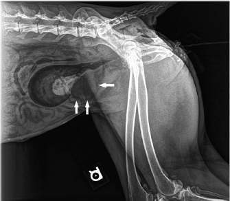
Fig. 1. Lateral radiograph of a double contrast cystogram in a maned wolf. Note the irregular mucosal and serosal outlines of the bladder caudoventrally (arrows). A mass involving the wall is highlighted.
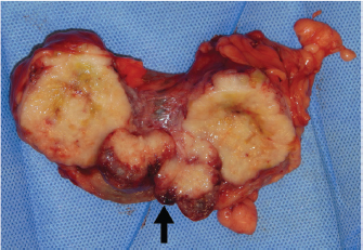
Fig. 2. Urinary bladder lumen of a maned wolf with multiple firm tumor nodules coalescing around the bladder neck (arrow). On histologic examination, the transitional cell epithelium of the urinary bladder was thickened and stretched over exophytic tumor tissue (Fig. 3); the epithelium formed an inverted papillomatous formation in one region. The majority of the sections of the mass were characterized by columnar epithelial cells forming tubules extending to, and occasionally into, the smooth muscle. The range of tissue patterns was much greater in a few sections leading to the final diagnosis of teratoma. Epithelial types recognized included stratified squamous epithelium with keratinization and columnar epithelium with goblet cell and ciliary development, including neural tube-like formations. Mesenchymal cell types included clusters of ring cells or adipocytes and areas of chondroid or osseous differentiation. Clusters of round cells, both lymphoid and hematopoietic, were seen. Tubular structures expanded to form cysts, and foci of hemorrhage and necrosis were seen throughout the mass. Mitotic activity in the tumor was not significant, but there was evidence of intravascular invasion.
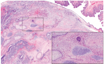
Fig. 3. Photomicrograph of the urinary bladder of a maned wolf. The urothelium is folded above and flattened over the teratoma below; the tumor expands into the lumen. Various cell populations are seen. Inset: stratified squamous and basaloid epithelium are present, as well as chondroid and smooth muscle mesenchymal tissue (H & E, Bar=120 μm). DiscussionThe mechanism that triggers abnormal migration of primordial germ cells leading to teratoma formation has not been confirmed. A study in zebrafish (Danio rerio) embryos concluded that hypoxia during embryonic development resulted in ectopic PGC (Lo et al., 2011). An early study in humans suggested that bladder teratomas develop from ectopic primordial germ cells in the caudally growing Wolffian duct during urogenital development (Pollack, 1936). Beta-1 integrin, a transmembrane receptor responsible for signal transduction, has been found to promote teratoma growth by stimulating angiogenesis (Bloch et al., 1997). Cystinuria is frequently reported in maned wolves and is a common cause for stranguria in the species (Bovee et al., 1981; Hammond, 2012). In this case, the wolf was initially treated for a urinary tract infection based on clinical signs and the presence of leukocytes and bacteria on urinalysis with an absence of cystinuria. The rapid return of clinical signs warranted further evaluation, including a double contrast cystogram, to diagnose the presence of a urinary bladder neoplasm. This case highlights the importance of evaluating beyond the expected diseases for a species and not making diagnostic assumptions based on signalment and clinical signs. Additionally, this is the first report of a teratoma originating in the urinary bladder of a non-human species. AcknowledgmentsThe authors wish to thank the animal care staff of the Endangered Wolf Center and the staff of the Saint Louis Zoo Department of Animal Health for their care and dedication to this animal. Conflict of InterestThe authors declare that there is no conflict of interest ReferencesAshley, D.J.B. 1971. Origin of teratomas. Cancer 31(2), 390–394. Bloch, W., Forsberg, W., Lentini, S., Brakebusch, C., Martin, K., Krell, H.W., Weidle, U.H., Addicks, K. and Fassler, R. 1997. Beta-1 integrin is essential for teratoma growth and angiogenesis. J. Cell Biol. 139(1), 265–278. Bovee, K.C., Bush, M., Dietz, J., Jezyk, P. and Segal, S. 1981. Cystinuria in the maned wolf of South America. Science 212(4497), 919–920. Gruys, E. and Van Dijk, J.E. 1976. Four canine ovarian teratomas and a nonovarian feline teratoma. Vet. Pathol. 13(6), 455–459. Gyimesi, Z.S., Forrester, J.W., Doering, D.L., Burns, R.B. and McManamon, R. 2015. Ovariectomy due to a dermoid cyst in an orangutan (Pongo pygmaeus). J. Zoo Wildl. Med. 46(1), 167–170. Hammond, E.E. 2012. Medical management of maned wolves (Chrysocyon brachyurus). In Zoo and wild animal medicine, Vol 7, Current therapy. Eds., Miller, R.E. and Fowler, M.E. Saint Louis, MO: Elsevier, pp: 451–457. Lanzkowsky, P. 2011. Germ cell tumors. In manual of pediatric hematology and oncology, 5th ed. Eds., Lanzkowsky, P. London, UK: Elsevier, pp: 776–795. Lo, K.H., Hui, M.N.Y., Yu, R.M.K., Wu, R.S.S. and Cheng, S.H. 2011. Hypoxia impairs primordial germ cell migration in zebrafish (Danio rerio) embryos. PLoS One. 6(9), e24540. Manasse, J.L. and Steinberg, H. 2015. “Teratocarcinomatosis” in an emu (Dromaius novaehollandiae). J. Zoo Wildl. Med. 46(3), 633–636. McKenney, J.K., Heerema-McKenney, A. and Rouse, R.V. 2007. Extragonadal germ cell tumors: a review with emphasis on pathologic features, clinical prognostic variables, and differential diagnostic considerations. Adv. Anat. Pathol. 14(2), 69–92. Munk, B.A., Turner, J.C. and Keel, M.K. 2013. Mediastinal teratoma in a free-ranging American black bear (Ursus americanus). J. Zoo Wildl. Med. 44(4), 1120–1122. Munson, L. and Montali, R.K. 1991. High prevalence of ovarian tumors in maned wolves (Chrysocyon brachyurus) at the National Zoological Park. J. Zoo Wildl. Med. 22(1), 125–129. Okeke, L.I., Ogun, G.O., Etukakpan, B.R., Iyama, A., Adeoye, A.O. and Duduyemi, B.M. 2007. Dermoid cyst of the urinary bladder as a differential diagnosis of bladder calculus: a case report. J. Med. Case Rep. 1(32), 1–3. Oosterhuis, J.W., Stoop, H., Honecker, F. and Looijenga, L.H.J. 2007. Why human extragonadal germ cell tumours occur in the midline of the body: old concepts, new perspectives. Int. J. Androl. 30(4), 256–263. Palmieri, C., Romanucci, M., Loi, P. and Salda, L.D. 2012. Intracoelomic teratoma in a domestic duck (Anas platyrhynchos domesticus): a case report including immunohistochemistry and electron microscopy. Res. Vet. Sci. 93(2), 862–864. Pollack, A.D. 1936. Malignant teratoma of the urinary bladder. Am. J. Pathol. 12(4), 561–568. Schelling, S.H. and Morton, D. 2015. An extragonadal teratoma in a female cynomolgus monkey (Macaca fascicularis). J. Med. Primatol. 44(2), 113–115. | ||
| How to Cite this Article |
| Pubmed Style Fox L, CSH, Padilla LR, Duncan M, . Bladder teratoma in a maned wolf (Chrysocyon brachyurus). Open Vet J. 2019; 9(3): 259-262. doi:10.4314/ovj.v9i3.11 Web Style Fox L, CSH, Padilla LR, Duncan M, . Bladder teratoma in a maned wolf (Chrysocyon brachyurus). https://www.openveterinaryjournal.com/?mno=34114 [Access: April 28, 2024]. doi:10.4314/ovj.v9i3.11 AMA (American Medical Association) Style Fox L, CSH, Padilla LR, Duncan M, . Bladder teratoma in a maned wolf (Chrysocyon brachyurus). Open Vet J. 2019; 9(3): 259-262. doi:10.4314/ovj.v9i3.11 Vancouver/ICMJE Style Fox L, CSH, Padilla LR, Duncan M, . Bladder teratoma in a maned wolf (Chrysocyon brachyurus). Open Vet J. (2019), [cited April 28, 2024]; 9(3): 259-262. doi:10.4314/ovj.v9i3.11 Harvard Style Fox, L. ., , C. S. H. ., Padilla, L. R., Duncan, M. . & (2019) Bladder teratoma in a maned wolf (Chrysocyon brachyurus). Open Vet J, 9 (3), 259-262. doi:10.4314/ovj.v9i3.11 Turabian Style Fox, Lana , Christopher S Hanley, Luis R Padilla, Mary Duncan, and . 2019. Bladder teratoma in a maned wolf (Chrysocyon brachyurus). Open Veterinary Journal, 9 (3), 259-262. doi:10.4314/ovj.v9i3.11 Chicago Style Fox, Lana , Christopher S Hanley, Luis R Padilla, Mary Duncan, and . "Bladder teratoma in a maned wolf (Chrysocyon brachyurus)." Open Veterinary Journal 9 (2019), 259-262. doi:10.4314/ovj.v9i3.11 MLA (The Modern Language Association) Style Fox, Lana , Christopher S Hanley, Luis R Padilla, Mary Duncan, and . "Bladder teratoma in a maned wolf (Chrysocyon brachyurus)." Open Veterinary Journal 9.3 (2019), 259-262. Print. doi:10.4314/ovj.v9i3.11 APA (American Psychological Association) Style Fox, L. ., , C. S. H. ., Padilla, L. R., Duncan, M. . & (2019) Bladder teratoma in a maned wolf (Chrysocyon brachyurus). Open Veterinary Journal, 9 (3), 259-262. doi:10.4314/ovj.v9i3.11 |

