| Original Article | ||
Open Vet. J.. 2022; 12(1): 124-128 Open Veterinary Journal, (2022), Vol. 12(1): 124–128 Original Research Prosthetic polyester-based hybrid mesh for repairing of perineal hernia in dogsAbdelhaleem Hamada Elkasapy1*, Mohamed M. Shokry2, Adel M. Alakraa1 and Olla A. Khalifa31Department of Surgery, Anesthesiology and Radiology, Faculty of Veterinary Medicine, Benha University, Banha, Egypt 2Department of Surgery, Anesthesiology and Radiology, Faculty of Veterinary Medicine, Cairo University, Giza, Egypt 3Genetics and Genetic engineering, Department of Animal Wealth Development, Faculty of Veterinary Medicine, Benha University, Benha, Egypt *Corresponding Author: Abdelhaleem Hamada Elkasapy. Department of Surgery, Anesthesiology and Radiology, Faculty of Veterinary Medicine, Benha University, Banha, Egypt. Email: Email: abdelhaleem_elkasapy [at] yahoo.com Submitted: 15/10/2021 Accepted: 05/02/2022 Published: 19/02/2022 © 2022 Open Veterinary Journal
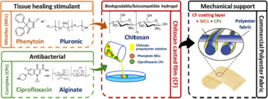
AbstractBackground: This study involves the use of a new multifunctional prosthetic mesh for treatment of the perineal hernia without complications. The prosthetic mesh is a hybrid platform of both synthetic and natural materials, mainly consisting of a synthetic commercial polyester fabric (CPF) to deliver the required mechanical integrity. The CPF mesh was coated by a natural biodegradable, biocompatible, and antimicrobial layer of chitosan incorporating phenytoin-loaded pluronic nanomicelles for healing promotion and ciprofloxacin-alginate polyelectrolyte complex-based microparticles as antibacterial agent. Aim: To evaluate the new developed multifunction polyester-based hybridmesh to repair perineal hernia clinical cases in dogs. Methods: The used multifunctional prosthetic mesh is a hybrid of the natural biodegradable, biocompatible, and antimicrobial material used in six cases with perineal hernia. Results: The prosthetic polyester-based hybrid mesh was found very helpful in repairing clinical cases of perineal hernias in six dogs without unwarranted other surgical procedures or complications. The developed mesh proved its feasibility in terms of efficient biocompatibility, stability, sterilizability, and low cost. Conclusion: The prosthetic commercial polyester-based mesh provided the ideal and feasible alternative prosthesis with many advantages. Keywords: Perineal, Hernia, Polyester-based hybrid mesh, Dogs. IntroductionPerineal hernia is the protrusion of the pelvic or abdominal contents through a defect in the pelvic diaphragm (Burrow and Harvey, 1973; Hayes et al., 1978). The pelvic diaphragm is the vertical closure of the pelvic cavity through which the rectum passes (Hermanson and de Lahunta, 2018). The muscular components are the levator ani and coccygeus muscles and sacrotuberous ligament laterally, the internal obturator and superficial gluteal muscles ventrally, and the external anal sphincter medially (Bellenger and Canfield, 2003). Perineal hernia occurs almost exclusively in middle aged to older entire male dogs (Bojrab and Toomy, 1981; Williams, 2014; Gill and Barstad, 2018). The pelvic diaphragm is stronger in female dogs than males (Hedlund, 1997). Approximately 59% of the perineal hernias are unilateral, while 41% are bilateral (Bellenger and Canfield, 2003). The etiology remains unclear but is associated with degenerative changes in the muscles of the pelvic diaphragm, which possibly resulted from hormonal imbalance, prostatic hyperplasia, chronic constipation, and docking (Merchav et al., 2005; Niebauer et al., 2005; Williams, 2014; Sprada et al., 2017). The common clinical signs are dyschezia, tenesmus, fecal incontinence, perineal swelling lateral to the anus, stranguria, and anuria (Hosgood et al., 1995; Sprada et al., 2017; Gill and Barstad, 2018). Diagnosis is confirmed by clinical examination, contrast radiography, and ultrasonography (Lee et al., 2012; Williams, 2014). All perineal hernias in dogs should be managed surgically by reconstruction of the pelvic diaphragm. There are many adopted surgical techniques for reconstruction of the pelvic diaphragm, including appositional herniorrhaphy by primary suture repair (Bojrab and Toomy, 1981; Orcher and Johnston, 1985; Anderson et al., 1998; Al-Akraa, 2015), the muscular transposition of the internal obturator muscle (Sjollema and Van Sluijs, 1989; Shokry et al., 1998; Bellenger and Canfield, 2003; Shaughnessy and Monnet, 2015), and the synthetic prosthetic mesh (Clarke, 1989; Vnuk et al., 2008). The objective of this study is to evaluate the newly developed multifunction polyester-based hybrid mesh which was successfully utilized for repairing abdominal wall hernias (Shokry et al., 2019) to repair perineal hernias in clinical cases in dogs. Materials and MethodsIn-vitro studyThe used multifunctional prosthetic mesh is a hybrid of the natural biodegradable, biocompatible, and antimicrobial materials [Chitosan (CS)], (Sigma-Aldrich, China and Germany), and pluronic nanomicelles for healing promotion [Phenytoin (PH)] (El-Nasr Pharmaceuticals, Egypt) and the synthetic antibacterial [ciprofloxacin (CPX)]-alignate polyelectrolyte complex-based microparticles (Epico Pharmaceuticals, Cairo, Egypt). The development and characterization of pH-loaded nanomicelles (Fig. 1) were according to Shokry et al. (2019). In-vivo studyDuring 2020, six male dog patients were referred to veterinary surgery teaching clinic of Benha University with signs of perineal swellings. All dogs are entire adult males. Their ages were between 4 and 13 years and the breeds were Pekinese (2), German Shepherd (1), Chihuahua (2), and Dachshund (1). History revolved around urination/defecation difficulties, tenesmus, fecal or urinary incontinence, anal sphincter ulcers, anorexia, and perineal swelling (five cases left side unilateral and one case bilateral). Radiography revealed distended retroverted bladder (one case bilateral), rectal diverticulum (three cases unilateral), and epiplocele (two cases unilateral) (Figs. 2 and 3). AnesthesiaPremedication consisted of IM xylazine HCl 2.0 mg/kg (Xylaject, Adwia, Egypt) and Ceftriaxone 500 mg (Rocefin, Switzerland) 30 minutes before induction of anesthesia. The anal glands were expressed; the rectum was manually evacuated; and the anus was closed with a purse string suture. The patient was placed in sternal recumbency ,while the table was tilted head down and the tail was tied over the back. The perineum and the areas lateral to the anus were surgically prepared. IV fluid therapy line with lactated ringers (10 ml/kg) was performed. Anesthesia was induced with IV propofol (Diprivan 1%, Fresenius Kabi USA) and maintained with isoflurane after intubation. Surgical techniqueA vertical incision through the skin and subcutaneous fascia just lateral to the anus from the base of the tail to the ischium was made. The subcutaneous tissues were bluntly carefully dissected to avoid the pudendal vessels and nerve, and the hernial sac contents (the bladder or rectum) were reduced intra-abdominal. The prolapsed omentum or the periprostatic fat was excised. At this stage, when the pelvic diaphragm defect was identified, the sterilized prosthetic polyester-based hybrid meshes were folded and trimmed to match the defects’ sizes. The mesh was then stitched along the perimeter of the defect with individual stitches of polypropylene 2/0 suture material (Prolene-Ethicon, Germany) for better fixation (Fig. 4). The subcutaneous tissues closed with continuous suture pattern with Polygalactin 2/0 (Vicryl-Germany) before closing the skin with simple interrupted suture with Prolene 2/0. Castration was carried out in only one case (13 years old), while the rest of owners denied the operation. All patients were discharged with postoperative care instructions comprising individual isolation, exercise restriction, using of an Elizabethan collar, and administration of Ceftriaxone 500 mg every 12 hours and the analgesic Carprofen 5% 50 mg (Adwia, Egypt) for 5 days. Feeding restricted low residue soft diet to half the amount for 7 days was also recommended. The operated dogs were kept under clinical observation by telecommunication with their owners for any complications and the recheck was after 10 days postoperation. Ethical approvalThe Institutional Animal Care and Use Committee (IACUC) approved this work (BUFTM 06-11-21), Faculty of Veterinary Medicine, Benha University. It was strictly designed under the consideration of animal welfare. ResultsSix adult male intact dogs were diagnosed with left-side unilateral (five cases) and bilateral (one case) perineal hernias. Pelvic radiography showed distended bladder retroflexion (two cases), rectal diverticulum (one case), and epiplocele (three cases). The technique of prosthetic polyester-based hybrid mesh hernioplasty was conducted on all patients with uneventful recovery without wound dehiscence or recurrence. Four cases informed edema with accumulation of inflammatory fluid during the first 3 days after surgery, which gradually reduced with administration of antibiotic and anti-inflammatory medicine. One case developed subcutaneous abscess after 1 week which were managed by daily dressing till complete recovery. Overall, the result was promising and satisfactory with uneventful repair.
Fig. 1. A schematic illustration of the study design (Shokry et al., 2019).
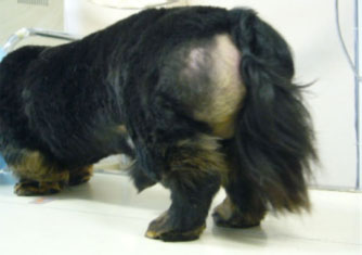
Fig. 2. Unilateral perineal hernia in a Dachshund.
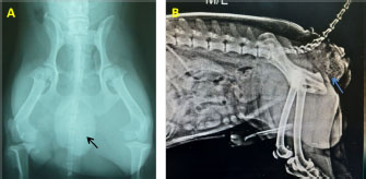
Fig. 3. (A): Pelvic AP radiograph showing the distended retroflexed bladder in a Chihuahua. (B): Lateral showing the rectal diverticulum in a Chihuahua. DiscussionIn this report, the presented cases were diagnosed as perineal hernias from the history, clinical, and radiographic examinations. All cases were entire adult males and most of them (five cases) had unilateral left-side hernias, while one case had bilateral hernia. The etiology was generally related to urinary and constipation problems. This agreed with previous reposts (Burrows and Harvey, 1973; Vnuk et al., 2008; Williams, 2014). However, many potential factors have been suggested in their development, which include congenital predisposition, hormonal imbalance, prostatic hyperplasia, and docked tails (Bojrab and Toomy, 1981; Bellenger and Canfield, 2003; Niebauer et al., 2005; Wallace et al., 2021). The most common signs reported by the owners were perineal swelling, constipation, tenesmus, fecal/urine incontinence, and anorexia. These signs were also observed by other authors (Bojrab and Toomy, 1981; Hosgood et al., 1995). The hernial contents of the presenting cases were urinary bladder retroflexion (two cases) associated with urinary complications, epiplocele (three cases) and rectal diverticulum (one case) associated with fecal impaction, and dyschezia. Many studies confirmed similar contents (Hosgood et al., 1995; Pekcan et al., 2010), while others reported prostatic hyperplasia (Vnuk et al., 2008; Sprada et al., 2017).
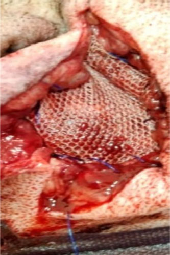
Fig. 4. Mesh implantation in the pelvic diaphragm defect. In the present study, the surgical management for repairing the pelvic diaphragm defects using the prosthetic polyester-based hybrid mesh with a natural biomaterial, antimicrobial layer of CS incorporating pH-loaded pluronic nanomicelles for healing promotion, and CPX provided efficient healing with excellent biocompatibility and limited complications. This agreed with previous comprehensive experimental study using the same prosthetic mesh for repairing abdominal hernias (Shokry et al., 2019). Many surgical procedures are used for repairing the pelvic diaphragm defects from simple suture herniorrhaphy (Bojrab and Toomy, 1981; Orcher and Johnston, 1985; Bellenger and Canfield, 2003), internal obturator muscle transposition (Hardie et al., 1983; Anderson et al., 1998; Shokry et al., 1998; Vnuk et al., 2008), and prosthetic polypropylene mesh (Clarke, 1989; Vnuk et al., 2006) with variable success outcomes. By reference to the owners’ information follow-ups for 3 months, recurrence did not occur in any of the presented cases. Moreover, adopting the prosthetic mesh avoids the potential of anatomical distortion after muscular transposition operation for repairing the pelvic diaphragm defects. ConclusionThe prosthetic commercial polyester-based mesh is a hybrid platform for natural biodegradable, biocompatible, and antimicrobial (CS) material. The healing promoter (pH) and synthetic antibacterial drug (CPX) provided the ideal and feasible alternative prosthesis with many advantages like low cost, mechanical stability, minimal infection, and biocompatibility. AcknowledgmentThis work was supported by the research project and the authors extend their appreciation to the scientific research fund (SRF) from Benha University (project no. M665). Conflict of interestThe authors declare that there is no conflict of interest. ReferencesAl-Akraa, A.M. 2015. Standard herniorrhaphy, polypropylene mesh and tension band for repair of perineal hernia in dogs. Int. J. Adv. Res. 3, 963–970. Anderson, M.A., Constantinescu, G.M. and Mann, F.A. 1998. Perineal hernia repair in the dog. In Current techniques in small animal surgery, 4th ed. Ed. Bojrab, MJ, Baltimore, ML: Williams & Wilkins, pp: 555–564. Bellenger, C.R. and Canfield, R.B. 2003. Perineal hernia. In Textbook of small animal surgery, 3rd ed. Ed. Slatter, D, Philadelphia, PL: Saunders, pp: 487–498. Bojrab, M.J. and Toomy, A.A. 1981. Perineal herniorrhaphy. Compend. Contin. Educ. Vet. 8, 8–15. Burrows, C.F. and Harvey, C.E. 1973. Perineal hernia in the dog. J. Small Anim. Pract. 14, 315–332. Clarke, R.E. 1989. Perineal herniorrhaphy in the dog using polypropylene mesh. Aust. Vet. Pract. 19, 8–14. Gill, S.S and Barstad RD. 2018. A review of the surgical management of perineal hernias in dogs. J. Am. Anim. Hosp. Assoc. 54, 179–187. Hardie, E.M., Kolata, R.J., Earley, T.D., Rawlings, C.A. and Gorgacz, E.J. 1983. Evaluation of internal obturator muscle transposition in treatment of perineal hernia in dogs. Vet. Surg. 12, 69–72. Hayes, H.M., Wilson, G.P. and Tarone, R.E. 1978. The epidemiologic features of perineal hernia 771 dogs. J. Am. Hosp. Assoc. 14, 703–707. Hedlund, C.S. 1997. Surgery of perineum, rectum and anus. In Animal surgery, 1st ed. Ed. Fossum, T.W., Maryland Heights, MI: Mosby Inc, pp: 325–356. Hermanson, J. and de Lahunta A. 2018. Miller’s anatomy of the dog, 5th ed. WB Saunders, Philedelphia, USA. Hosgood, G., Hedlund, C.S., Pechman, R.D. and Dean, P.W. 1995. Perineal herniorrhaphy: perioperative data from 100 dogs. J. Am. Hosp. Assoc. 31, 331–342. Lee, A.J., Chung, W.H., Kim, D.H., Lee, K.P., Suh, H.J., Do, S.H., Eom, K.D. and Kim, H.Y. 2012. Use of canine small intestinal mucosa allograft for treating perineal hernias in two dogs. J. Vet. Sci. 13, 327–330. Merchav, R., Feuermann, Y., Shamay, A., Ranen, E., Stein, U., Johnston, D.E. and Shahar, R. 2005. Expression of relaxin receptor LRG7, canine relaxin-like factor in the pelvic diaphragm musculature of dogs with and without perineal hernia. Vet. Surg. 35, 476–481. Niebauer, G.W., Shibly, S., Seltenhammer, M., Piker, A. and Brandt, S. 2005. Relaxin of prostatic origin might be linked to perineal hernia formation in dogs. Anny. N. Y. Acad. Sci. 1041, 415–422. Orcher, R.J. and Johnston, D.E. 1985. The surgical treatment of perineal hernia in dogs by transposition of the obturator muscle. Comp. Cont. Educ. 7, 233–239. Pekcan, Z., Besalti, O., Sirin, Y.S. and Caliskan, M. 2010. Clinical and surgical evaluation of perineal hernia in dogs: 41 cases. Kafkas. Univ. Vet. Fak. Derg. 16, 573–578. Shaughnessy, M. and Monnet, E. 2015. Internal obturator muscle transposition for treatment of perineal hernia in dogs: 34 cases (1998-2012). J. Am. Vet. Med. Assoc. 246, 321–326. Shokry, M.M., Khalil, I., El-Kasapy, A., Osman, A., Mostafa, A., Salah, M. and El-Sherbiny, I.M. 2019. Multifunctional prosthetic polyester-based hybrid mesh for repairing of abdominal wall hernias and defects. Carbohydr. Polym. 223, 115027. Shokry, M., El-Keiey, M. and Gadallah, S. 1998. Perineal hernioplasty by muscular transposition in dogs. Vet. Med. J. Giza 46, 599–601. Sjollema, B.E. and Van Sluijs, F.J. 1989. Perineal hernia repair in the dog by transposition of the internal obtrurator muscle. II. Complications and results in 100 patients. Vet. Q. 11, 18–23. Sprada, A.G., Huuppes, R.R., Feranti, J.P.S., de Souza, F.W., Coelho, L.P., Moraes, P.C. and Minto, B.W. 2017. Perineal hernia in dogs: which technique should we use? Acta Sci. Vet. 45, 244. Vnuk, D., Lipar, M., Maticic, D., Smoled, O., Pecin, M. and Brkie, A. 2008. Comparison of standard herniorrhaphy and transposition of the internal obturator muscle for perineal hernia in the dog. Vet. Arch. 78, 197–207. Vnuk, D., Maticic, D., Kreszinger, M., Radisic, B., Kos, J., Lipar, M. and Babic, T.A. 2006. Modified salvage technique in surgical repair of perineal hernia in dogs using polypropylene mesh. Vet. Med. 51, 111–117. Wallace, M.L., Grimes, J.A., Duffy, D.J., Kindra, C., MacIver, M., Lin, S., Scharf, V.F. and Schmiedt, C.W. 2021. Evaluation of concurrent perineal hernia in adult male dogs presenting with non traumatic, acquired inguinal hernias. Can. Vet. J. 62(6), 617–620. Williams, J. 2014. Perineal hernia. WSAVA. Congress-VIN. Available via http://www.vin.com/apputil/content/defaultadv1.aspx?pld=12886&catld=57122&id=7054726 | ||
| How to Cite this Article |
| Pubmed Style Elkasapy AH, Shokry MM, Alakraa AM, Khalifa OA. Prosthetic polyester-based hybrid mesh for repairing ofperineal hernia in dogs. Open Vet. J.. 2022; 12(1): 124-128. doi:10.5455/OVJ.2022.v12.i1.15 Web Style Elkasapy AH, Shokry MM, Alakraa AM, Khalifa OA. Prosthetic polyester-based hybrid mesh for repairing ofperineal hernia in dogs. https://www.openveterinaryjournal.com/?mno=130544 [Access: January 25, 2026]. doi:10.5455/OVJ.2022.v12.i1.15 AMA (American Medical Association) Style Elkasapy AH, Shokry MM, Alakraa AM, Khalifa OA. Prosthetic polyester-based hybrid mesh for repairing ofperineal hernia in dogs. Open Vet. J.. 2022; 12(1): 124-128. doi:10.5455/OVJ.2022.v12.i1.15 Vancouver/ICMJE Style Elkasapy AH, Shokry MM, Alakraa AM, Khalifa OA. Prosthetic polyester-based hybrid mesh for repairing ofperineal hernia in dogs. Open Vet. J.. (2022), [cited January 25, 2026]; 12(1): 124-128. doi:10.5455/OVJ.2022.v12.i1.15 Harvard Style Elkasapy, A. H., Shokry, . M. M., Alakraa, . A. M. & Khalifa, . O. A. (2022) Prosthetic polyester-based hybrid mesh for repairing ofperineal hernia in dogs. Open Vet. J., 12 (1), 124-128. doi:10.5455/OVJ.2022.v12.i1.15 Turabian Style Elkasapy, Abdelhaleem H., Mohamed M. Shokry, Adel M. Alakraa, and Olla A. Khalifa. 2022. Prosthetic polyester-based hybrid mesh for repairing ofperineal hernia in dogs. Open Veterinary Journal, 12 (1), 124-128. doi:10.5455/OVJ.2022.v12.i1.15 Chicago Style Elkasapy, Abdelhaleem H., Mohamed M. Shokry, Adel M. Alakraa, and Olla A. Khalifa. "Prosthetic polyester-based hybrid mesh for repairing ofperineal hernia in dogs." Open Veterinary Journal 12 (2022), 124-128. doi:10.5455/OVJ.2022.v12.i1.15 MLA (The Modern Language Association) Style Elkasapy, Abdelhaleem H., Mohamed M. Shokry, Adel M. Alakraa, and Olla A. Khalifa. "Prosthetic polyester-based hybrid mesh for repairing ofperineal hernia in dogs." Open Veterinary Journal 12.1 (2022), 124-128. Print. doi:10.5455/OVJ.2022.v12.i1.15 APA (American Psychological Association) Style Elkasapy, A. H., Shokry, . M. M., Alakraa, . A. M. & Khalifa, . O. A. (2022) Prosthetic polyester-based hybrid mesh for repairing ofperineal hernia in dogs. Open Veterinary Journal, 12 (1), 124-128. doi:10.5455/OVJ.2022.v12.i1.15 |


