| Case Report | ||
Open Vet. J.. 2024; 14(8): 2092-2096 Open Veterinary Journal, (2024), Vol. 14(8): 2092–2096 Case Report Lower lip avulsion: Surgery and management using stem cell metabolites preparation in a domestic catLina Susanti1*, Suryo Kuncorojakti2, Shafira Oktaviani3 and Mardina Girada Simarmata3*Corresponding Author: Lina Susanti. Veterinary Clinical Division, Department of Veterinary Science, Faculty of Veterinary Medicine, Airlangga University, Surabaya, Indonesia. Email: linavetoph_ua [at] fkh.unair.ac.id Submitted: 10/05/2024 Accepted: 22/07/2024 Published: 31/08/2024 © 2024 Open Veterinary Journal
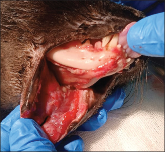
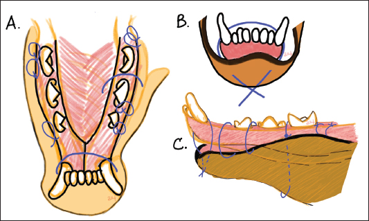
ABSTRACTBackground: Lower lip avulsion is a separation between the lip and the associated soft tissue from the mandible. The degree of these types of injuries varies and heavily affects the outcome of the case. Case Description: This study reported an extensive lower lip avulsion managed by surgery and stem cell metabolite preparation. A one year and nine month-old domestic cats was referred for lower lip avulsion surgery to the Veterinary Teaching Hospital Airlangga University. Owing to the limited amount of tissue, immediate successful results cannot be achieved after the first surgery. Furthermore, tissue necrosis and lack of physical restraint to the cat at home contributed to the delayed union between the soft tissue and mandible, resulting in repeated surgery. Stem cell metabolites preparation was applied at the surgical site and was incorporated into the therapy to support tissue growth. Conclusion: The combination of surgical treatment and stem cell metabolite preparation resulted in good wound healing in the present case. Keywords: Lower lip avulsion, Stem cell, Wound healing. IntroductionThe separation between the lip and the associated soft tissue from the mandible is known as the lower lip avulsion and is quite common in companion animals (Stanley et. al., 2020). This type of injury is caused by caudal force against gingiva and labial mucosa (Vicari and Stepaniuk, 2014) and is commonly reported in young animals (White, 2010; Saverino and Reiter, 2018). Several events are known to cause lip avulsion including injuries such as vehicular trauma, falling from a height, and bite wounds (Pope, 2006; Vicari and Stepaniuk, 2014; Saverino and Reiter, 2018). Avulsion of the lower lip can range from minor unilateral soft tissue separation to major separation of tissue involving the bilateral part of the mandible and extending to the oral commissures (Stanley et. al., 2020). In cats, lip avulsions have also been reported to involve fractured bones and are sometimes accompanied by luxated teeth (White, 2010; Stanley et. al., 2020). Several surgical techniques have been described to repair lip avulsion (Pope, 2006; White, 2010; Vicari and Stepaniuk, 2014; Stanley et. al., 2020). The extent of the injury usually dictates the type and technique of surgery to repair the case (Stanley et. al., 2020). Depending on the severity of the case, reattaching the soft tissue can be accomplished by suturing or wiring, and sometimes, drilling the mandible was also done to facilitate the attachment of the soft tissue to the mandible (Pope, 2006; White, 2010; Vicari and Stepaniuk, 2014; Stanley et. al., 2020). This study described a severe lower lip avulsion in a domestic cat managed by surgery combined with stem cell metabolite application to enhance wound healing. Case DetailsA one year and nine months-old intact male domestic cat was referred to the Veterinary Teaching Hospital, Universitas Airlangga for avulsion of the lower lips. The cat was missing for several days before returning with the injury and was brought to the local animal clinic where the cat was hospitalized for two days before being referred for surgery. Medications given at the previous clinic including vitamins, antibiotics, and non-steroidal anti-inflammatory drugs (NSAIDs). No abnormalities were found during the physical examination other than those involving the oral cavity. The bilateral lip avulsion extended through the lower mandibular tissue all the way to the oral commissures (Fig. 1). The rectal temperature was 38.8oC. The owner reported good appetite, normal activity, and also normal urination and defecation. The cat was scheduled for surgery the next day. Surgical managementBefore the soft tissue was surgically reattached, scaling was performed using hand-held ultrasonic scaler (UDS-K LED, Woodpecker, China). The wound was thoroughly cleaned and lavaged using normal saline and 20% ampicillin (Vicillin®1000, Meiji Indonesian, Bangil, Indonesia). Tissue debridement was performed before the reattachment using gauze and surgical iris scissors. After the necrotic tissue was removed and hemorrhage was present, the attachment surgery was performed using several techniques as described previously (Pope, 2006; White, 2010; Vicari and Stepaniuk, 2014; Stanley et. al., 2020). However, due to limited tissue in the oral cavity of the cat, coupled with the extent of injury, several types of sutures were applied as permitted by the injured tissue (Fig. 2A). These sutures are including: 1) a single large horizontal mattress suture, passed through the chin and around the canine teeth, tied at the outer part of the skin (Fig. 2B), 2) major matrass suture placed from the detached mucosal tissue, passing the gingiva between the teeth, passing near the ventral midline between the mandibular area to the outer skin before the needle was passed back into the soft tissue on the inside part of the lip to join the other end of the suture and ligated (Fig. 2A, C), 3) several simple interrupted sutures opposing the labial mucosa to the gingiva.
Fig. 1. Photograph showing the extend of lower lip avulsion through the lower mandibular all the way to the oral commissures. Both mandibular dexter and sinister barely had any tissue attached to them.
Fig. 2. Stitches made during the surgery. (A) The sutures from the dorsal view. (B) A single large horizontal mattress suture, passed through the chin and around the canine teeth, and was tied at the outer part of the skin. (C) The scheme of the sutures depicted from the lateral view of the mandibula sinister.
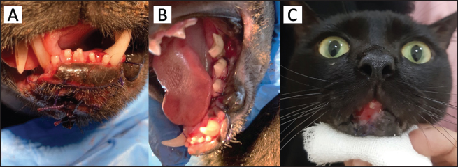
Fig. 3. Photograph of the cat after the surgery and at the last follow-up. (A) Due to tissue necrosis, some part of labial dexter was removed. Several simple interrupted stitches were made along the midline of the skin to hold the skin and the soft tissue that were separated due to necroses. (B) Simple interrupted stitches joining the labial sinister and the gingiva were visible. (C) The appearance of the cat after 30 days. The wound was substantially healed. The cat was discharged. Surgery was repeated three times: the second surgery was performed three days after the first surgery, and the third surgery was done two days following the second. For the second and third surgery, stem cell metabolites preparation was applied to the surface of the wound after the tissue was debrided. Owing to necrotic tissue on part of the lips, during the second and the third surgery, several simple interrupted sutures were also performed on the skin along the midline to join the remaining tissue and keep the tissue together (Fig. 3A). Suture materials used in this surgery were 4-0 absorbable braided polyglycolic acid (RTMED®, Shandong, China) and 4-0 monofilament poly (glycolideco-caprolactone) (PGA-PCL) (Monovet, Hidup Mitra Cahaya, Indonesia). Dexamethasone (Glucortin-20, TMC, Indonesia) was injected on the day after the first surgery as an anti-inflammatory medication and was repeated as needed throughout the treatment. Antibiotics used were amoxicillin/clavulanic acid (62.5 mg/cat, twice a day for two weeks; Claneksi, Sanbe, Indonesia). Stem cell metabolites preparation (a cellulose-based hydrogel of adipose mesenchymal stem cell metabolites; patent owned by Research Center for Vaccine Technology and Development/RCVTD Airlangga University) was applied to the wound twice daily to stimulate the growth of the tissue and support the healing process. Post-surgical diet was restricted to a soft diet and was strictly administered using a syringe to prevent contamination of the wound. The Elizabethan collar was also used to prevent self-trauma. DiscussionThe surgical repair technique of lip avulsion depends greatly on the degree and severity of the tissue separation (Stanley et. al., 2020). In a less complicated case of lower lip avulsion, several surgical techniques had been described to reattach the lower lip including a single horizontal mattress in minor avulsion and interrupted horizontal mattress sutures through lip mucosa looped around the incisor teeth on moderate avulsion (Stanley et. al., 2020). In addition, Vicari and Stepaniuk (2014) also described a series of horizontal mattress sutures passed through mucosa and gingiva on the opposite side of the tooth to repair minor mandibular avulsion. Drilling the mandibles to facilitate the passing of the suture material has also been described (Stanley et. al., 2020). In the present case, the degree of avulsion was really severe in which the separation of the tissue extended to the oral commissures (Fig. 1). Vicari and Stepaniuk (2014) demonstrated soft tissue attachment of a similar case in a dog where the soft tissue of the lower lip was sutured to the midline fascia ventral to the mandibular symphysis. However, differ with dog, the tissue in the midline area of the cat’s mandible is limited and fairly thin, rendering the technique impossible. During the first surgery, when such a technique was attempted, it resulted in the tearing of the tissue. The limited tissue at the injured site and the severity of avulsion are the major challenges in the present case. Generally, in lip avulsion, opposing the labial mucosa to the gingiva can be performed regardless of the severity of tissue separation (Vicari and Stepaniuk, 2014; Stanley et. al., 2020) assuming that healthy gingival tissue is sufficient to allow such a procedure. In the present case, such attempts were barely possible and were only successful in some parts of the tissue (Fig. 3B). The gingival tissue in cats was especially fragile and was easily torn when pierced by the needle. Consequently, to attach the soft tissue back to the mandible, a major matrass suture was placed from the detached tissue, passing the mucosa and gingiva on the opposite side of the tooth. In addition, a single large horizontal mattress suture was passed through the chin and around the canine teeth as described by Vicari and Stepaniuk (2014; Fig. 2A and B). In the present case, owing to the substantial amount of time that passed between the injury to the first surgery, some parts of the lower lip underwent tissue necrosis, probably owing to the severed blood vessels resulting from the avulsion itself. This resulted in failure in the union of the soft tissue and the mandible. This fact contributes to the complexity of the present case. Three days post-surgery, the tissue necrosis on the left lip caused the wound to reopen. The second surgery was attempted and this time the owner requested for the cat to be discharged one day post-surgery. Unfortunately, due to lack of restrain at home, several sutures were undone and the lower lips were separated from the mandible. The third surgery was attempted the next day. This surgery was agreed to be the last surgery attempted and in the event of another failure, the wound would be managed as semi open wound. After the third surgery, the cat was hospitalized to ensure the strict postoperative management and the stitches stayed until the last follow up (30 days; Fig. 3C). Postoperative management recommended for lip avulsion surgery includes soft diet, at least for 2 weeks (Vicari and Stepaniuk, 2014; Saverino and Reiter, 2018). This helps to minimize the pressure and movement on the wound which was important to ensure that the suture will stay in place. Restricting the cat’s general movement is also important during wound healing including after lip avulsion surgery. Often, most owner is less compliance to apply such restrain as they might considered it to be against animal welfare. After the second surgery, the cat was discharged the following day through the request from the owner. The suture immediately break as the cat was allowed to roam free at home. This highlights the importance of measured restrain on post-surgical patients in lip avulsion. In the present case, stem cell metabolite preparation was applied throughout the exposed tissue before the suture was attempted during the second surgery and the third surgery. In addition, stem cell metabolite preparation was applied twice daily on the surgical wound as postoperative management. This supportive treatment was established to enhance the granulation process of the tissue especially since many parts of the tissue underwent necrosis, leaving the frontal area of the mandibula dexter to be exposed (Fig. 3A). Growth factors such as vascular endothelial growth factor (VEGF) and fibroblast growth factor—2 (FGF-2) in stem cell metabolites were able to promotes wound healing by increasing the level of angiogenesis. Furthermore, the role of FGF-2 was not limited to angiogenesis induction only but also promoted the proliferation of fibroblast which was important for the formation of tissue granulation (Wicaksono et al., 2023). Three weeks after the third surgery, the wounds on the frontal part of the lower lips had healed substantially (Fig. 3C). The cat was discharged and the owner was advised to apply the stem cell daily. Since meloxicam had been injected twice at the previous clinic, we refrained from using NSAIDs and opted to use dexamethasone injection as an anti-inflammatory drug to avoid possible kidney injury. Repeated use of meloxicam in cats is controversial as repeated doses have been associated with renal failure and death (Plumb, 2018). However, meloxicam is among the recommended NSAID for oral and maxillofacial surgery in cats (Pascoe, 2020) and previous case reports of lip avulsion also use meloxicam perioperatively (White, 2010). Perioperative analgesic in dental procedures in general had been recommended as it is considered better in managing pain associated with dental procedures (Pascoe, 2020). Analgesic medication utilized perioperatively ranging from local anesthesia, opioid, and NSAID (Saverino and Reiter, 2018; Pascoe, 2020). Steroidal inflammatory agents, such as dexamethasone, do not by itself have a direct effect on nociception and are not primary analgesic, but since many painful conditions are the result of inflammation, steroidal anti-inflammatory agents could be beneficial in the treatment of such cases (Papich, 2015). On one study by Saverino and Reiter (2018), complications following lip avulsion repair in cats and dogs included, wound dehiscence, postoperative infection, tooth discoloration, neurapraxia, and dyspnea. Lip avulsion wounds often are contaminated and therefore a thorough debridement and rinsing is important before surgery is performed (Pope, 2006). In the present case, the wound was thoroughly rinsed with natural saline and ampicillin combination. Postoperative antibiotics given in the present case were amoxicillin and clavulanic acid which were among the common antibiotic choices in the previous reports (White, 2010; Vicari and Stepaniuk, 2014; Saverino and Reiter, 2018). Furthermore, since the cause of mandibular lip avulsion including vehicular trauma (Vicari and Stepaniuk, 2014), an X-ray was suggested but was declined by the owner due to financial restrictions. Ideally, an x-ray should be performed in such cases in order to evaluate the possible extent of internal injuries such as bone fractures and internal bleeding. Fortunately, no signs of internal bleeding were observed during the treatment period. In conclusion, successful surgical management of lip avulsion depends on the severity of the cases, which also dictates the technique used to opposed the tissue back to its place. Owing to the limited amount of tissue, severe mandibular lip avulsion can be hard to be surgically managed in cats. The use of stem cell metabolite preparation was considered to contribute substantially to wound healing in the present case. AcknowledgmentNot applicable. Conflict of interestThe authors declare no conflict of interest. Authors’ contributionsSusanti L: Evaluation, surgical management, follow-up, conceptualization, manuscript drafting, and finalization. Kuncorojakti S: stem cell metabolites preparation, manuscript finalization. Oktaviani S., Simarmata M.G.: Evaluation, management, and follow-up. All authors discussed this patient and approved this manuscript. FundingThis research received no specific grant. Data availabilityAll data supporting the findings of this study are available within the manuscript. ReferencesPapich, M.G. 2015. Glucocorticoids. In: Mosby: Handbook of veterinary pain management. 3rd edition. Linn, MO: Elsevier, pp: 266-279. Pascoe, P.J. 2020. Anaesthesia and pain management. In Oral and maxillofacial surgery in dogs and cats. Eds., Verstraete, F.J.M., Lommer, M.J. Edinburgh, Scotland: Saunders, pp: 22–43. Plumb, D.C. 2018. Plumb’s veterinary handbook. 9th ed. USA: Wiley-Blackwell, pp: 1036–1040. Pope, E.R. 2006. Head and facial wounds in dogs and cats. Vet. Clin. North Am. Small Anim. 36: 793–817. Saverino, K.M. and Reiter, A.M. 2018. Clinical presentation, causes, treatment, and outcome of lip avulsion injuries in dogs and cats: 24 cases (2001–2017). Front. Vet. Sci. 5: 1–11. Stanley, B.J., Campbell, B.G. and Swaim, S.F. 2020. Facial soft tissue injuries. In Oral and maxillofacial surgery in dogs and cats. Eds., Verstraete, F.J.M., Lommer, M.J., 2nd edition. Edinburgh, Scotland: Saunders, pp: 262–280. Tsugawa, A.J. and Verstraete, F.J.M. 2020. Suture materials and biomaterials. In Verstraete, F.J.M., Lommer, M.J. (Ed) Oral and maxillofacial surgery in dogs and cats. 2nd edition. Edinburgh, Scotland: Saunders, pp: 79–91. Vicari, E. and Stepaniuk, K. 2014. Mandibular lip avulsion repair in the dog and cat. J. Vet. Dent. 31(3), 212–217. White, T.L. 2010. Lip avulsion and mandibular symphyseal separation repair in an immature cat. J. Vet. Dent. 27(4), 228–233. Wicaksono, S., Nugraha, A.P., Rahmahani, J., Rantam, F.A., Kuncorojakti, S., Susilowati, H., Riawan, W., Arundina, I., Lestari, P., Masya, R.N., Surboyo, M.D.C. and Ernawati, D.S. 2023. Adipose mesenchymal stem cell metabolites oral gel enhance pro-angiogenic factors expression, angiogenesis, and clinical outcome of oral ulcer rat model. Eur. J. Dent. 18(1), 117–123. | ||
| How to Cite this Article |
| Pubmed Style Susanti L, Kuncorojakti S, Oktaviani S, Simarmata MG. Lower lip avulsion: Surgery and management using stem cell metabolites preparation in a domestic cat. Open Vet. J.. 2024; 14(8): 2092-2096. doi:10.5455/OVJ.2024.v14.i8.39 Web Style Susanti L, Kuncorojakti S, Oktaviani S, Simarmata MG. Lower lip avulsion: Surgery and management using stem cell metabolites preparation in a domestic cat. https://www.openveterinaryjournal.com/?mno=180408 [Access: January 25, 2026]. doi:10.5455/OVJ.2024.v14.i8.39 AMA (American Medical Association) Style Susanti L, Kuncorojakti S, Oktaviani S, Simarmata MG. Lower lip avulsion: Surgery and management using stem cell metabolites preparation in a domestic cat. Open Vet. J.. 2024; 14(8): 2092-2096. doi:10.5455/OVJ.2024.v14.i8.39 Vancouver/ICMJE Style Susanti L, Kuncorojakti S, Oktaviani S, Simarmata MG. Lower lip avulsion: Surgery and management using stem cell metabolites preparation in a domestic cat. Open Vet. J.. (2024), [cited January 25, 2026]; 14(8): 2092-2096. doi:10.5455/OVJ.2024.v14.i8.39 Harvard Style Susanti, L., Kuncorojakti, . S., Oktaviani, . S. & Simarmata, . M. G. (2024) Lower lip avulsion: Surgery and management using stem cell metabolites preparation in a domestic cat. Open Vet. J., 14 (8), 2092-2096. doi:10.5455/OVJ.2024.v14.i8.39 Turabian Style Susanti, Lina, Suryo Kuncorojakti, Shafira Oktaviani, and Mardina Girada Simarmata. 2024. Lower lip avulsion: Surgery and management using stem cell metabolites preparation in a domestic cat. Open Veterinary Journal, 14 (8), 2092-2096. doi:10.5455/OVJ.2024.v14.i8.39 Chicago Style Susanti, Lina, Suryo Kuncorojakti, Shafira Oktaviani, and Mardina Girada Simarmata. "Lower lip avulsion: Surgery and management using stem cell metabolites preparation in a domestic cat." Open Veterinary Journal 14 (2024), 2092-2096. doi:10.5455/OVJ.2024.v14.i8.39 MLA (The Modern Language Association) Style Susanti, Lina, Suryo Kuncorojakti, Shafira Oktaviani, and Mardina Girada Simarmata. "Lower lip avulsion: Surgery and management using stem cell metabolites preparation in a domestic cat." Open Veterinary Journal 14.8 (2024), 2092-2096. Print. doi:10.5455/OVJ.2024.v14.i8.39 APA (American Psychological Association) Style Susanti, L., Kuncorojakti, . S., Oktaviani, . S. & Simarmata, . M. G. (2024) Lower lip avulsion: Surgery and management using stem cell metabolites preparation in a domestic cat. Open Veterinary Journal, 14 (8), 2092-2096. doi:10.5455/OVJ.2024.v14.i8.39 |


