| Research Article | ||
Open Vet. J.. 2024; 14(11): 2995-3003 Open Veterinary Journal, (2024), Vol. 14(11): 2995-3003 Research Article Morphological and genetic demonstration of Cephalopina titillator in dromedary camelsIsraa M. Essa1, Mohammed H. Al-Saadi2*, Akram M. Amanah3, Monyer Abdulamier Abd3 and Mansour J. Ali31Department of Parasitology, College of Veterinary Medicine, University of Basrah, Basra, Iraq 2Department of Internal and Preventive Medicine, College of Veterinary Medicine, University of Al-Qadisiyah, Al Diwaniyah, Iraq 3Department of Parasitology, College of Veterinary Medicine, University of Al-Qadisiyah, Al Diwaniyahs, Iraq *Corresponding Author: Mohammed H. Al-Saadi. Department of Internal and Preventive Medicine, College of Veterinary Medicine, University of Al-Qadisiyah, Al Diwaniyah, Iraq. Email: mohammed.alsaadi [at] qu.edu.iq Submitted: 22/08/2024 Accepted: 22/10/2024 Published: 30/11/2024 © 2024 Open Veterinary Journal
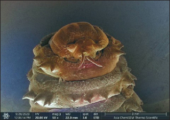
AbstractBackground: Cephalopina titillator is one of the most important parasites, which infests the upper respiratory tract of camels leading to deteriorating health effects, substantial economic losses, and even death. Aim: This study aimed to detect the prevalence rate of C. titillator in slaughtered camels, determining its morphology using the electron microscope, and confirming its species by molecular phylogeny. Methods: A total of 200 slaughtered camels at different areas in Al Muthanna province (Iraq) were inspected visually to collect the parasite samples that were identified initially based on their morphological characteristics. To confirm the parasite species, molecular phylogeny was conducted targeting the COX1 gene. Results: An overall 19.5% of study camels were found infested with C. titilator. Based on light and electron microscopes, the larval stage of C. titilator was shown numerous posterior spiracular pores, cephalo-pharyngeal skeleton, abdominal segments, spinulation in anterior ventral portion, no spines on the final segment of abdomen, and rounded dorsal surface. Dorsoventrally, a slender and flattened shape with the presence of 12 segments as well as widely separated antennal lobes and obligate mouth hooks were seen. Molecularly, all the tested samples were found positive by polymerase chain reaction (PCR). Additionally, some positive PCR products were sequenced, and reported in the NCBI-GenBank under the access numbers OP218846, OP218847, and OP218845, OP218839, OP218840, OP218841, OP218842, OP218843, and OP218844. Sequence analysis revealed the obvious identity between the local isolates and the global NCBI-GenBank Iran isolate (MW136151.1). Conclusion: This study described precisely the morphology of C. titillator using the light and electron microscopes suggesting its role in appropriate identification and classification. Molecular examination demonstrated the importance of COX1 gene in the identification of C. titillator and sequencing of the local isolates; however, additional molecular phylogenetic studies are needed to establish the evolutionary relationships among the oestrid group of insects with specialized habits and habitats. Keywords: Nsopharyngeal myiasis, Cytochrome C oxidase subunit 1, Camelus dromedarius, Electron microscopy, Polymerase chain reaction, Sequence. IntroductionIn Iraq, dromedary camels (Camelus dromedarius) are a vital domestic animal species that plays an important role as the primary source of milk and meat in many areas (Al-Graibawi et al., 2021). Although, camels best adapted to harsh environments and fluctuating nutritional conditions of semi-arid and arid areas, infectious bacterial, parasitic, and viral diseases appear to be the major health problems that are potentially hampering the performance of camels (Al-Abedi et al., 2020; Al-Bayati et al., 2023; Al-Taee et al., 2023). Cephalopina titillator, (Clark, 1797) belongs to the Oestridae Family in the Diptera Order of Insecta Class, and is an obligate parasite of camelids only (Yao et al., 2022). Directly at the nostrils of camels, the female ostrid fly deposits the first larval instars that crawl up to the nasopharynx and paranasal sinuses and remain attached to the mucous membrane for approximately 11 months, throughout, they moult twice (Yousef et al., 2016; Hassan et al., 2022). Hence, the infested camels mostly suffer from head shaking and anxiety, loss of appetite, and decreased in milk production in addition to respiratory distress that is characterized by nasal discharge, labored breathing, recurrent sneezing, expelling the larvae from their nostrils, and snoring (Al-Jindeel and Jasim, 2018; Chalchisa and Bersissa, 2023). Also, mechanical injuries caused by the penetrating larvae may help in invading bacterial and viral infections leading to nervous neurological disorders, secondary complications, and even death (Abu El Ezz et al., 2018). For the sensitive and specific diagnosis of early infestation in living camels, different serological techniques were developed such as indirect enzyme-linked immunosorbent assay based on the utilization of specific purified protein fraction extracted from the parasite larvae; however, no commercially available kits for detecting parasites in camels (Rahman and Mohammed, 2021; Toaleb and Abdel-Rahman, 2021). The DNA-based molecular techniques, such as polymerase chain reaction (PCR), are the most developed diagnostics that are characterized by their high sensitivity and specificity in the detection of a broad spectrum of pathogens, evaluation of emerging novel infections, surveillance, early identification of biothreat agents and profiling the therapeutic resistance (Wolk et al., 2012; Walper et al., 2018). Targeting different DNA regions, different sequencing methods have been applied in the classification and comparison of the same taxonomic groups. The DNA barcoding system using the mitochondrial cytochrome C oxidase subunit 1 (COX1) gene is a highly efficient method and standardized single molecular marker for the identification of known and new species (Duman et al., 2015; Rodrigues et al., 2017). Therefore, this study aims to detect the morphology of C. titillator using the electronic microscope, and then, molecular sequencing of the suspected isolates to be aligned with the global NCBI-GenBank isolates. Materials and MethodsSample collectionTotally, 200 slaughtered camels were examined to collect the parasitic samples that existed in their heads in Al Muthanna province (Iraq). The samples were kept into plastic labeled Petri dishes containing 70% ethanol until be tested morphologically and molecularly. Morphological characterizationPreparation and general examination of morphology and larval identification using the light microscope were performed according to information from other studies (Zumpt, 1965; Hussein et al., 1984; Fatani and Hilali, 1994; Schauff, 2001; Oryan et al., 2008); whereas, preparation and ultrastructural examination of the freshly larval stages by the scanning electron microscopy was carried out as described by others (Colwell, 1998; El-Bassiony and Awad, 2007; Hilali et al., 2015). Molecular assayGenomic DNA was extracted using a kit from AddBio (Korea) according to their recommendations as each worm was washed with distal water three times before being processed for DNA extraction. About 20 mg of tissue from each worm was utilized for the extraction of DNAs following the manufacturer’s instructions (Intron Biotechnology, Korea). Post measurement the concentration and purity of the extracted DNAs using the Nanodrop system (Thermo-scientific, UK). Targeting COX1 gene, one set of primers was designed (Nelson et al., 2007) and recruited (Li et al., 2020) for Oestridae; in which, the forward primer includes (LCO1490-L) 5’ GGT CWA CWA ATC ATA AAG ATA TTG G3’ and reverse primer (HCO2198-LR) 5’ RAA ACT TCW GGR TGW CCA AAR AAT CA 3’ that manufactured by Macrogen Company (Korea). The hot start Taq-polymerase Mastermix (AddBio, Korea) was utilized to prepare the Mastermix tubes at a final volume of 20 μl, and the thermal conditions were conducted using a thermocycler (BioRad, USA), (Table 1). All the generated amplicons were electrophoresed using 1.5 % agarose gel stained with Ethidium Bromide at 100 Volt and 80 Am for 90 minutes. The positive amplicon size was detected at 650 bp using the UV transilluminator. For genetic analysis, nine samples were selected and sent for sequencing in the Macrogen Company (Korea) by the Sanger method. Statistical analysisThe obtained data were documented using Microsoft Office Excel (version 2016) and analyzed statistically using the GraphPad Prism Software (version 6.0.1) at a significant difference of p < 0.05. Ethical approvalThis study was licensed by the Scientific Committees of the Department of Parasitology in the College of Veterinary Medicine (University of Basrah) and the College of Veterinary Medicine (University of Al-Qadisiyah). Table 1. The thermal conditions for PCR assay.
Table 2. Prevalence of C. tittlator among a total of 200 slaughtered camels.
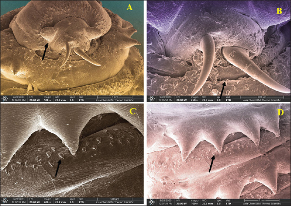
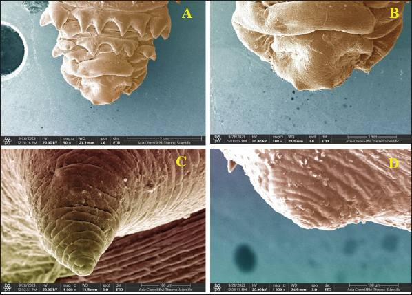
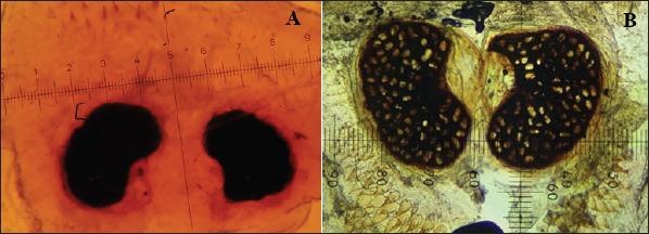
Fig. 1. (A) Ventral aspects of three larval stages of C. tittlator larval stage. (B) Anatomy of the cephalic segment of a second-stage larvae. ResultsThe results showed that 29.5% (59/200) of study camels were infested with C. titilator (Table 2). Morphologically, the characteristics of the larval instars of C. titilator using the light and scanning electron microscopes were showed the anterodorsal incomplete rings of spines, spinulation of anterventral part, lack of spines on last abdominal segment, several posterior spiracular pores and cephalopharngeal skeleton. Dorsally, there was a rounded surface; while abdominally, cuticular pits, inter-segmental grooves, obvious spines, and oval-shaped preitremes. Dorsoventrally, the larvae appeared as a slender flattened-shape with the presence of 12 segments with widely separated antennal lobes and obligate mouth hooks were observed (Figs. 1 and 2). The spinulation of the anterior ventral portion was noticed. However, no spines were found on the final segment of the abdomen. In the dorsal part, the surface exhibited a rounded surface; whereas in the abdominal portion, numerous cuticular pits and spines with intersegmental grooves and oval-shaped preitermes were seen. Dorsoventrally, the larvae appeared as a slender and flattened shape with the presence of 12 segments as well as widely separated antennal lobes and obligate mouth hooks. The cephalic segment observed as curved shaped in the second-stage larvae. Similar findings with more magnification were obtained by scanning electron microscopy. Importantly, the larval stages presented the following morphological characteristics: a rounded surface, numerous posterior spiracular pores, a cephalopharyngeal skeleton, spinulation of the anterior region, an absence of spines on the final segment of the abdomen, cuticular pits, intersegmental grooves, and prominent spines on the dorsal and anterodorsal surfaces. The preitremes display an elliptical shape. The larvae exhibited a dorsoventral orientation and had a thin, flattened form. They possessed 12 segments, with antennal lobes that were widely spaced apart. Additionally, the larvae were observed to have obligatory mouth hooks (Figs. 1–5).
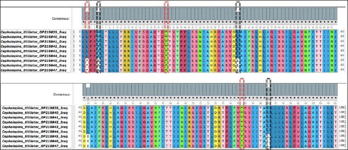
Fig. 2. The ventral view by scanning electron microscopy magnification shows the cephalic, thoracic, and abdominal portions of second larval stage. Molecular assayAll the tested samples were found to be positive by PCR. This indicates successful amplification of the designed primers targeting the COX1 gene (Fig. 6). Furthermore, some positive PCR products were sequenced and reported in NCBI GenBank, and the specific accession numbers were OP218846, OP218847, OP218845, OP218839, OP218840, OP218841, OP218842, OP218843, and OP218844. Sequence analysis revealed that numerous mutations were found within this genetic region. These mutations were found as substitutions. Importantly, some of these effects were caused by the cessation of translation (Fig. 7). However, other amino acids can change from alanine (A) to serine (S), leucine (L) to isoleucine (I), or isoleucine (I) to leucine (L).
Fig. 3. The cephalic segment shows (A) the separated bases of antennal lobes (scale bar=1 mm), (B) (scale bar=500 µm). (C) numerous spines (scale bar=500 µm), (D) (scale bar=1mm).
Fig. 4. (A)The terminal part of the abdomen (scale bar=3 mm) (B) (scale bar=1mm). (C) Larval papillae on the abdominal segment (100 µm) (D) the magnified papillae that appears as button shape (scale bar=100 µm).
Fig. 5. The terminal part of the abdominal segment. (A) is the first larval stage. (B) is the third larval stage (bar=1000 um).
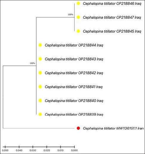
Fig. 6. Agarose-gel electrophoresis (1.5%) targeting the COX1 gene of some positive PCR products to C. titillator at 650 bp. Evolutionary analysis via the maximum likelihood method (500 bootstrap values) was performed via analysis of the partial sequence of the COX1 gene of C. titillator, which clearly diverges from the local isolates (yellow circles) and has significant identity with the global NCBI-GenBank Iran (red circle) isolate (MW136151.1) (Fig. 8). DiscussionThe study revealed that C. titillator was present in 29.5% of the population. Compared with other studies, there were 42.43% in Al-Qadisiyah Province (Atiyah et al., 2011) and 40.07% in Al-Muthanna Province (Al-Jindeel et al., 2018). In contrast, there were 33% in Japan (Al-Rawashdeh et al., 2000), 41% in Saudi Arabia (Alahmed, 2002), 79% in Libya (Abd El-Rahman, 2010), 41.67% in Egypt (Khater et al., 2013), 55.86% in Sudan (Makin, 2015), 46.39% in Jordan (Al-Ani and Amr, 2016), 52.3% in Iran (Jalali et al., 2016), 54.2% in China (Yao et al., 2022), and 23.9%–82.6% in Ethiopia (Chalchisa and Bersissa, 2023). Animal husbandry practices, geographic location, and sample variables could all play a role in explaining this substantial variation in C. titillator prevalence. In fact, Iraq is endemic with numerous parasites not only worms but also protozoa and pathogens (Al-Ubaidy and Alsultan, 2023; Alsaadi et al., 2023). These findings are consistent with earlier (Guitton et al., 1997) and more recent (Toaleb and Abdel-Rahman, 2021; Hamed et al., 2022; Yao et al., 2022) investigations. Our results provide a comprehensive and accurate description of the outwards morphological features of the C. titillator larval stages in infected camels. Modern molecular biology has considerably improved parasitological methods for investigating parasites that affect both humans and animals (Gunn and Pitt, 2022). According to several studies (Chan et al., 2021; Elyasigorji et al., 2023), mitochondrial DNA is the preferred marker for classifying organisms at various levels of taxonomy, from phylum to species. The current analysis validated all morphologically suspected C. titillator samples molecularly targeting the COX1 gene. The findings of the PCR experiment highlighted this finding. Various parasites, such as Enterobius vermicularis (Hagh et al., 2014), Plasmodium falciparum (Salman and Goldring, 2022), Trypanosoma spp. (Gnipová et al., 2012; Rodrigues et al., 2017), Sarcocystis spp. (Gjerde, 2013), Ancylostoma ceylanicum (Ngui et al., 2013), Diplostomum (Kudlai et al., 2017), Posthodiplostomum (Landeryou et al., 2020), Ornithodiplostomum (García-Varela et al., 2016), and C. titillator (Hafedh et al., 2021), have been detected and sequenced worldwide. Hafedh et al. (2021), Shamsi et al. (2023), and Shaalan et al. (2024) reported that the COX1 gene, which is a mitochondrial gene in Animalia that is well conserved, is a valuable tool for research on evolution and taxonomy. Nevertheless, the phylogenetic links of the Oestridae family remain a mystery, and there have been few efforts to incorporate molecular data into the taxonomy of this family in recent years (Shaalan et al., 2024). This work used light and ultrastructural electron microscopy to characterize the morphological characteristics of C. titillator in great detail, which could be useful for proper identification and categorization. Molecular analysis revealed that the COX1 gene is critical for C. titillator identification, and the sequences of local isolates presented a relatively high degree of similarity to those of Iranian isolates. To fill the gaps in our understanding of the evolutionary connections among the oestrid group of insects, further molecular phylogenetic investigations are needed. Not only is it crucial to conduct extensive genomic research on the family owing to the absence of clear identification criteria, but it is also necessary to use the collected molecular data to address this mystery and understand the larger evolutionary path.
Fig. 7. Multiple sequence alignment targeting the COX1 gene of C. titillator shows numerous mutations encoding stop codons, as depicted with red dashed-line rectangles, whereas other mutations leading to substitutions, as shown by black dashed-line rectangles.
Fig. 8. Phylogenetic tree analysis of local C. titillator isolates with global NCBI-GenBank C. titillator isolates. ConclusionThis study described precisely the morphological features of C. titillator using light and ultrastructural electron microscopes, which might aid in the appropriate identification and classification. Molecular examination demonstrated the importance of COX1 gene in the identification of C. titillator and sequencing of the local isolates that revealed a higher identity to Iranian isolates. However, additional molecular phylogenetic studies are needed to establish the evolutionary relationships among the oestrid group of insects with specialized habits and habitats. Also, the lack of unambiguous identification criteria creates an essential need not only for producing intensive molecular studies on the family but also for utilizing the accumulated molecular data in an attempt to resolve the enigma and decipher the broader evolutionary route. AcknowledgmentsThe authors thank all workers in abattoirs who contributed to the examination and collection of parasite samples. Conflicts of interestThere are no conflicts of interest. FundingNo external funds were received (private funding). Authors’ contributionsIsraa M. Essa: Morphological examination via light and electron microscopes. Mohammed Al-Saadi: Molecular testing and analysis of the parasite and sequencing of the local isolates. Akram M. Amanah: collection of parasite samples and documentation of obtained data. Monyer Abdulamier Abd: Writing draft and data validation. Mansour J Ali: Data validation and morphological identification. All authors contributed equally to writing of the manuscript and approved the final copy of it. Data availabilityAll the obtained data were utilized within the manuscript. ReferencesAbd El-Rahman, S. 2010. Prevalence and pathology of nasal myiasis in camels slaughtered in El-Zawia Province-Western Libya: with reference to thyroid alteration and renal lipidosis. Global Vet. 4(2), 190–197. Abu El Ezz, N.M., Hassan, N.M., El Namaky, A.H. and Abo-Aziza, F. 2018. Efficacy of some essential oils on Cephalopina titillator with special concern to nasal myiasis prevalence among camels and its consequent histopathological changes. J. Parasit. Dis. 42, 196–203. Al-Abedi, G.J., Sray, A.H., Hussein, A.J. and Gharban, H.A. 2020. Detection and bloody profiles evaluation of naturally, infected camels with subclinical Trypanosoma evansi, Iraq. Ann. Trop. Medic. Pub. Health. 23, 232–243. Al-Saadi, M., Al-Sallami, D. and Alsultan, A. 2023. Molecular identification of Anaplasma platys in cattle by nested PCR. Iran. J. Microbiol. 15(3), 433. Alahmed, A.M.I. 2002. Seasonal prevalence of Cephalopina titillator larvae in camels in Riyadh Region, Saudi. Arab Gulf J. Sci. Res. 20(3), 161–164. Al-Ani, F. and Amr, Z. 2016. Seasonal prevalence of the larvae of the nasal fly (Cephalopina titillator) in camels in Jordan. Revue d’élevage et de médecine vétérinaire des pays tropicaux 69(3), 125–127. Al-Graibawi, M.A., Yousif, A.A., Gharban, H.A. and Zinsstag, J. 2021. First serodetection and molecular phylogenetic documentation of Coxiella burnetii isolate from female camels in Wasit governorate, Iraq. Iraq. J. Vet. Sci. 35, 47–52. Al-Jindeel, T.J., Jasim, H.J., Alsalih, N.J. and Al-Yasari, A.M.R. 2018. Clinical, immunological and epidemiological studies of nasopharyngeal myiasis in camels slaughtered in al-Muthanna province. Adv. Anim. Vet. Sci. 6(7), 299–305. Al-Rawashdeh, O.F., Al-Ani, F.K., Sharrif, L.A., Al-Qudah, K.M., Al-Hami, Y. and Frank, N. 2000. A survey of camel (Camelus dromedarius) diseases in Jordan. J. Zoo. Wildl. Med. 31(3), 335–338. Al-Saadi, M., Al-Sallami, D. and Alsultan, A. 2023. Molecular identification of Anaplasma platys in cattle by nested PCR. Iranian J. Microbiol. 15(3), p.433. Al-Taee, H.S., Sekhi, A.A., Gharban, H.A. and Biati, H.M. 2023. Serological identification of MERS-CoV in camels of Wasit province, Iraq. Open Vet. J. 13(10), 1283–1289. Al-Ubaidy, Y. and Alsultan, A. 2023. Using of integrons as biomarker to assess dissemination and diversity of antimicrobial resistance genes in farm animal manure. J. Pure Appl. Microbiol. 17(3), 1708–1714. Atiyah, W.R., Dawood, K.A. and Dagher, A.L. 2011. Prevalence of infestation by camels nasal myiasis fly larvae in Al-Diwania city abattoir. AL-Qadisiyah J. Vet. Med. Sci. 10(2), 20–26. Bekele, T. and Molla, B. 2001. Mastitis in lactating camels (Camelus dromedarius) in Afar Region, north-eastern Ethiopia. Berl Munch Tierarztl. Wschr. 114(5-6), 169–172. Chalchisa, A. and Bersissa, E. 2023. Ectoparasites and their effect on camels (Camelus dromedarius) in Ethiopia. J. Vet. Res. Adv. 5(1), 55–69. Chan, A.HE., Chaisiri, K., Saralamba, S., Morand, S. and Thaenkham, U. 2021. Assessing the suitability of mitochondrial and nuclear DNA genetic markers for molecular systematics and species identification of helminths. Parasit. Vectors 14(1), 233. Colwell, D.D. 1986. Cuticular sensilla on newly hatched larvae of Cuterebra fontinella Clark (Diptera: Cuterebridae) and Hypoderma spp. (Diptera: Oestridae). Inter. J. Insect. Morphol. Embryol. 15(5-6), 385–392. Duman, M., Guz, N. and Sertkaya, E. 2015. DNA barcoding of sunn pest adult parasitoids using cytochrome c oxidase subunit I (COI). Bioch. Sys. Ecol. 59, 70–77. El-Bassiony, G.M. and Awad, H.H. 2007. Surface ultrastructure of the third-instar larva of Cephalopina titillator (Diptera: Oestridae). J. Egypt. German Soc. Zool. 52(E), 20. Elyasigorji, Z., Izadpanah, M., Hadi, F. and Zare, M. 2023. Mitochondrial genes as strong molecular markers for species identification. Nucleus 66(1), 81–93. Fatani, A. and Hilali, M. 1994. Prevalence and monthly variations of the second and third instars of Cephalopina titillator (Diptera: Oestridae) infesting camels (Camelus dromedarius) in the Eastern Province of Saudi Arabia. Vet. Para. 53(1-2), 145–151. García-Varela, M., Sereno-Uribe, A.L., Pinacho-Pinacho, C.D., Domínguez-Domínguez, O. and de León, G.P.P. 2016. Molecular and morphological characterization of Austrodiplostomum ostrowskiae Dronen, 2009 (Digenea: Diplostomatidae), a parasite of cormorants in the Americas. J. Helminthol. 90(2), 174–185. Gjerde, B. 2013. Phylogenetic relationships among Sarcocystis species in cervids, cattle and sheep inferred from the mitochondrial cytochrome c oxidase subunit I gene. Int. J. Parasitol. 43(7), 579–591. Gnipová, A., Panicucci, B., Paris, Z., Verner, Z., Horváth, A., Lukeš, J. and Zíková, A. 2012. Disparate phenotypic effects from the knockdown of various Trypanosoma brucei cytochrome c oxidase subunits. Mol. Biochem. Parasitol. 184(2), 90–98. Guitton, C., Dorchies, P. and Morand, S. 1997. Morphological comparison of second-stage larvae of Oestrus ovis (Linnaeus, 1758), Cephalopina titillator (Clark, 1816) and Rhinoestrus usbekistanicus Gan, 1947 (Oestridae) by scanning electron microscopy. Parasit. 4(3), 277–282. Gunn, A. and Pitt, S.J. 2022. Parasitology: an integrated approach. Newyork, NY: John Wiley and Sons. Hafedh, A.A., Humide, A.O. and Alrikaby, N.J.A. 2021. Cytochrome oxidase subunit I (COI) gene sequencing for identification of Cephalopina titillator local isolates from camels in Thi-Qar Province/Iraq. Annal. Roman. Soc. Cell Biol. 25(4), 3146–3152. Hagh, V.R.H., Oskouei, M.M., Bazmani, A., Miahipour, A. and Mirsamadi, N. 2014. Genetic classification and differentiation of Enterobius vermicularis based on mitochondrial cytochrome c oxidase (cox1) in northwest Iran. J. Pure Appl. Microbiol. 8(5), 3995–3999. Hamed, S., Shaalan, M.G., Khater, E.I., Kenawy, M.A. and Ghallab, E.H. 2022. Prevalence and morphological characterization of the camel nasal botfly, Cephalopina titillator (Diptera: Oestridae) collected from abattoirs in Egypt. Egypt. Acad. J. Biol. Sci. E. Med. Entomol. Parasitol. 14(2), 87–107. Hassan, N.M., Sedky, D., El Ezz, N.M.A. and El Shanawany, E.E. 2022. Seroprevalence of nasal myiasis in camels determined by indirect enzyme-linked immunosorbent assay utilizing the most diagnostic Cephalopina titillator larval antigens. Vet. World 15(12), 2830. Hilali, M.A., Mahdy, O.A. and Attia, M.M. 2015. Monthly variations of Rhinoestrus spp. (Diptera: Oestridae) larvae infesting donkeys in Egypt: morphological and molecular identification of third stage larvae. J. Adv. Res. 6(6), 1015–1021. Hussein, M.F., Elamin, F.M., El-Taib, N.T. and Basmaeil, S.M. 1982. The pathology of nasopharyngeal myiasis in Saudi Arabian camels (Camelus dromedarius). Vet. Parassit. 9(3-4), 179–183. Jalali, M.H.R., Dehghan, S., Haji, A. and Ebrahimi, M. 2016. Myiasis caused by Cephalopina titillator (Diptera: Oestridae) in camels (Camelus dromedarius) of semiarid areas in Iran: distribution and associated risk factors. Comp. Clin. Pathol. 25, 677–680. Khater, H.F., Ramadan, M.Y. and Mageid, A.D.A. 2013. In vitro control of the camel nasal botfly, Cephalopina titillator, with doramectin, lavender, camphor, and onion oils. Parasitol. Res. 112, 2503–2510. Kudlai, O., Oros, M., Kostadinova, A. and Georgieva, S. 2017. Exploring the diversity of Diplostomum (Digenea: Diplostomidae) in fishes from the River Danube using mitochondrial DNA barcodes. Parasit. Vectors 10, 1–21. Landeryou, T., Kett, S.M., Ropiquet, A., Wildeboer, D. and Lawton, S.P. 2020. Characterization of the complete mitochondrial genome of Diplostomum baeri. Parasitol. Inter. 79, 102166. Li, X., Yan, L., Pape, T., Gao, Y. and Zhang, D. 2020. Evolutionary insights into bot flies (Insecta: Diptera: Oestridae) from comparative analysis of the mitochondrial genomes. Int. J. Biol. Macromole. 149, 371–380. Makin, E.M.M. 2015. Detection and prevalence of nasal bot fly (Cephalopina titillator) larvae in camels in slaughterhouses in Ombadda and Tamboul localities, (Doctoral dissertation, University of Khartoum), Khartoum, Sudan. Nelson, L.A., Wallman, J.F. and Dowton, M. 2007. Using COI barcodes to identify forensically and medically important blowflies. Med. Vet. Entomol. 21, 44–52. Ngui, R., Mahdy, M.A., Chua, K.H., Traub, R. and Lim, Y.A. 2013. Genetic characterization of the partial mitochondrial cytochrome oxidase c subunit I (cox 1) gene of the zoonotic parasitic nematode, Ancylostoma ceylanicum from humans, dogs and cats. Acta Trop. 128(1), 154–157. Oryan, A., Valinezhad, A. and Moraveji, M. 2008. Prevalence and pathology of camel nasal myiasis in eastern areas of Iran. Trop. Biomed. 25(1), 30–36. Rahman, A. and Mohammed, M. 2021. Assessment of Cephalopina titillator, different prepared antigens in diagnosis by indirect ELISA in serum and mucus of camels. Egypt. Vet. Med. Soc. Parasitol. J. 17(1), 92–108. Rodrigues, M.S., Morelli, K.A. and Jansen, A.M. 2017. Cytochrome c oxidase subunit 1 gene as a DNA barcode for discriminating Trypanosoma cruzi DTUs and closely related species. Parasit. Vectors 10, 1–18. Salman, A.A. and Goldring, J.D. 2022. Expression and copper binding characteristics of Plasmodium falciparum cytochrome c oxidase assembly factor 11, Cox11. Malaria J. 21(1), 173. Schauff, M.E. 2001. Collecting and preserving insects and mites: techniques and tools. In: systematic entomology laboratory, USDA, National Museum of Natural History, NHB 168. Eds., Schauff, M.E. Washington, DC, USA. Shaalan, M.G., Farghaly, S.H., Khater, E.I., Kenawy, M.A. and Ghallab, E.H. 2024. Molecular characterization of the camel nasal botfly, Cephalopina titillator (Diptera: Oestridae). Beni-Suef University J. Basic Appl. Sci. 13(1), 8. Shamsi, E., Radfar, M.H., Nourollahifard, S.R., Bamorovat, M., Nasibi, S., Fotoohi, S. and Kheirandish, R. 2023. Nasopharyngeal myiasis due to Cephalopina titillator in Southeastern Iran: a prevalence, histopathological, and molecular assessment. J. Parasit. Dis. 47(2), 369–375. Toaleb, N.I. and Abdel-Rahman, E.H. 2021. Accurate diagnosis of camel nasal myiasis using specific second instar larval fraction of cephalopods titillator extract. Adv. Anim. Vet. Sci. 9(5), 674–681. Walper, S.A., Lasarte Aragonés, G., Sapsford, K.E., Brown III, C.W., Rowland, C.E., Breger, J.C. and Medintz, I.L. 2018. Detecting biothreat agents: from current diagnostics to developing sensor technologies. ACS Sensors 3(10), 1894–2024. Wolk, D.M., Kaleta, E.J. and Wysocki, V.H. 2012. PCR–electrospray ionization mass spectrometry: the potential to change infectious disease diagnostics in clinical and public health laboratories. J. Mol. Diagnost. 14(4), 295–304. Yao, H., Liu, M., Ma, W., Yue, H., Su, Z., Song, R. and Yang, J. 2022. Prevalence and pathology of Cephalopina titillator infestation in Camelus bactrianus from Xinjiang, China. BMC Vet. Res. 18(1), 360. Yousef, H.A., Abdel Meguid, A., Afify, A. and Hassan, H.M. 2016. Analysis of larval antigens of Cephalopina titillator in the camel mucus for diagnosis of infestation. Biologia 71(4), 438–443. Zumpt, F. 1965. Myiasis in man and animals in the Old World. A textbook for physicians, veterinarians and zoologists. London, UK: Butterworths. | ||
| How to Cite this Article |
| Pubmed Style Essa IM, Al-saadi MH, Amanah AM, Alfatlawi MAA, Ali MJ. Morphological and genetic demonstration of Cephalopina titillator in dromedary camels. Open Vet. J.. 2024; 14(11): 2995-3003. doi:10.5455/OVJ.2024.v14.i11.28 Web Style Essa IM, Al-saadi MH, Amanah AM, Alfatlawi MAA, Ali MJ. Morphological and genetic demonstration of Cephalopina titillator in dromedary camels. https://www.openveterinaryjournal.com/?mno=216719 [Access: January 25, 2026]. doi:10.5455/OVJ.2024.v14.i11.28 AMA (American Medical Association) Style Essa IM, Al-saadi MH, Amanah AM, Alfatlawi MAA, Ali MJ. Morphological and genetic demonstration of Cephalopina titillator in dromedary camels. Open Vet. J.. 2024; 14(11): 2995-3003. doi:10.5455/OVJ.2024.v14.i11.28 Vancouver/ICMJE Style Essa IM, Al-saadi MH, Amanah AM, Alfatlawi MAA, Ali MJ. Morphological and genetic demonstration of Cephalopina titillator in dromedary camels. Open Vet. J.. (2024), [cited January 25, 2026]; 14(11): 2995-3003. doi:10.5455/OVJ.2024.v14.i11.28 Harvard Style Essa, I. M., Al-saadi, . M. H., Amanah, . A. M., Alfatlawi, . M. A. A. & Ali, . M. J. (2024) Morphological and genetic demonstration of Cephalopina titillator in dromedary camels. Open Vet. J., 14 (11), 2995-3003. doi:10.5455/OVJ.2024.v14.i11.28 Turabian Style Essa, Israa M., Mohammed H. Al-saadi, Akram M. Amanah, Monyer Abdulamier Abd Alfatlawi, and Mansour J. Ali. 2024. Morphological and genetic demonstration of Cephalopina titillator in dromedary camels. Open Veterinary Journal, 14 (11), 2995-3003. doi:10.5455/OVJ.2024.v14.i11.28 Chicago Style Essa, Israa M., Mohammed H. Al-saadi, Akram M. Amanah, Monyer Abdulamier Abd Alfatlawi, and Mansour J. Ali. "Morphological and genetic demonstration of Cephalopina titillator in dromedary camels." Open Veterinary Journal 14 (2024), 2995-3003. doi:10.5455/OVJ.2024.v14.i11.28 MLA (The Modern Language Association) Style Essa, Israa M., Mohammed H. Al-saadi, Akram M. Amanah, Monyer Abdulamier Abd Alfatlawi, and Mansour J. Ali. "Morphological and genetic demonstration of Cephalopina titillator in dromedary camels." Open Veterinary Journal 14.11 (2024), 2995-3003. Print. doi:10.5455/OVJ.2024.v14.i11.28 APA (American Psychological Association) Style Essa, I. M., Al-saadi, . M. H., Amanah, . A. M., Alfatlawi, . M. A. A. & Ali, . M. J. (2024) Morphological and genetic demonstration of Cephalopina titillator in dromedary camels. Open Veterinary Journal, 14 (11), 2995-3003. doi:10.5455/OVJ.2024.v14.i11.28 |


