| Short Communication | ||
Open Vet. J.. 2024; 14(11): 3108-3112 Open Veterinary Journal, (2024), Vol. 14(11): 3108-3112 Short Communication Fracture toughness of cancellous iliac boneCalin-Daniel Stefan1,2, Liviu Marsavina1* and Catalin Adrian Miu2,31Department of Mechanics and Strength of Materials, Politehnica University Timisoara, Timisoara, Romania 2Orthopedics Unit, “Dr. Victor Popescu” Emergency Military Hospital, Timisoara, România 3Department XV, Discipline of Orthopedics, “Victor Babeș” University of Medicine and Pharmacy Timișoara, Timisoara, România *Corresponding Author: Liviu Marsavina. Department of Mechanics and Strength of Materials, Politehnica University Timisoara, Timisoara, Romania. Email: liviu.marsavina [at] upt.ro Submitted: 27/08/2024 Accepted: 24/10/2024 Published: 30/11/2024 © 2024 Open Veterinary Journal
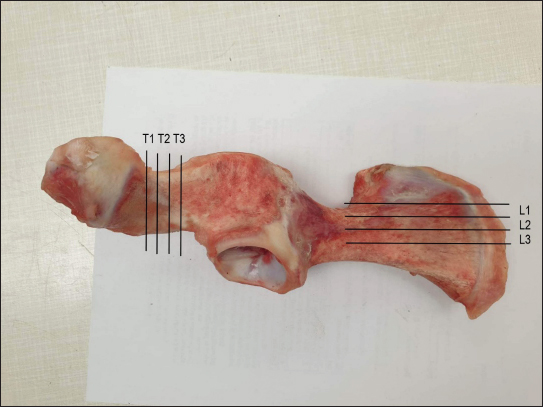
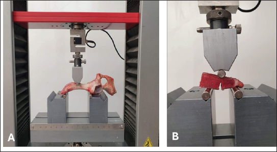
AbstractBackground: Cancellous bone mechanical properties are directly linked to structural integrity, which is a result of bone quantity, the quality of its bone matrix, and its microarchitecture. Several studies highlighted the bone behavior under specific loads, contributing to understanding risk factors and developing more effective therapeutic strategies. The anatomy and stability of iliac bone fractures, providing insight into pelvic trauma management. Aim: To evaluate the anisotropic behavior of fracture toughness of porcine iliac bone. Methods: Longitudinal and transversal specimens from pork iliac bone were cut and tested in three-point bending fixtures at room temperature, in order to determine the fracture toughness of the cancellous iliac bone. Results: The tests on the entire iliac bone reveal a brittle behavior. The tests on notched specimens cut on longitudinal and transversal directions show different fracture toughness values. Also, a brittle behavior occurs. The obtained results are at the lower limit of other fracture toughness results of cancellous bones. Conclusion: Our results point out the anisotropic behavior of the cancellous iliac bone. Higher fracture toughness values were obtained in the longitudinal direction comparing with the transversal direction with 36.5 %. Keywords: Iliac bone fractures, Cancelous bone, Fracture toughness. IntroductionThe ilium or the iliac bone is a part of the hip bone together with the ischium and the pubic bone. It consists of the body (corpus ossis ilii), which belongs to the acetabulum, and the wing (alla ossis ilii). The lateral/external face of the ilium represents the gluteal surface which is slightly concave in the middle and presents three distinct curved lines: anterior gluteal line, posterior gluteal line, and inferior gluteal line. The three lines delimit three areas that give attachment to the gluteus maximus, gluteus medius, and gluteus minimus muscles; they also represent strength arches in the architecture of the bone. The lateral face of the ilium contains the iliac fossa (fossa iliaca) which gives attachment to the iliac muscle. The anterior border of the ilium represents the antero superior iliac spine (spina iliaca anterior superior), which serves as the insertion point for the inguinal ligament, the tensor fasciae latae and the sartorius muscles, the antero inferior iliac spine (spina iliaca anterior inferior), which provides attachment to the rectus femoris (right femoral) muscle and the iliopubic eminence, which leads to the iliopubic part of the pelvis. The posterior border consists of the postero superior iliac spine (spina iliaca posterior superior) and the postero inferior iliac spine (spina iliaca posterior inferior), providing attachment to the ligaments of the sacroiliac joint. The superior border of the ilium is called iliac crest (crista iliaca) and provides attachment to the gluteus maximus, gluteus medius, iliac, transverse, quadratus lumborum, internal oblique, and external oblique muscles (Papilian, 2011). The fractures of the iliac bone have to be approached as any fracture of the pelvis, taking into account the stability, i.e., the integrity of the pelvic rings. If we consider stability to be the intrinsic ability of the fractured pelvis to support forces without any significant movement, and having to quantify stability from the point of view of treatment and prophylaxis of complications, trauma practice widely uses the Tile classification of pelvic fractures. Thus, fractures are classified into: type A (stable) fractures: A1—the pelvic ring is not affected, A2—fracture with minimal displacement, it does not impair the stability of the pelvic ring; type B fractures (rotationally unstable/vertically stable): B1 –“open-book” fracture, B2 – ipsilateral pelvic overlapping fracture, B3 – contralateral “bucket handle” fracture; type C (rotationally and vertically unstable) fractures: C1 – vertical shear fracture, C2 – vertical bilateral shear fracture, C3 – association with acetabular, joint fracture. The more a fracture is comminuted and the greater the bone fragment displacement in iliac bone fractures, the higher the risk of complications. According to their occurrence, there may be vascular, urologic, neurological, digestive damage, gynecological, or associated fractures. Fractures of the iliac bone are more common in cases of multiple trauma, secondary to high-kinetic-energy traumatic factors, and have a high morbidity and mortality rate (Antonescu, 2009; Stoica and Dragosloveanu, 2023). Assumptions were performed that bone is a linear-elastic material, respectively, plasticity is limited. For these materials, the stress and displacement fields local to the tip of a pre-existing crack can be described according to Linear Elastic Fracture Mechanics using the stress-intensity factor (Lucksanasombool et al., 2001; Taylor, 2003; Launey et al., 2010). The fracture toughness of bovine femur cortical bone was determined by Bonfield (1987) obtaining values in the range of 2.5 to 6.3 MPa m0.5. Zioupos and Currey (1998) investigated the influence of age on stiffness, strength, and toughness of human cortical bone. They observed that the fracture toughness is decreasing from 7 MPa m0.5 at 30 years to approximately 5 MPa m0.5 for older people (90 years). Fewer values are provided in the literature for the fracture toughness of cancellous bone. Cook and Zioupos (2009) found a correlation between fracture toughness and the apparent and relative density of cancellous bone tissue. Their values range from 0.2 to 1.75 MPa m0.5, for apparent densities from 300 to 1,000 kg/m3. Greenwood et al. (2015) liked the microarchitecture and mechanical properties of cancellous bone specimens collected from 37 patients who had suffered fractures of the neck of femur. Using single-edge notched beam specimens obtained from trabeculae (stronger/tougher behavior) and along/in-between the trabeculae (softer/weaker behavior) the fracture toughness ranges from 0.1 to 0.5 MPa m0.5, respectively, from 0.1 to 0.4 MPa m0.5. Craciun et al. 2018 investigated analytically the crack propagation in bones. This paper investigates the fracture toughness of pork iliac cancellous bone. Materials and MethodsFor this research, we utilized specimens of porcine iliac bone obtained from a local abattoir, Figure 1. Once prepared by cutting into standardized dimensions, the specimens were secured onto a universal testing machine (Zwick Roell Z005). Two tests were carried on the entire iliac bone loaded in bending, Figure 2.a. Second test consists of the determination of fracture toughness using Single Edge Notched Bend specimens cut longitudinally, respective transversally, Figure 2.b. After cutting the iliac bone the density of the cancellous part of the bone was determined on cubic specimens with a length of 12.5 mm and weighted with an electronic balance. The average result of the density was 1044.6 kg/m3. A gradual force was applied to each specimen until fracture using the aforementioned testing machine. Tests were performed at room temperature (20°C) with a loading speed of 2 mm/minute. The testing machine automatically recorded the force required to fracture each specimen. The fracture toughness of cancelous iliac bone was determined according to standard test methods for plane-strain fracture toughness and strain energy release rate of plastic materials (ASTM 5045–14 standard):
where
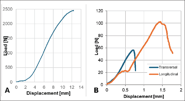
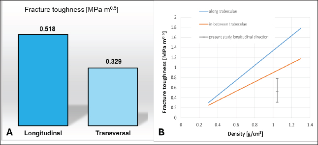
For this study, we choose to perform Mode I fracture toughness tests because the fracture of bending test of the entire iliac bone highlighted a Mode I fracture (opening crack mode). On the other hand, the dimensions of the specimens, which could be obtained from the pork iliac bone, are relatively small, while the mode II and mixed mode fracture tests required a sufficient amount of material for Brazilian disk, semi-circular bend, compact edge notch specimens used for mode II or mixed mode tests. However, the Mode I fracture is the most important, because even if the cracks are initiated in Mode II or mixed mode will propagate in Mode I. Results and DiscussionsThe study presents the fracture toughness results of fracture toughness of pork iliac bone. A brittle fracture was observed on testing of the entire iliac bone and a mean fracture load of 2470 N was obtained. A typical load-displacement curve is presented in Figure 3.a. The fracture toughness of cancelous iliac bone was evaluated in two directions longitudinal and transversal to the bone. Load—displacement curves from testing of cancelous iliac bone are shown in Figure 3.b. The results on the fracture toughness are plotted in Figure 4. The anisotropy of fracture toughness is highlighted by different values obtained on longitudinal (0.518 MPa m0.5, standard deviation 0.11 MPa m0.5), respectively, transversal directions (0.329 MPa m0.5, standard deviation 0.09 MPa m0.5), Figure 4.a. The obtained values are lower than those presented by Greenwood et al. (2015), which presents a correlation of fracture toughness of cancellous bone with volumetric bone mineral density, from 37 human patients (males and females), Figure 4.b. Interpolating by liner regression they obtained higher values of fracture toughness along the trabeculae and lower values in-between the trabeculae. Our maximum results of fracture toughness on the longitudinal direction being closer to in-between trabeculae. The degree of anisotropy in this case is KIC,transversal / KIC,longitudinal=0.63, which is in good agreement with the degree of anisotropy of bovine cancelous bone between 0.34–0.6 reported by Frayssinet et al. (2022). The anisotropic behavior of cancelous bone could be explained based on the architecture of the cellular structure, which is influenced by the trabeculae orientation. Lucksanasombool et al. (2001), also observed the anisotropy of fracture toughness for cortical bovine bone.
Fig. 1. Cutting of specimens from the iliac bone. (T1,T2,T3—transverse samples L1,L2,L3—longitudinal samples).
Fig. 2. Test specimens (samples) prepared for testing. A: Bending testing of entire bone. B: Fracture toughness testing.
Fig. 3. Typical load—displacement curves. A: Testing of entire bone. B: Fracture toughness tests.
Fig. 4. Fracture toughness of the cancellous iliac bone. A: Comparison between longitudinal and transversal directions. B: Comparison with other studies. AcknowledgmentsThe authors acknowledge the access to the St. Nadasan Strength of Materials Laboratory from University Politehnica Timisoara. Conflict of interestThe authors declare no conflicts of interest. FundingThis research was partially funded by grant number FDI 2024-0695. Authors’ contributionsConceptualization L.M. and C.D.S., methodology C.D.S. and L.M., investigation C.D.S. and C.D.M., data curation L.M. and C.D.S., writing—original draft preparation C.D.S. and C.D.M., writing - review and editing C.D.M., C.D.S., and L.M. All authors have read and agreed to the published version of the manuscript. Data availabilityData will be made available on request. ReferencesAntonescu, D. 2009. Fracturile bazinului. In: Tratat de chirurgie Vol. X Ortopedie—Traumatologie, Popescu I. Ed. Bucharest, Romania: Academiei Române, pp: 238–240, 245–250. Bonfield, W. 1987. Advances in the fracture mechanics of cortical bone. J. Biomech. 20(11-12), 1071–1081. Cook, R.B. and Zioupos, P. 2009. The fracture toughness of cancellous bone. J. Biomech. 42, 2054–2060. Craciun, E.M., Marin, M. and Rabaea, A. 2018. Anti-plane crack in human bone. I. Mathematical modelling. An. St. Univ. Ovidius. Constanta. 26(1), 81–90. Greenwood, C., Clement, J.G., Dicken, A.J., Evans, J.P.O., Lyburn, I.D., Martin, R.M., Rogers, K.D., Stone, N., Adams, G. and Zioupos, P. 2015. The micro-architecture of human cancellous bone from fracture neck of femur patients in relation to the structural integrity and fracture toughness of the tissue. Bone Rep. 3, 67–75. Frayssinet, E.E., Colabella, L. and Cisilino, A.P. 2022. Design and assessment of the biomimetic capabilities of a Voronoi-based cancellous microstructure. J. Mech. Behav. Biomed. Mater. 130, 105186. Launey, M.E., Buehler M.J. and Ritchie R.O. 2010. On the mechanistic origins of toughness in bone. Annu. Rev. Mater. Res. 40, 25–53. Lucksanasombool, P., Higgs, W.A.J., Higgs, R.J.E.D. and Swain, M.V. 2001. Fracture toughness of bovine bone: influence of orientation and storage media. Biomater. 22, 3127–3132. Papilian, V. 2011. Coxalul. Anatomia omului. Vol. I Aparatul locomotor. Bucharest, Romania: Editura All, pp: 68–70. Stoica, C.I. and Dragosloveanu S. 2023. Fracturile de pelvis si acetabulum. Patologia aparatului locomotor, Vol. 3. Ed., Antonescu D. Bucharest, Romania: Medicală Amaltea, pp: 1489–1491. Taylor, D. 2003. How does bone break? Nat. Mater. 2, 133–134. Wang, X., Bank, R.A., TeKoppele J.M. and Mauli Agrawal, C.M. 2001. The role of collagen in determining bone mechanical properties. J. Orthop. Res. 19, 1016–1021. Zioupos, P. and Currey J.D. 1998. Changes in the stiffness, strength, and toughness of human cortical bone with age. Bone, 22(1), 57–66. | ||
| How to Cite this Article |
| Pubmed Style Stefan C, Marsavina L, Miu C. Fracture toughness of cancellous iliac bone. Open Vet. J.. 2024; 14(11): 3108-3112. doi:10.5455/OVJ.2024.v14.i11.40 Web Style Stefan C, Marsavina L, Miu C. Fracture toughness of cancellous iliac bone. https://www.openveterinaryjournal.com/?mno=216864 [Access: December 15, 2025]. doi:10.5455/OVJ.2024.v14.i11.40 AMA (American Medical Association) Style Stefan C, Marsavina L, Miu C. Fracture toughness of cancellous iliac bone. Open Vet. J.. 2024; 14(11): 3108-3112. doi:10.5455/OVJ.2024.v14.i11.40 Vancouver/ICMJE Style Stefan C, Marsavina L, Miu C. Fracture toughness of cancellous iliac bone. Open Vet. J.. (2024), [cited December 15, 2025]; 14(11): 3108-3112. doi:10.5455/OVJ.2024.v14.i11.40 Harvard Style Stefan, C., Marsavina, . L. & Miu, . C. (2024) Fracture toughness of cancellous iliac bone. Open Vet. J., 14 (11), 3108-3112. doi:10.5455/OVJ.2024.v14.i11.40 Turabian Style Stefan, Calin-daniel, Liviu Marsavina, and Catalin-adrian Miu. 2024. Fracture toughness of cancellous iliac bone. Open Veterinary Journal, 14 (11), 3108-3112. doi:10.5455/OVJ.2024.v14.i11.40 Chicago Style Stefan, Calin-daniel, Liviu Marsavina, and Catalin-adrian Miu. "Fracture toughness of cancellous iliac bone." Open Veterinary Journal 14 (2024), 3108-3112. doi:10.5455/OVJ.2024.v14.i11.40 MLA (The Modern Language Association) Style Stefan, Calin-daniel, Liviu Marsavina, and Catalin-adrian Miu. "Fracture toughness of cancellous iliac bone." Open Veterinary Journal 14.11 (2024), 3108-3112. Print. doi:10.5455/OVJ.2024.v14.i11.40 APA (American Psychological Association) Style Stefan, C., Marsavina, . L. & Miu, . C. (2024) Fracture toughness of cancellous iliac bone. Open Veterinary Journal, 14 (11), 3108-3112. doi:10.5455/OVJ.2024.v14.i11.40 |


