| Case Report | ||
Open Vet J. 2024; 14(11): 3127-3131 Open Veterinary Journal, (2024), Vol. 14(11): 3127-3131 Case Report Temporary circular external fixation for effective management of bilateral radial non-union in a Toy PoodleByoungho An, Bokyung Song, Yehyeon Jang, Dongwook Kim and Gonhyung Kim*Department of Veterinary Surgery, College of Veterinary Medicine, Chungbuk National University, Cheongju, Republic of Korea *Corresponding Author: Gonhyung Kim. Department of Veterinary Surgery, College of Veterinary Medicine, Chungbuk National University, Cheongju, Republic of Korea. Email: ghkim [at] cbu.ac.kr Submitted: 23/08/2024 Accepted: 23/10/2024 Published: 30/11/2024 © 2024 Open Veterinary Journal
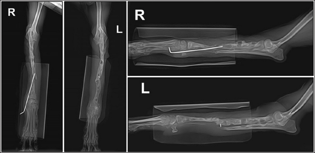
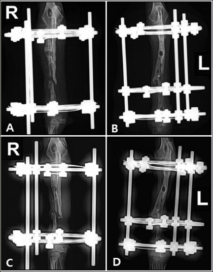
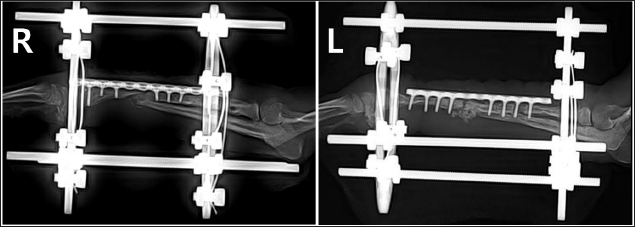
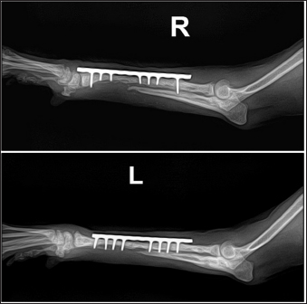
AbstractBackground: Distal radius fractures are prevalent in small and toy-breed dogs, presenting significant treatment challenges due to complications such as delayed union or non-union. These complications are often exacerbated by reduced vascular density at the distal diaphyseal-metaphyseal junction of the radius, which is vital for bone healing, particularly in toy and small breed dogs. Circular external fixation (CEF) is known for its effectiveness in managing acute and chronic fractures and providing temporary stabilization in humans. This study documented the use of temporary CEF in a Toy Poodle with non-union fractures of the radius and ulna, addressing bone atrophy and resorption caused by repeated implant failures. Case Description: A 3-year-old, 4.2 kg, neutered male Toy Poodle was referred for treatment after multiple attempts to fix radial and ulnar fractures failed to achieve union over 1 year, leaving the dog barely using its forelimbs. In the first surgery, CEF was applied to heal holes in the bone caused by previous fixation devices and encourage forelimb use through rehabilitation. In the second surgery, a combination of cancellous bone grafting, plate fixation, and CEF was used, resulting in successful bone union and restoration of normal gait after 10 weeks. Conclusion: In conclusion, CEF is a valuable option for temporary fixation and fracture reduction in toy-breed dogs and offers a promising approach to managing challenging cases. Keywords: Circular external fixation, Fracture, Non-union, Small animal, Toy-breed dog. IntroductionFractures in small and toy-breed dogs, especially involving the radius and ulna, present unique challenges in veterinary orthopedics (Rudd and Whitehair, 1992; Welch et al., 1997; Piermattei et al., 2006). These fractures, often caused by minor trauma, have a higher risk of delayed union or non-union compared to similar fractures in larger breeds (Welch et al., 1997; Aikawa et al., 2018; Zeki et al., 2022). The distal diaphyseal-metaphyseal junction of the radius in small-breed dogs shows decreased vascular density, particularly in areas associated with a poor prognosis for fracture healing (Welch et al., 1997). Consequently, fractures in toy-breed dogs, such as Toy Poodle, Maltese, Pomeranian, Chihuahua, and Yorkshire Terrier can be clinically challenging because of bone resorption during the healing, which increases the likelihood of refracture. Traditional methods for managing fractures in small-breed dogs involve the use of bone plates to facilitate early weight-bearing (Larsen et al., 1999; Aikawa et al., 2018). However, in small breeds, particularly in distal limbs, the small size of bones and limited space make it challenging to apply appropriate bone plates. Consequently, this approach can lead to complications such as implant failure. The primary cause of implant failure is loosening of screws and pins, followed by plate failure (Dos Santos et al., 2016). Microfractures around the screws, caused by excessive loads on the screws, are recognized as the main cause (Feng et al., 2019). Additionally, excessive stress protection can lead to a decrease in the load transmitted through the bone tissue, potentially leading to disuse atrophy (Field, 1997; Chen et al., 2005; Andrzejowski and Giannoudis, 2019; Hirashima et al., 2021). These complications elevate the risk of delayed union, malunion, or non-union (Dos Santos et al., 2016). Circular external fixation (CEF) is a valuable method for fracture fixation that can be combined with other techniques to provide adequate stability (Lewis et al., 2001). It is effective in managing acute and chronic fractures and also provides temporary stabilization for humans and animals (Lewis et al., 2001; Diwan et al., 2018; Bose and Piper, 2021; van Leeuwen et al., 2022). However, the application of CEF in toy-breed dogs is challenging (Hamilton and Langleyhobbs, 2005). This case demonstrates the use of CEF for temporary stabilization and treatment of non-union radius and ulnar fractures in a toy-breed dog. Case DetailsA 3-year and 6-month-old Toy Poodle presented with a history of failed treatment for bilateral radius and ulnar fractures over the course of 1 year. The initial treatment involved plate fixation and linear external fixation; however, repeated implant failure resulted in a non-union fracture (Fig. 1). At the time of presentation, radiographs revealed that most plates, screws, and pins had been removed from both limbs. The prolonged non-union resulted in continued bone resorption at the fracture site without any signs of healing. Loosening was evident around the bone holes where the fixation devices had been applied, and some pins were still visible on the radiographs, indicating they had not been removed. The dog mainly used its hind limbs and exhibited a plantigrade gait when moving the forelimb. Overall systemic conditions were satisfactory. No specific abnormalities were found in the skin surrounding the fracture site or pin insertions, and blood tests showed normal results, except for a slight increase in alkaline phosphatase. The diagnosis was confirmed as non-union fractures of the radius and ulna in both forelimbs. A two-step surgical plan was developed for managing the fractures. The remaining pin was removed in the first surgery and a CEF was applied for temporary fixation (Fig. 2A and B). Pins used included two 0.9 mm K-wires placed proximally and distally in a crossed manner, and one 1.2 mm olive wire placed in a single location. Anticipation of bone regeneration around areas of bone lysis near screw holes, and physical rehabilitation focused on inducing forelimb use until the second surgery. During the 6-week maintenance period of the temporary external fixation device, partial bone regeneration was observed in some areas of bone loss (Fig. 2C and D). Although forelimb ambulation remained plantigrade, there was an improvement in utilizing the limb smoothly with weight-bearing. In the second surgery, the CEF device was removed, and a surgical approach was made to apply the bone plate. The atrophied fracture sites on both sides showed no signs of bone regeneration and were partially debrided to expose the bone marrow cavities. The fracture was then fixed with a long bone plate and 1.5 mm screws. Autografts were harvested from the iliac wing and transplanted around the fracture site. This procedure was performed identically on both limbs with the fascia and skin closed in a standard manner. Additionally, CEF methods involved applying 0.9 mm K-wires, the same size as those used in the first surgery, above and below the plate on the fractured radius to provide further stability (Fig. 3). Preoperative medications for both surgeries included tramadol (5 mg/kg, IV), cefazolin (30 mg/kg, IV), famotidine (0.5 mg/kg, IV), and midazolam (0.2 mg/kg, IV). General anesthesia was induced with propofol (5 mg/kg, IV) and maintained with 1.5%–2% isoflurane in oxygen. Intraoperative analgesia was provided by a constant-rate infusion of ketamine and lidocaine. The forelimbs were then prepared for routine aseptic surgery. The entire surgery lasted 2 hours and 50 minutes. Postoperatively, cefazolin was administered for 5 days, along with tramadol and meloxicam (0.1 mg/kg, SC) for pain management. Amoxicillin syrup was prescribed for an additional 3 weeks as an antibiotic. Sutures were removed in the second postoperative week, and ongoing physical rehabilitation was implemented. External fixation devices were removed in the fourth postoperative week. Radiographic assessments showed a union of the right radius by the eighth postoperative week and the left radius by the tenth postoperative week (Fig. 4). Although forelimb ambulation remained plantigrade, the dog’s overall gait improved, enabling consistent quadrupedal movement.
Fig. 1. Bilateral radiographs of the radius and ulna, showing osteolytic bone holes at previous screw sites and nonunion at the fracture sites.
Fig. 2. Radiographs taken immediately after the first surgery and 6 weeks postoperatively, showing the effects of temporary circular external fixation on bone remodeling. (A) Immediate postoperative view of right radius and ulna. (B) Immediate postoperative view of left radius and ulna. (C) 6-week postoperative view of right radius and ulna. (D) 6-week postoperative view of left radius and ulna. DiscussionThis study demonstrated the effectiveness of CEF in managing non-union fractures of the radius and ulna in toy-breed dogs. Non-union is a common complication in the fracture healing process of toy-breed dogs (Rudd and Whitehair, 1992; Welch et al., 1997; Piermattei et al., 2006; Aikawa et al., 2018; Zeki et al., 2022). It typically manifests in two forms: hypertrophic and atrophic, each requiring a distinct therapeutic approach. Hypertrophic non-union is marked by excessive but ineffective callus formation that fails to bridge the fracture gap, while atrophic non-union involves sclerotic and lysed fracture sites with minimal callus formation (Elliott et al., 2016; Reahl et al., 2020). Differentiating between these types is crucial because specific treatment is required, to enhance mechanical stability or promote biological healing (Andrzejowski and Giannoudis, 2019; Saul et al., 2023). In this case, the dog underwent multiple surgeries using plates and screws. Radiographic examination revealed bone holes and reduced bone thickness at the screw sites, leading to the diagnosis of atrophic non-union. A targeted approach, such as improving blood flow, is necessary to enhance biological healing. Recently, there has been an increasing trend in using temporary stabilization with external fixation techniques for managing patients with polytrauma and as initial interventions for open fractures, periarticular fractures, and implant-related infections (Diwan et al., 2018; Bose and Piper, 2021). Staged treatment through temporary stabilization has the advantage of providing time for the recovery of both surrounding soft tissues and the bone prior to open reduction and internal fixation (van Leeuwen et al., 2022). Additionally, temporary fixators are employed intra-operatively to reduce and stabilize fractures and deformities and prepare for subsequent definitive internal fixation (Bose and Piper, 2021). Advancements in external fixation techniques include the application of CEF (Lewis et al., 2001). CEF has proven effective in treating acute and chronic fractures, managing bone deformities, stabilizing joints while preserving range of motion, and performing arthrodesis and limb-sparing procedures in dogs (McCartney et al., 2010).
Fig. 3. Lateral radiograph of bilateral radius and ulna fracture reduced using circular external fixation along with plate and screw fixation.
Fig. 4. Lateral radiograph 10 weeks after the second surgery showing bone union progression. Previous studies have highlighted challenges in using external fixation in toy-breed dogs due to their size, particularly in the radius (Hamilton and Langleyhobbs, 2005). However, based on the author’s clinical experience with successful CEF applications, this study initially applied CEF for temporary fixation to promote the reconstruction of lysed bone and stabilize surrounding soft tissue. Rehabilitation was performed to maintain joint function, promote vascularization, and enhance muscle mass until a second surgery was conducted. Although radiographic evaluation revealed no signs of union at the fracture site, the frequency of forelimb use increased after temporary fixation. Six weeks after the first surgery, radiography showed increased bone thickness and a decrease in the areas of lysis. In the second surgery, the fracture was reduced using plates and screws, and CEF was applied simultaneously for 4 weeks. During the 10 weeks of CEF application, no complications such as infections or implant loosening were observed, and it provided the necessary stability to maintain alignment and regenerate the bone. In conclusion, CEF is an effective method for treating fractures in toy-breed dogs. Using CEF for temporary fixation offers significant advantages, especially when direct fracture reduction is challenging. This approach stabilizes the anatomical structures around the fracture site, preparing them for any necessary subsequent surgical intervention. AcknowledgmentsNone. Conflict of interestThe authors declare that there is no conflict of interest. FundingThis research received no specific grant. Authors’ contributionsAn B. and Kim G. conceptualized, wrote, and supervised the article. Song B., Jang Y., and Kim D. did practical work and revised the manuscript. All authors revised and approved the final manuscript. Data availabilityAll data supporting the findings of this study are available within the manuscript. ReferencesAikawa, T., Miyazaki, Y., Shimatsu, T., Iizuka, K. and Nishimura, M. 2018. Clinical outcomes and complications after open reduction and internal fixation utilizing conventional plates in 65 distal radial and ulnar fractures of miniature- and toy-breed dogs. Vet. Comp. Orthop. Traumatol. 31(3), 214–217. Andrzejowski, P. and Giannoudis, P.V. 2019. The ‘diamond concept’ for long bone non-union management. J. Orthop. Traumatol. 20(1), 21. Bose, D. and Piper, D. 2021. Temporary external fixation in the management of orthopaedic trauma. Orthop. Trauma. 35(2), 80–83. Chen, J.C., Carter, D.R. and Williams, L. 2005. Important concepts of mechanical regulation of bone formation and growth. Curr. Opin. Orthop. 16(5), 338–345. Diwan, A., Eberlin, K.R. and Smith, R.M. 2018. The principles and practice of open fracture care, 2018. Chin. J. Traumatol. 21(4), 187–192. Dos Santos, J.F., Ferrigno, C.R.A., Dos Santos Dal-Bó, Í. and Caquías, D.F.I. 2016. Nonunion fractures in small animals—a literature review. Semin. Cienc. Agrar. 37(5), 3223–3230. Elliott, D.S., Newman, K.J.H., Forward, D.P., Hahn, D.M., Ollivere, B., Kojima, K., Handley, R., Rossiter, N.D., Wixted, J.J. and Moran, C.G. 2016. A unified theory of bone healing and nonunion. Bone Joint J. 98B(7), 884–891. Feng, X., Lin, G., Fang, C.X., Lu, W.W., Chen, B. and Leung, F.K.L. 2019. Bone resorption triggered by high radial stress: the mechanism of screw loosening in plate fixation of long bone fractures. J Orthop Res. 37(7):1498–1507. Field, J.R. 1997. Bone plate fixation: Its relationship with implant induced osteoporosis. Vet. Comp. Orthop. Traumatol. 10(02), 88–94. Hamilton, M.H. and Langleyhobbs, S.J. 2005. Useofthe AO veterinary mini ’T’-plate for stabilisation of distal radius and ulna fractures in toy breed dogs. Vet. Comp. Orthop. Traumatol. 18(1), 18–25. Hirashima, T., Matsuura, Y., Suzuki, T., Akasaka, T., Kanazuka, A. and Ohtori, S. 2021. Long-term evaluation using finite element analysis of bone atrophy changes after locking plate fixation of forearm diaphyseal fracture. J. Hand Surg. Glob. Online. 3(5), 240–244. Larsen, L.J., Roush, J.K. and Mclaughlin, R.M. 1999. Bone plate fixation of distal radius and ulna fractures in small-and miniature-breed dogs. J. Am. Anim. Hosp. Assoc. 35(3), 243–250. Lewis, D.D., Cross, A.R., Carmichael, S. and Anderson, M.A. 2001. Recent advances in external skeletal fixation. J. Small Anim. Pract. 42(3), 103–112. McCartney, W., Kiss, K. and Robertson, I. 2010. Treatment of distal radial/ulnar fractures in 17 toy breed dogs. Vet. Rec. 166(14), 430–432. Piermattei, D., Flo, G. and DeCamp, C. 2006. Brinker, Piermattei, and Flo’s handbook of small animal orthopedics and fracture repair, 4th ed. Amsterdam, The Netherlands: Elsevier. Reahl, G.B., Gerstenfeld, L. and Kain, M. 2020. Epidemiology, clinical assessments, and current treatments of nonunions. Curr. Osteoporos. Rep. 18(3), 157–168. Rudd, R.G. and Whitehair, J.G. 1992. Fractures of the radius and ulna. Vet. Clin. North Am. Small Anim. Pract. 22(1), 135–148. Saul, D., Menger, M.M., Ehnert, S., Nüssler, A.K., Histing, T. and Laschke, M.W. 2023. Bone healing gone wrong: pathological fracture healing and non-unions—overview of basic and clinical aspects and systematic review of risk factors. Bioengineering 10(1), 85. van Leeuwen, R.J.H., van de Wall, B.J.M., van Veleen, N.M., Hodel, S., Link, B.C., Knobe, M., Babst, R. and Beeres, F.J.P. 2022. Temporary external fixation versus direct ORIF in complete displaced intra-articular radius fractures: a prospective comparative study. Eur. J. Trauma. Emerg. Surg. 48(6), 4349–4356. Welch, J.A., Boudrieau, R.J., DeJardin, L.M. and Spodnick, G.J. 1997. The intraosseous blood supply of the canine radius: Implications for healing of distal fractures in small dogs. Vet. Surg. 26(1), 57–61. Zeki, M., Deveci, Y., İşler, C.T., Altuğ, E., Kırgız, Ö., Yurtal, Z., Alakuş, H., Alakuş, İ., Nur Ağyar, Z. and Öründü, N. 2022. Treatment of radius and ulna fractures in toy and miniature breed dogs (22 cases). J. Am. Vet. Med. Assoc. 5(2), 66–72. | ||
| How to Cite this Article |
| Pubmed Style An B, Song B, Jang Y, Kim D, Kim G. Temporary circular external fixation for effective management of bilateral radial non-union in a Toy Poodle. Open Vet J. 2024; 14(11): 3127-3131. doi:10.5455/OVJ.2024.v14.i11.43 Web Style An B, Song B, Jang Y, Kim D, Kim G. Temporary circular external fixation for effective management of bilateral radial non-union in a Toy Poodle. https://www.openveterinaryjournal.com/?mno=216894 [Access: January 15, 2025]. doi:10.5455/OVJ.2024.v14.i11.43 AMA (American Medical Association) Style An B, Song B, Jang Y, Kim D, Kim G. Temporary circular external fixation for effective management of bilateral radial non-union in a Toy Poodle. Open Vet J. 2024; 14(11): 3127-3131. doi:10.5455/OVJ.2024.v14.i11.43 Vancouver/ICMJE Style An B, Song B, Jang Y, Kim D, Kim G. Temporary circular external fixation for effective management of bilateral radial non-union in a Toy Poodle. Open Vet J. (2024), [cited January 15, 2025]; 14(11): 3127-3131. doi:10.5455/OVJ.2024.v14.i11.43 Harvard Style An, B., Song, . B., Jang, . Y., Kim, . D. & Kim, . G. (2024) Temporary circular external fixation for effective management of bilateral radial non-union in a Toy Poodle. Open Vet J, 14 (11), 3127-3131. doi:10.5455/OVJ.2024.v14.i11.43 Turabian Style An, Byoungho, Bokyung Song, Yehyeon Jang, Dongwook Kim, and Gonhyung Kim. 2024. Temporary circular external fixation for effective management of bilateral radial non-union in a Toy Poodle. Open Veterinary Journal, 14 (11), 3127-3131. doi:10.5455/OVJ.2024.v14.i11.43 Chicago Style An, Byoungho, Bokyung Song, Yehyeon Jang, Dongwook Kim, and Gonhyung Kim. "Temporary circular external fixation for effective management of bilateral radial non-union in a Toy Poodle." Open Veterinary Journal 14 (2024), 3127-3131. doi:10.5455/OVJ.2024.v14.i11.43 MLA (The Modern Language Association) Style An, Byoungho, Bokyung Song, Yehyeon Jang, Dongwook Kim, and Gonhyung Kim. "Temporary circular external fixation for effective management of bilateral radial non-union in a Toy Poodle." Open Veterinary Journal 14.11 (2024), 3127-3131. Print. doi:10.5455/OVJ.2024.v14.i11.43 APA (American Psychological Association) Style An, B., Song, . B., Jang, . Y., Kim, . D. & Kim, . G. (2024) Temporary circular external fixation for effective management of bilateral radial non-union in a Toy Poodle. Open Veterinary Journal, 14 (11), 3127-3131. doi:10.5455/OVJ.2024.v14.i11.43 |


