


| Case Report | ||
Open Vet. J.. 2024; 14(12): 3649-3655 Open Veterinary Journal, (2024), Vol. 14(12): 3649-3655 Case Report First case report of fibropapillomatosis tumor regression identified through photoidentification and histopathology in a Chelonia mydas in Itapirubá, Santa Catarina, BrazilYohany Arnold Alfonso Pérez1,2*, Samara Rosolem Lima1, Gustavo Martinez-Souza2,3, Thayana Gião2,4, Marina Galindo Chenard1, Michel José Abdalla Helayel1,5, Eliane Teixeira Mársico5, Kássia Valéria Gomes Coelho da Silva5 and Nayro Xavier de Alencar1,51Graduate Program in Veterinary Medicine (Clinical and Animal Reproduction), Faculty of Veterinary, Federal Fluminense University, Rio de Janeiro, Brazil 2Caminho Marinho Project, Instituto Socioambiental de Cominicação, Santa Catarina, Brazil 3Biometrics and Conservation Laboratory, Institute of Mathematics, Statistics and Physics, Federal University of Rio Grande, Rio Grande do Sul, Brazil 4Graduate Program in Oceanography, Federal University of Santa Catarina, Santa Catarina, Brazil 5Faculty of Veterinary, Fluminense Federal University, Rio de Janeiro, Brazil *Corresponding Author: Yohany Arnold Alfonso Pérez. Graduate Program in Veterinary Medicine (Clinical and Animal Reproduction), Faculty of Veterinary, Federal Fluminense University, Rio de Janeiro, Brazil. Email: alfonsoyohany [at] gmail.com; yohanyperez [at] id.uff.br Submitted: 26/09/2024 Accepted: 07/11/2024 Published: 31/12/2024 © 2024 Open Veterinary Journal
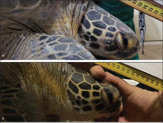
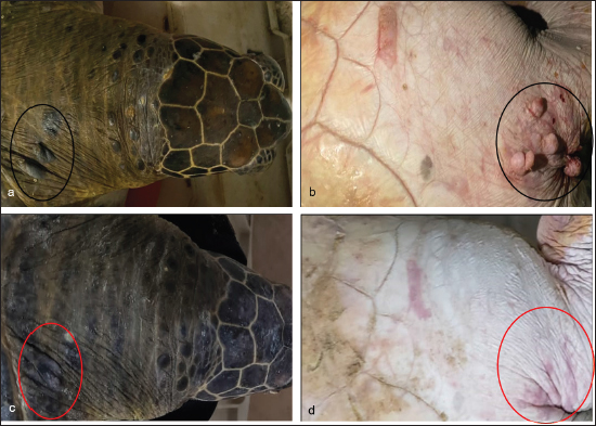
AbstractBackground: Fibropapillomatosis (FP) is a tumor disease primarily affecting juvenile sea turtles, often characterized by external growths that can regress spontaneously. This study reports the first documented case of total tumor regression in a free-living green turtle (Chelonia mydas) with FP in southern Brazil. Case Description: A juvenile green sea turtle (Chelonia mydas) was captured and recaptured on Itapirubá Beach, Santa Catarina, Brazil, showing signs of tumor regression with a period of 302 days between captures. At the first capture, photographs of the head and other regions were taken for photoidentification, along with documentation of fibropapilloma sites and tumor scoring. Tumor samples from the initial capture were histopathologically confirmed as fibropapillomas. At the recapture, the turtle showed a slight increase in carapace length, remaining classified as a juvenile. Tumors observed during the first capture were absent at recapture, with only scars remaining in the affected areas. Photoidentification confirmed the recapture, facilitating case monitoring. Tumor regression in this turtle is likely linked to various environmental and ecological factors. Conclusion: Spontaneous regression of FP tumors remains a crucial indicator in the health monitoring of sea turtle populations. This is the first documented case of FP regression in this region of Brazil, suggesting that the turtle’s increasing age and reduced exposure to anthropogenic pressure may have contributed to the tumor’s regression. Although clinical follow-up of free-living sea turtles is challenging, reports of FP tumor regression are vital for understanding the health dynamics of sea turtle populations. Keywords: Marine turtle, Skin tumor, Follow-up, Coast, Diagnosis. IntroductionFibropapillomatosis (FP) in sea turtles is an epidermal neoplasm characterized by the presence of arborized papillary projections, supported by an abundant fibrovascular base, which presents with variable external morphology, location, color, and texture (Work et al., 2017; Rossi et al., 2021). The development of these tumors is proven to be related to chelonian herpesvirus type 5 (ChHV5), but its transmission process and specific role in oncogenesis and tumor progression are still unclear (Farrell et al., 2021). There are environmental factors such as variations in sea surface temperature, salinity, and the discharge of effluent nutrients from surrounding rivers, which are correlated with a higher incidence of the disease (Manes et al., 2022), with pollution being one of the main factors. However, the role of these factors in the development of the disease is not yet well established (Lawrance et al., 2018). The diagnosis of FP is performed through histopathological analysis and identification of the virus by the Polymerase Chain Reaction (PCR) technique (Work et al., 2017). Some studies carried out both in Brazil and in other countries report spontaneous regression of FP (Hirama and Ehrhart, 2007; Guimarães et al., 2013; Tagliolatto et al., 2016); however, such findings are still scarce, especially in southern Brazil. This is believed to be due to the difficulty of capturing and recapturing free-living sea turtles (Tagliolatto et al., 2016). Therefore, this study aimed to report the first case of total spontaneous regression of FP in a free-living green turtle captured and recaptured on the beach of Itapirubá, Santa Catarina, Brazil. Case DetailsThe capture area was at Itapirubá beach in Santa Catarina, Brazil, using a 50 m long, 3.2 m high holding and/or trawl net with 0.30 m mesh for capture procedures (Martinez-Souza et al., 2013). In August 2022, a juvenile green sea turtle weighing 32 kg with a 63 cm curved carapace length (CCL) was captured (Fig. 1), showing seven small skin tumors, two on the neck (Fig. 2a), and five on the right front fin (Fig. 2b), all under 4 cm with mild scores, and photographs were taken for identification purposes (Reisser et al., 2008). Tumor presence sites were recorded, and scores were determined according to established protocols, based on the following tumor size categories: A (<1 cm), B (1–4 cm), C (4–10 cm), and D (>10 cm) (Work and Balazs, 1999; Rossi et al., 2016). The fibropapilloma tumor index (FPI) was then calculated using the formula FPI=0.1 × NA + 1 × NB + 20 × NC + 40 × ND (Rossi et al., 2016). Based on this index, tumors were categorized using the Fibropapillomatosis Score Southwest Atlantic system: mild (FPI < 40), moderate (40 < FPI < 120), or severe (FPI ≥ 120). Biopsies were taken from larger, less adherent tumors, fixed in 10% formalin for 48 hours, and routinely processed for histopathological evaluation, assessing criteria such as anatomical location, size, color, texture, consistency, surface characteristics, and skin integrity (Page-Karjian, 2019). In May 2023, the same turtle was recaptured in the same area, confirmed through photo-comparison of post-orbital scales (Fig. 1), weighing 31.8 kg with a 63.4 cm CCL, showing no FP tumors but scar tissue in their place, with reddish scars on the fin region and black scars on the neck, maintaining a similar shape to the previous FP sites, indicating total regression of FP tumors (Fig. 2c and d). The tumors presented as smooth and whitish on the fin, while appearing black on the neck, measuring approximately 1.8 × 0.7 cm. They featured a white base with a blackened apex, soft and irregular surfaces that were predominantly flat, along with areas of ulceration. Microscopic examination revealed significant connective tissue proliferation in the dermis (Fig. 3a and b), forming a cellular mantle interspersed with foci of neovascularization (Fig. 3a, c, d), and surrounded by a moderate inflammatory infiltrate consisting of lymphocytes, heterophils, and macrophages (Fig. 3b–d). Notable findings included intense inflammation, mild liquefactive necrosis, and edema at the epidermal junction. The stratum corneum exhibited both orthokeratotic and parakeratotic hyperkeratosis, while the stratum spinosum demonstrated papillomatous acanthosis, edema, liquefactive necrosis, and lymphocyte exocytosis.
Fig. 1. Green sea turtle (Chelonia mydas), captured (a) and recaptured (b) after 302 days, at Itapirubá beach (SC), with tumor regression. Photoidentification of the post-orbital scales on the right side, confirming that it was the same animal.
Fig. 2. At capture: presence of two tumors on the right cervical dorsum (a) and five tumors in the ventro-cranial region of the right anterior fin (b). Recapture: black scars in the neck where there were tumors (c) and reddish scars approximately maintaining the shape of the insertion of FP tumors. Ethical approvalThe procedures for capturing and collecting biological samples were carried out with prior federal authorization by the Biodiversity Authorization and Information System (SISBIO), under numbers 7860 and 51465, and by the Ethics Committee on the Use of Animals of the Federal Fluminense University (CEUA/UFF) under No. 8308100221. DiscussionThe comparison of photographs enabled the confirmation of both the capture and recapture of the green turtle, as well as clear documentation of FP tumor regression. Photoidentification, a validated technique for systematically identifying individual sea turtles based on their unique markings and features (Reisser et al., 2008), has proven to be an invaluable tool in the study of FP, allowing for accurate detection and evaluation of tumor growth and regression (Guimarães et al., 2013). To the best of the authors’ knowledge, this is the first documented case of total FP regression in a green sea turtle along the southern coast of Brazil. In contrast, two cases of FP regression were previously documented using photoidentification in the state of Rio de Janeiro, both occurring within 188 days, a shorter timeframe than observed in this study (Guimarães et al., 2013; Tagliolatto et al., 2016). Nonetheless, identifying cases of FP regression in turtles remains challenging due to the difficulties in monitoring these animals in the wild. The macroscopic and microscopic characteristics of the tumors were consistent with those previously described in the initial report by Smith and Coates (1938) and closely resembled findings from other studies on FP in green turtles, loggerhead turtles, and olive ridley turtles (Page-Karjian et al., 2014; Rossi et al., 2015; Reséndiz et al., 2022). Based on these evaluations, along with their epithelial and stromal proliferation, the cutaneous tumors collected were determined to be consistent with FP. Although there are limited studies characterizing the anatomo-histopathology of regressing FP tumors, previous research on green turtles has indicated a more favorable prognosis for those with only flat FP lesions compared to those exhibiting verrucous morphology, with over 50% of the former group experiencing spontaneous regression (Page-Karjian et al., 2014). However, it should be noted that flat tumors cannot be definitively interpreted as a sign of tumor regression (Bennett et al., 1999; Page-Karjian et al., 2014).
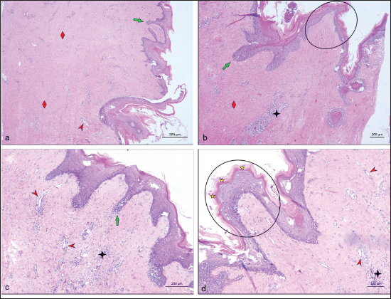
Fig. 3. Microscopy of the FP tumors showing proliferation of fibrous connective tissue (red rhombi), neovascularization (red arrow head), papillary projections towards the interior of the dermis (green arrow), papillary projections (rounded areas), inflammatory infiltrate (4-pointed black star), and parakeratosis (yellow start). The tumor score serves as a reliable indicator for predicting potential regression of FP. Supporting this, research indicates that 72% of cases showing tumor regression were classified as mild (Bennett et al., 1999). Furthermore, it has been suggested that turtles with less severe FP may undergo spontaneous tumor regression, as seen in the current case (Hirama and Ehrhart, 2007). However, we advise caution in generalizing this observation, as lighter FP tumors might also suggest that the animal is still in the early stages of disease progression. The turtle in this report was classified as juvenile, nearing the sub-adult phase (Chaloupka and Limpus, 2005), a critical stage when turtles begin to migrate to new feeding areas (Bolten, 2003). The 302-day interval between captures supports the notion that the turtle was transitioning from the juvenile neritic zone into the sub-adult phase. Despite this extended interval, the turtle’s return to the same location highlights its fidelity to the study area, likely as it prepares for migration to deeper waters (Bolten, 2003). This behavior could influence tumor regression, as reduced exposure to coastal stressors and pollutants may occur when the turtle moves away from these areas (Bolten, 2003; Shaver et al., 2019). Research indicates that sub-adult and adult turtles migrating into deeper zones, which typically have lower pollutant densities, show lower incidences of FP (Jones et al., 2016). As turtles move farther from the coast, their decreased exposure to environmental pollutants may enhance the likelihood of tumor regression. Size and age also play significant roles in FP regression; sub-adults and adults rarely exhibit FP, indicating either recovery through acquired immunity before migration or a lack of exposure to the disease during their juvenile stages (Jones et al., 2016; Kelley et al., 2022). Recent studies reinforce this, showing higher FP rates in smaller turtles and lower rates with increased carapace length (Jones et al., 2016; Kelley et al., 2022). Although the study area is near ports with dredging activities and known pollution in adjacent regions (Righetti et al., 2019; Ferreira et al., 2021), it is designated as an Environmental Preservation Area (EPA), likely maintaining relatively low pollution levels that could contribute to the regression of FP tumors. Supporting this hypothesis, recent studies on trace elements in sea turtle blood from this area indicate low concentrations of these elements in the local population (Pérez et al., 2023). However, research on organic compounds in Sotalia guianensis dolphins and in oysters (Crassostrea gasar and Crassostrea rhizophorae) has revealed significant concentrations of these compounds along the coast of Santa Catarina, including southern areas close to the study site (Vidal et al., 2023; Bastolla et al., 2024; Guerreiro et al., 2024), which may contribute to the prevalence of FP (Vilca et al., 2018). Thus, it is very important to investigate the potential long-term impact of these compounds on the health of free-living sea turtles in this region. The observed tumor regression may also be linked to the low incidence of FP in sea turtles from this region (Rossi et al., 2019; Capri, 2022; Pérez et al., 2023), which could facilitate the turtle’s recovery. A lower presence of sick animals reduces the shedding of the ChHV5 virus in the water and the risk of infection and tumor development (Farrell et al., 2021). This low occurrence is likely due to the study area’s inclusion within the EPA and its overall environmental quality. The case of FP regression reported here is essential for a better understanding of this disease. The mechanisms of FP regression are not yet well determined (Guimarães et al., 2013) and it is not clear whether the virus is lytic or latent in tumors (Farrell et al., 2021). It demonstrates that exposure of data and reports on cases of this type are extremely necessary for the elucidation and understanding of the mechanisms involved in regression, as well as the factors that could help in the spontaneous recovery of FP, a disease so serious and common in sea turtles. Several factors may be related to the regression of these tumors and the environmental and immunological factors of the turtles are probably the most relevant. In addition, free-living sea turtles offer a great challenge to carry out clinical follow-up, which makes this study a valuable information tool. ConclusionThis case of FP regression in a green sea turtle illustrates the importance of monitoring environmental conditions and biological factors influencing tumor recovery. The findings highlight the potential for spontaneous regression in less severe cases, particularly in areas with low pollutant exposure. This study contributes valuable insights into the dynamics of FP in sea turtles and emphasizes the necessity for ongoing research to understand the underlying mechanisms of tumor regression. Ultimately, it underscores the role of environmental quality in the health of marine species and the need for conservation efforts to protect these ecosystems. AcknowledgmentsThe authors thank Caminho Marinho Project as well as all volunteers involved in this work, and Coordenação de Aperfeiçoamento de Pessoal de Nível Superior – Brasil (CAPES). Conflict of interestThe authors declare that there is no conflict of interest. FundingThis work was supported in part by Coordenação de Aperfeiçoamento de Pessoal de Nível Superior – Brasil (CAPES). Authors’ contributionsYAAP carried out conceptualization, methodology, investigation, data curation, writing—original draft, writing—review and editing, visualization. SRL and MGC: carried conceptualization, validation, writing—review and editing. GMS: carried field methodologies for capture and data collection from the animal, investigation, resources, supervision, project administration, and funding acquisition. MJAH and TG: methodology, resources, project administration, funding acquisition. ETM, KVGCS, and NXA: conceptualization, methodology, validation, resources, supervision, project administration, and funding acquisition. All authors read and approved the final manuscript. Data availabilityAll data were provided in the manuscript. ReferencesBastolla, C.L.V., Guerreiro, F.C., Saldana-Serrano, M., Gomes, C.H.A.M., Lima, D., Rutkoski, C.F., Mattos, J.J., Dias, V.H.V., Righetti, B.P.H, Ferreira, C.P., Martim, J., Alves, T.C., Melo, C.M.R., Marques, M.R.F., Lüchmann, K.H., Almeida, E.A. and Bainy, A.C.D. 2024. Emerging and legacy contaminants on the Brazilian southern coast (Santa Catarina): a multi-biomarker approach in oysters Crassostrea gasar (Adanson, 1757). Sci. Total Environ. 925, 171679. Bennett, P., Keuper-Bennett, U. and Balazs, G.H. 1999. Photographic evidence for the regression of fibropapilloma afflicting green turtles at Honokawai, Maui, in the Hawaiian Islands. In the Proceedings of the Nineteenth Annual Symposium on Sea Turtle Biology and Conservation, Anais, NOAA Tech Memo. Bolten, A. 2003. Variation in sea turtle life history patterns: neritic vs. oceanic developmental stages. In The biology of sea turtles Vol II. Eds., Lutz, P.L., Musick, J.A. and Wyneken, J. Boca Raton, FL: CRC Press, pp: 243–254. Capri, A. 2022. Análise de Biomarcadores de estresse oxidativo em juvenis de tartaruga-verde (Chelonia mydas) na região sul do Brasil. Trabalho de Conclusão de Curso apresentado como requisito parcial para obtenção do título de bacharel em engenharia ambiental e sanitaria. Florianópolis, Brazil: Universidade do Estado de Santa Catarina. Chaloupka, M. and Limpus, C. 2005. Estimates of sex- and age-class-specific survival probabilities for a southern Great Barrier Reef green sea turtle population. Mar. Biol. 146, 1251–1261. Farrell, J.A., Yetsko, K., Whitmore, L., Whilde, J., Eastman, C.B., Ramia, D.R., Thomas, R., Linser, P., Creer, S., Burkhalter, B., Schnitzler, C. and Duffy, D.J. 2021. Environmental DNA monitoring of oncogenic viral shedding and genomic profiling of sea turtle fibropapillomatosis reveals unusual viral dynamics. Commun. Biol. 4, 565. Ferreira, C.P., Piazza, T.B., Souza, P., Lima, D., Mattos, J.J., Saldaña-Serrano, M., Piazza, R.S., Jorge, M.B., Bianchini, A., Taniguchi, S., Sasaki, S.T., Montone, R.C., Bícego, M.C., Bainy, A.C.D. and Lüchmann, K.H. 2021. Integrated biomarker responses in oysters Crassostrea gasar as an approach for assessing aquatic pollution of a Brazilian estuary. Mar. Environ. Res. 165, 105252. Guerreiro, F.C., Alves, T.C., Saldana-Serrano, M., Gomes, C.H.A.M., Lima, D., Bastolla, C.L.V., Ferreira, C.P., Bitschinski, D., Rutkoski, C.F., Grott, S.C., Israel, N.G., Lüchmann, H., Marques, M.R.F., Melo, C.M.R., Bainy, C.D. and Almeida, E.A. 2024. Integrating pollutant levels and biochemical biomarkers in oysters (Crassostrea rhizophorae and Crassostrea gasar) indicates anthropic impacts on marine environments along the coast of Santa Catarina state, Brazil. Mar. Environ. Res. 194, 106309. Guimarães, S., Mas Gitirana, H., Vidal Wanderley, A., Monteiro-Neto, C. and Lobo-Hajdu, G. 2013. Evidence of regression of fibropapillomas in juvenile green turtles Chelonia mydas caught in Niterói, southeast Brazil. Dis. Aquat. Organ. 102, 243–247. Hirama, S. and Ehrhart, L.M. 2007. Description, prevalence and severity of green turtle fibropapillomatosis in three developmental habitats on the east coast of Florida. Flor. Sci. 70, 435–480. Jones, K., Ariel, E., Burgess, G. and Read, M. 2016. A review of fibropapillomatosis in green turtles (Chelonia mydas). Vet. J. 212, 48–57. Kelley, J.R., Kelley, K.L., Savage, A.E. and Mansfield, K.L. 2022. Novel disease state model finds most juvenile green turtles develop and recover from fibropapillomatosis. Ecosphere 13, 1–13. Lawrance, M.F., Mansfield, K.L., Sutton, E. and Savage, A.E. 2018. Molecular evolution of fibropapilloma-associated herpesviruses infecting juvenile green and loggerhead sea turtles. Virology 521, 190–197. Manes, C., Pinton, D., Canestrelli, A. and Capua, I. 2022. Occurrence of fibropapillomatosis in green turtles (Chelonia mydas) in relation to environmental changes in coastal ecosystems in Texas and Florida: a retrospective study. Animals 12, 1236. Martinez-Souza, G., Bortolotto, J., Steigleder, K.M., Filho, P.R.G., Vélez - Rubio, G. and Kinas, P. 2013. Ocorrência anual de tartaruga—verde Chelonia mydas no sul de Santa Catarina, Brasil. In the Proceedings of the VI Jornada Y VII Reunión de Conservación e Investigación de Tortugas Marinas en el Atlántico Sur Occidental (ASO). Page-Karjian, A. 2019. Fibropapillomatosis in Marine Turtles. In Fowler’s Zoo and Wild Animal Medicine Current Therapy, Vol 9, Eds., Miller, R.E., Lamberski, N. and Calle, P.P. St. Louis, MI: Elsevier, pp: 398–403. https://doi.org/10.1016/B978-0-323-55228-8.00057-6 Page-Karjian, A., Norton, T.M., Krimer, P., Groner, M., Nelson, S.E. and Gottdenker, N.L. 2014. Factors influencing survivorship of rehabilitating green sea turtles (Chelonia mydas) with fibropapillomatosis. J. Zoo. Wildl. Med. 45, 507–519. Pérez, Y.A.A., Lima, S.R., Souza, G.M., Gião, T., Bispo, F.J.S., Reis, A.M.F., da Silva Leite, J. and da Silva, K.V.G.C. 2023. Evaluation of biometry and blood concentration of heavy metals in free-living Chelonia mydas with and without fibropapillomatosis in southern Brazil. Mar. Pollut. Bull. 190, 114879. Reisser, J., Proietti, M., Kinas, P. and Sazima, I. 2008. Photographic identification of sea turtles: method description and validation, with an estimation of tag loss. End. Species Res. 5, 73–82. Reséndiz, E., Fernández-Sanz, H., Espinoza, J. and Cedillo-Peláez, C. 2022. Fibropapilomatosis en tortugas marinas: una visión de conjunto. Rev. Invest. Mar. 42, 115–137. Righetti, B.P.H., Mattos, J.J., Siebert, M.N., Daura-Jorge, F.G., Bezamat, C., Fruet, P.F., Genoves, R.C., Taniguchi, S., da Silva, J., Montone, R.C., Simões-Lopes, P.C. de A., Bainy, A.C.D. and Lüchmann, K.H. 2019. Biochemical and molecular biomarkers in integument biopsies of free-ranging coastal bottlenose dolphins from southern Brazil. Chemosphere 225, 139–149. Rossi, A.S., Gattamorta, M.A., Prioste, F.E.S., Lima, E.H.S.M., Melo, M.T.D., Brandão, P.E., Silva, S.O. de S., da Silveira, F.M. and Matushima, E.R. 2015. Fibropapillomas in a Loggerhead sea turtle (Caretta caretta) caught in Almofala, Ceará, Brazil: histopathological and molecular characterizations. Mar. Turtle News. 147, 13–16. Rossi, S., Sánchez-Sarmiento, A.M., Santos, R.G. dos, Zamana, R.R., Prioste, F.E.S., Gattamorta, M.A., Ochoa, P.F.C., Grisi-Filho, J.H.H. and Matushima, E.R. 2019. Monitoring green sea turtles in Brazilian feeding areas: relating body condition index to fibropapillomatosis prevalence. J. Mar. Biol. Assoc. UK. 99, 1879–1887. Rossi, S., Sánchez-Sarmiento, A.M., Vanstreels, R.E.T., dos Santos, R.G., Prioste, F.E.S., Gattamorta, M.A., Grisi-Filho, J.H.H. and Matushima, E.R. 2016. Challenges in evaluating the severity of fibropapillomatosis: a proposal for objective index and score system for green sea turtles (Chelonia mydas) in Brazil. PLoS One 11, e0167632. Rossi, S., Zamana, R.R., De Andrade-Santos, P.P., Bomfim, A.D.C., De Farias, D.S.D., Freire, A.C.D.B., De Oliveira, R.M., Gattamorta, M.A., Matushima, E.R., Pires, J.M.D.L., Sacristán, C., Da Silva-Júnior, E.S., de Lima Silva, F.J. and Gavilan, S.A. 2021. Neoplasias viscerais e Chelonid alphaherpesvirus 5 em tartarugas verdes com fibropapilomatose. Arch. Vet. Sci. 2021, 26. Shaver, D., Walker, J. and Backof, T. 2019. Fibropapillomatosis prevalence and distribution in green turtles Chelonia mydas in Texas (USA). Dis. Aquat. Organ. 136, 175–182. Smith, G.M. and Coates, C.W. 1938. Fibro-epithelial growths of the skin in large marine turtles, Chelonia mydas (Linnaeus). Zool. NY. Zoo. Soc. 23, 93–98. Tagliolatto, A., Guimarães, S., Lobo-Hajdu, G. and Monteiro-Neto, C. 2016. Characterization of fibropapillomatosis in green turtles Chelonia mydas (Cheloniidae) captured in a foraging area in southeastern Brazil. Dis. Aquat. Organ. 121, 233–240. Vidal, L.G. Oliveira-Ferreira, N., Torres, J.P.M, Azevedo, A.F., Meirelles, A.C.O., Flach, L., Domit, C., Fragoso, A.B.L., Silva, F.J.L., Carvalho, V.L., Marcondes, M., Barbosa, L.A., Cremer, M.J., Malm, O., Lailson-Brito, J. and Eljarrat, E. 2023. Brominated flame retardants and natural organobrominated compounds in a vulnerable delphinid species along the Brazilian coast. Sci. Total Environ. 905, 167704. Vilca, F.Z., Rossi, S., de Olinda, R.A., Sánchez-Sarmiento, A.M., Prioste, F.E.S., Matushima, E.R. and Tornisielo, V.L. 2018. Concentrations of polycyclic aromatic hydrocarbons in liver samples of juvenile green sea turtles from Brazil: can these compounds play a role in the development of fibropapillomatosis? Mar. Pollut. Bull. 130, 215–222. Work, T.M. and Balazs, G.H. 1999. Relating neoplasm score to hematology in green turtles with fibropapillomatosis in Hawaii. J. Wildlife Dis. 35, 804–807. Work, T.M., Dagenais, J., Weatherby, T.M., Balazs, G.H. and Ackermann, M. 2017. In vitro replication of chelonid herpesvirus 5 in organotypic skin cultures from Hawaiian green turtles (Chelonia mydas). J. Virol. 91, e00404–e00417. | ||
| How to Cite this Article |
| Pubmed Style Pérez YAA, Lima SR, Martinez-souza G, Gião T, Chenard MG, Helayel MJA, Mársico ET, , Alencar NXD. First case report of fibropapillomatosis tumor regression identified through photoidentification and histopathology in a Chelonia mydas in Itapirubá, Santa Catarina, Brazil. Open Vet. J.. 2024; 14(12): 3649-3655. doi:10.5455/OVJ.2024.v14.i12.46 Web Style Pérez YAA, Lima SR, Martinez-souza G, Gião T, Chenard MG, Helayel MJA, Mársico ET, , Alencar NXD. First case report of fibropapillomatosis tumor regression identified through photoidentification and histopathology in a Chelonia mydas in Itapirubá, Santa Catarina, Brazil. https://www.openveterinaryjournal.com/?mno=222109 [Access: June 27, 2026]. doi:10.5455/OVJ.2024.v14.i12.46 AMA (American Medical Association) Style Pérez YAA, Lima SR, Martinez-souza G, Gião T, Chenard MG, Helayel MJA, Mársico ET, , Alencar NXD. First case report of fibropapillomatosis tumor regression identified through photoidentification and histopathology in a Chelonia mydas in Itapirubá, Santa Catarina, Brazil. Open Vet. J.. 2024; 14(12): 3649-3655. doi:10.5455/OVJ.2024.v14.i12.46 Vancouver/ICMJE Style Pérez YAA, Lima SR, Martinez-souza G, Gião T, Chenard MG, Helayel MJA, Mársico ET, , Alencar NXD. First case report of fibropapillomatosis tumor regression identified through photoidentification and histopathology in a Chelonia mydas in Itapirubá, Santa Catarina, Brazil. Open Vet. J.. (2024), [cited June 27, 2026]; 14(12): 3649-3655. doi:10.5455/OVJ.2024.v14.i12.46 Harvard Style Pérez, Y. A. A., Lima, . S. R., Martinez-souza, . G., Gião, . T., Chenard, . M. G., Helayel, . M. J. A., Mársico, . E. T., & Alencar, . N. X. D. (2024) First case report of fibropapillomatosis tumor regression identified through photoidentification and histopathology in a Chelonia mydas in Itapirubá, Santa Catarina, Brazil. Open Vet. J., 14 (12), 3649-3655. doi:10.5455/OVJ.2024.v14.i12.46 Turabian Style Pérez, Yohany Arnold Alfonso, Samara Rosolem Lima, Gustavo Martinez-souza, Thayana Gião, Marina Galindo Chenard, Michel José Abdalla Helayel, Eliane Teixeira Mársico, Kássia Valéria Gomes Coelho Da Silva, and Nayro Xavier De Alencar. 2024. First case report of fibropapillomatosis tumor regression identified through photoidentification and histopathology in a Chelonia mydas in Itapirubá, Santa Catarina, Brazil. Open Veterinary Journal, 14 (12), 3649-3655. doi:10.5455/OVJ.2024.v14.i12.46 Chicago Style Pérez, Yohany Arnold Alfonso, Samara Rosolem Lima, Gustavo Martinez-souza, Thayana Gião, Marina Galindo Chenard, Michel José Abdalla Helayel, Eliane Teixeira Mársico, Kássia Valéria Gomes Coelho Da Silva, and Nayro Xavier De Alencar. "First case report of fibropapillomatosis tumor regression identified through photoidentification and histopathology in a Chelonia mydas in Itapirubá, Santa Catarina, Brazil." Open Veterinary Journal 14 (2024), 3649-3655. doi:10.5455/OVJ.2024.v14.i12.46 MLA (The Modern Language Association) Style Pérez, Yohany Arnold Alfonso, Samara Rosolem Lima, Gustavo Martinez-souza, Thayana Gião, Marina Galindo Chenard, Michel José Abdalla Helayel, Eliane Teixeira Mársico, Kássia Valéria Gomes Coelho Da Silva, and Nayro Xavier De Alencar. "First case report of fibropapillomatosis tumor regression identified through photoidentification and histopathology in a Chelonia mydas in Itapirubá, Santa Catarina, Brazil." Open Veterinary Journal 14.12 (2024), 3649-3655. Print. doi:10.5455/OVJ.2024.v14.i12.46 APA (American Psychological Association) Style Pérez, Y. A. A., Lima, . S. R., Martinez-souza, . G., Gião, . T., Chenard, . M. G., Helayel, . M. J. A., Mársico, . E. T., & Alencar, . N. X. D. (2024) First case report of fibropapillomatosis tumor regression identified through photoidentification and histopathology in a Chelonia mydas in Itapirubá, Santa Catarina, Brazil. Open Veterinary Journal, 14 (12), 3649-3655. doi:10.5455/OVJ.2024.v14.i12.46 |

