


| Research Article | ||
Open Vet. J.. 2025; 15(3): 1157-1165 Open Veterinary Journal, (2025), Vol. 15(3): 1157-1165 Research Article Anatomical study and distribution of taste buds in the palatal organs of various types of local koi fish (Cyprinus rubrofuscus) in IndonesiaSynthia Regita Noor Mahesti1, Aris Haryanto2, Krisna Noli Andrian2 and Tri Wahyu Pangestiningsih3*1Master Student of Veterinary Science, Faculty of Veterinary Medicine, Universitas Gadjah Mada, Yogyakarta, Indonesia 2Department of Biochemistry and Molecular Biology, Faculty of Veterinary Medicine, Universitas Gadjah Mada, Yogyakarta, Indonesia 3Department of Anatomy, Faculty of Veterinary Medicine, Universitas Gadjah Mada, Yogyakarta, Indonesia *Corresponding Author: Tri Wahyu Pangestiningsih. Department of Anatomy, Faculty of Veterinary Medicine, Universitas Gadjah Mada, Yogyakarta, Indonesia. Email: estifkh [at] ugm.ac.id Submitted: 13/11/2024 Accepted: 19/02/2025 Published: 31/03/2025 © 2025 Open Veterinary Journal
AbstractBackground: Koi fish (Cyprinus rubrofuscus), which are celebrated for their vibrant colors and cultural significance, are also valued in the ornamental fish industry. Despite extensive research on fish sensory biology, specific studies on taste buds in koi, especially among different ornamental varieties, are limited. Aim: This study aimed to analyze the anatomical structure and distribution of taste buds in the palatal organs of six popular koi varieties in Indonesia: Kohaku, Shiro, Showa, Sanke, Platinum, and Ogon. Method: Eighteen koi fish, three of each variety and aged 4–6 months, were examined using histological techniques and scanning electron microscopy. Results: Although no macroscopic or histological differences were observed in the palatal organs across varieties, significant variation in taste bud density was noted. The Ogon variety exhibited the highest average taste bud density (23.95 ± 7.03), whereas the Shiro variety had the lowest (17.03 ± 5.26). Conclusion: Statistical analysis revealed that taste bud density significantly differed among varieties, with the Ogon variety having a notably higher density than the Shiro and Sanke. These findings suggest that selective breeding of ornamental traits may affect sensory function. Keywords: Cyprinus rubrofuscus, Taste buds, Palatal organ, Koi fish, Aquaculture. IntroductionKoi fish, a domesticated variety of the common carp (Cyprinus carpio), have become an iconic species within the ornamental fish industry because of their vibrant colors, intricate patterns, and symbolic cultural significance, particularly in Japan (de Kock and Gomelsky, 2015; Andrian et al., 2023 and 2024). The cultivation of koi fish, known as Nishikigoi, has evolved into a specialized field with a rich history that dates back over two centuries. Their striking appearance and adaptability to various environments have made them a favorite among aquarists worldwide. However, behind their ornamental value lies a complex biological system that governs their survival and success in aquaculture. Among the key aspects of their biology is their feeding behavior, which is closely linked to the functionality and efficiency of their sensory organs. The ability of fish to detect, select, and consume food is crucial not only for their growth and health, reproductive success, and overall quality as an ornamental species (Kasumyan, 2019). This makes the study of the sensory organs, particularly the taste buds, essential for optimizing aquaculture practices and ensuring the sustainability of koi farming. The sensory biology of fish, especially the role of taste buds in feeding behavior, has been extensively studied in various species due to its implications for understanding ecological adaptations and improving aquaculture efficiency (Doosey and Bert, 2011; Hansen et al., 2014; Atkinson et al., 2016; Kasumyan, 2019; Assan et al., 2021; Wang et al., 2023). The taste buds, located in the palatal organ of koi fish, are integral to their ability to detect and respond to chemical stimuli in water. These chemosensory organs allow koi fish to distinguish between edible and non-edible particles, a function vital for their survival, especially in environments where visual cues may be limited (Cohen and Hernandez, 2018). The distribution and density of taste buds in the palatal organ are not uniform across all fish species or even among different varieties of the same species. These changes often reflect the fish’s natural habitat, dietary habits, and evolutionary pressures. In koi fish that have been selectively bred for specific esthetic traits, it is particularly important to understand how these breeding practices may have inadvertently affected their sensory capabilities, including the distribution of taste buds in the palatal organ. This knowledge is crucial for developing targeted feeding strategies that can enhance growth rates and overall health in koi aquaculture. Despite the importance of sensory organs in fish biology, limited research has focused specifically on the taste buds of koi fish, especially in relation to the different varieties bred for ornamental purposes. This study sought to fill this knowledge gap by providing a detailed analysis of the anatomical structure and distribution of taste buds in the palatal organs of six popular koi varieties in Indonesia. By examining the differences in taste bud density and distribution, we aim to understand the potential implications for feeding behavior and how this knowledge can be applied to improve breeding and feeding practices in koi aquaculture. The findings of this study are expected to contribute not only to the scientific understanding of koi fish biology but also to the practical aspects of managing koi farms, ensuring the health and vitality of these prized ornamental fish in the growing aquaculture industry. Materials and MethodsExperimental design and sample selectionThis experiment was designed to investigate the anatomical structure and distribution of taste buds in the palatal organs of six local koi fish varieties: Showa, Shiro, Sanke, Platinum, and Ogon. A total of 18 koi fish (three from each variety) were selected as the animal model. The fish were collected from reputable breeders in Indonesia, ensuring that they were representative of the typical phenotypes of each variety. Euthanasia and dissectionPrior to dissection, the fish were euthanized using diethyl ether at a concentration of 0.5 ml/l of water. This method was chosen to minimize stress and ensure quick, humane death. After euthanasia, the palatal organs were carefully dissected from the roof of the mouth using fine surgical instruments. The organs were immediately fixed in 10% formalin solution to preserve tissue integrity for subsequent histological and scanning electron microscopy (SEM) analyses. Histological preparationOne palatal organ from each variety was selected for histological analysis. The tissues were dehydrated using a graded series of ethanol, cleared in xylene, and embedded in paraffin wax. Serial sections of 5 µm thickness were cut using a microtome and mounted on glass slides. The sections were stained with hematoxylin and eosin (HE), a standard staining technique that highlights cellular structures and allows for a detailed examination of tissue morphology. The stained sections were observed under a light microscope at various magnifications to identify the presence and distribution of taste buds. Scanning electron microscopyThe remaining palatal organs were prepared for SEM analysis to obtain high-resolution images of the taste bud surface and surrounding tissues. The samples were dehydrated in a graded ethanol series, critical-point dried, and coated with a thin layer of gold using a sputter coater. The coated samples were then examined using a SEM to obtain detailed images of the external morphology of the taste buds. SEM was particularly useful for observing the spatial arrangement and density of taste buds across different regions of the palatal organ. Data collection and analysisThe distribution of taste buds was quantified by counting the number of taste buds per unit area in different regions of the palatal organ, categorized as A1, A2, A3 (anterior to posterior regions) and B1, B2, B3, B4, B5 (lateral regions) (Fig. 1). The data were analyzed using statistical software to assess the variability in taste buds among the different koi varieties. ANOVA was used to determine the significance of differences in taste bud density, followed by Post Hoc (Tukey HSD) tests to identify specific pairwise differences between varieties. Ethical approvalAll procedures involving the handling and euthanasia of fish were conducted in accordance with ethical guidelines for the use of animals in research and were approved by the Institutional Animal Care and Use Committee (IACUC) Faculty of Veterinary Medicine, Gadjah Mada University (No: 043/EC-FKH/Int/2022). ResultsMacroscopic structure of the palatal organThe palatal organ of koi fish used in this study was measured between 1.3 and 1.4 cm in length (with an average of 1.6 cm) (Fig. 1). Macroscopically, there were no structural or locational differences in the palatal organ among the six koi variants examined. The palatal organ is located on the dorsal side of the mouth and extends posteriorly toward the chewing pad (CP), with the anterior part being wider than the posterior. This organ consists of thick muscles located posterior to the koi fish’s mouth (Fig. 1, PO). Its function is to trap food particles on the gill arches and gill rakers, which form the floor of the mouth cavity. The palatal organ is divided into two parts, dexter, and sinister, by a median groove (MG) (Fig. 2). Histological structure of taste budsHE staining revealed that the palatal organ in Cyprinus rubrofuscus consists of two layers: the tunica mucosa and the tunica muscularis. Microscopically, there were no histological differences in the palatal organ structure among the six koi variants studied. The tunica mucosa of the palatal organ contains taste buds and mucus cells, which are visible as the cells epithelium that covers the surface of the palatal organ (Fig. 3A), whereas the tunica muscularis is composed of striated muscle fibers (Fig. 3B). There were no microscopic histological differences in the palatal organ structures of the kohaku, shiro, showa, sanke, platinum, and ogon variants.
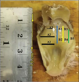
Fig. 1. Macroscopic image of eight palatal organ areas in C. rubrofuscus. mediolateral anterior (A1), mediolateral medial (A2), mediolateral posterior (A3), medial anterior (B1), medial posterior (B2), intermediate anterior (B3), intermediate posterior (B4), and lateral (B5) regions.
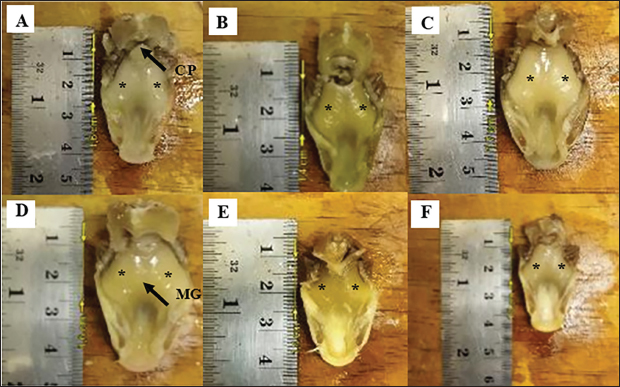
Fig. 2. A macroscopic image of the palatal organ of C. rubrofuscus is shown from a ventral view. The photo highlights the palatal organ (PO, marked with *), the MG, and the CP across different koi fish variants: kohaku (A), shiro (B), showa (C), sanke (D), platinum (E), and ogon (F).
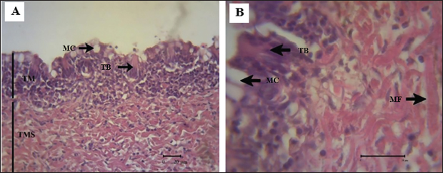
Fig. 3. Microscopic images of the palatal organ of C. rubrofuscus are shown. The palatal organ is composed of the tunica mucosa (TM) and the tunica muscularis (TMS). (A) illustrates that the tunica mucosa is composed of two types of cells: taste buds and mucous cells, including taste buds (TB) and mucus cells (MC). (B) shows that the tunica muscularis is composed of striated muscle fibers (MF).
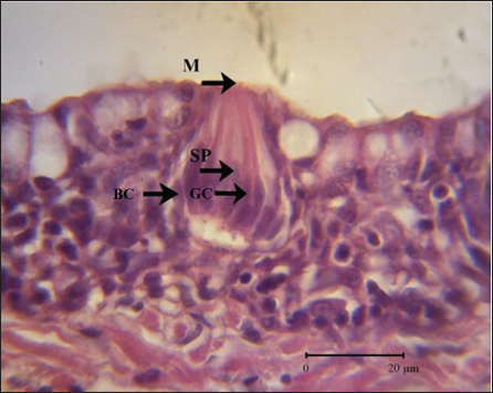
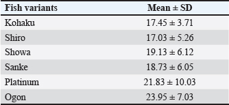
Fig. 4. Microscopic features of the taste bud on the palatal organ of C. rubrofuscus. The taste bud is composed of gustatory cells (GC), supporting cells (SP) with apical microvilli (M) that form the taste pores, and basal cells (BC). The morphology of the taste buds looks like an onion or pear. These taste buds are composed of three types of cells: gustatory cells, supporting cells, and basal cells. Gustatory cells have dense cytoplasm and vesicular nuclei, whereas supporting cells have vacuolated cytoplasm and dense nuclei. Basal cells are small and are located along the base of the taste buds (Fig. 4). Histological analysis revealed that the taste buds in the palatal organs of koi fish are consistently pear-shaped, a characteristic feature of teleost fish. Each taste bud was composed of three distinct cell types: (1) gustatory cells, which are the primary sensory receptors responsible for detecting chemical stimuli; (2) supporting cells, which provide structural and metabolic support to gustatory cells; and (3) basal cells, which are progenitor cells involved in the regeneration and maintenance of the taste bud. The taste buds were embedded within the epithelial tissue of the palatal organ, with their apical ends protruding slightly into the oral cavity. Palatal organ ultrastructureSurface observation of the palatal organ in koi fish was conducted in three areas: anterior, medial, and posterior. The results obtained using SEM revealed that the surface of the palatal organ is covered with small protrusions of taste buds and mucus cells. The posterior area of the palatal organ showed numerous ducts, which were absent in the anterior and medial areas (Fig. 5). Upon closer examination of the surface of the taste buds at higher magnification, the protrusions displayed the taste pores of the taste buds, and the epithelial cells exhibited a distinct fingerprint-like pattern. The taste buds of fish are shaped like onions or pears (Fig. 6). Variation in taste bud distributionThe statistical analysis of the average proportion of taste buds across eight areas of the palatal organ in six koi fish variants showed the following results: the kohaku variant had 17.45 ± 3.71, the shiro variant had 17.03 ± 5.26, the showa variant had 19.13 ± 6.12, the sanke variant had 18.73 ± 6.05, the platinum variant had 21.83 ± 10.03, and the ogon variant had 23.95 ± 7.03. Based on these results, the ogon variant had the highest average proportion of taste buds (23.95 ± 7.03), whereas the shiro variant had the lowest (17.03 ± 5.26) (Table 1). The distribution of taste buds varies among the koi fish variants in different areas. For the kohaku variant, the highest proportion of taste buds was found in area A1, whereas the lowest proportion was found in area A2. In the shiro variant, taste buds are most abundant in area B3 and least abundant in area B2. For the showa variant, the highest concentration of taste buds is observed in area A3, whereas the fewest are in area B3. The sanke variant had the highest number of taste buds in area B4 and the lowest number of taste buds in area B1. In the platinum variant, the most taste buds are present in area B4, and the fewest are found in area A1. Lastly, the ogon variant had the highest number of taste buds in area B5 and the lowest number of taste buds in area B1 (Fig. 7).
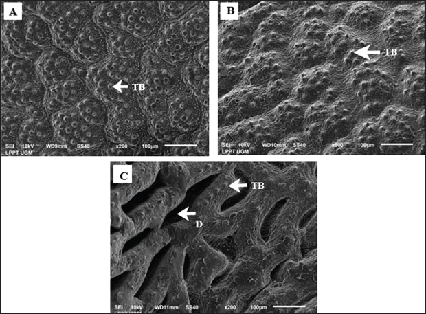
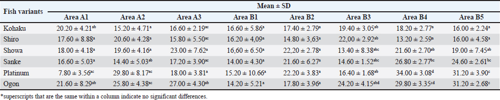
Fig. 5. SEM images of the palatal organ surface in C. rubrofuscus at three areas: anterior (A), medial (B), and posterior (C) at 200× magnification. The images show taste buds (TB) present on the surface of the palatal organ. The posterior area of the palatal organ contains numerous ducts (D). The proportion of taste buds of different areas and koi variants shows distinct patterns (Table 2). In Area A1, the ogon variant had the highest proportion of taste buds, significantly differing from the platinum variant, which had the lowest proportion. The ogon and kohaku variants did not significantly differ, while the shiro, showa, and sanke variants did not show significant differences among themselves or with others. In Area A2, the platinum variant has the highest proportion, with no significant difference from the ogon variant, while the sanke variant has the lowest proportion and significantly differs from both the ogon and platinum variants. In Area A3, the ogon variant has the highest proportion, significantly differing from the shiro, kohaku, and sanke variants, which have lower proportions; the shiro, kohaku, and sanke variants do not differ significantly from each other. Areas B1 and B2 showed no significant differences in taste bud proportions across all variants. In Area B3, the ogon variant has the highest proportion, with no significant difference from the shiro variant, while the showa and sanke variants have the lowest proportions, with no significant differences between them. The kohaku and platinum variants were also not significantly different from the other variants. In Area B4, the platinum variant exhibits the highest proportion, significantly differing from the shiro variant, which has the lowest proportion. The shiro variant did not significantly differ from the kohaku variant, and the showa variant significantly differed from the shiro, platinum, and ogon variants. The sanke variant did not differ significantly from the showa and ogon variants. In Area B5, the platinum and ogon variants had high proportions with no significant difference between them, while the kohaku and shiro variants had the lowest proportions, also showing no significant difference between them. The showa variant differs significantly from the platinum and ogon variants, and the sanke variant differs significantly from the kohaku and shiro variants.
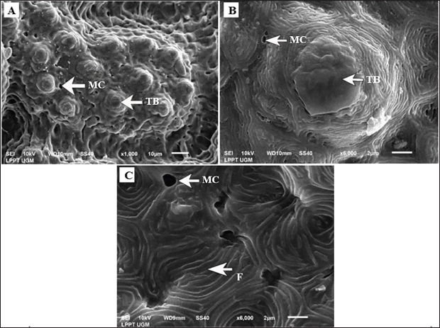
Fig. 6. SEM images of the taste bud surface on the palatal organ of C. rubrofuscus show protrusions indicating the taste pores of the taste buds (TB) and mucus cells (MC). The epithelial cells display a fingerprint-like pattern (F). (A) is shown at 1,000× magnification, while (B and C) are shown at 6,000× magnification. Table 1. Average proportion of taste buds across eight areas in six koi fish variants.
Statistical analysisANOVA indicated significant differences in taste bud density among the different koi varieties (p < 0.05). Post Hoc (Tukey HSD) tests further revealed that the ogon variety had a significantly higher taste bud density than the shiro and sanke varieties, while the differences between kohaku, showa, and platinum were not statistically significant. These results highlight the influence of genetic and environmental factors on taste bud development and distribution in koi fish. DiscussionThis study provides valuable insights into the sensory biology of koi fish, focusing on the distribution and density of taste buds in the palatal organs of six ornamental varieties. Although macroscopic and histological structures of the palatal organs did not differ significantly among the varieties, the distribution of taste buds exhibited notable variation. The analysis revealed significant differences in taste buds between the koi varieties. The ogon variety had the highest density of taste buds, particularly in areas B4 and B5 of the palatal organ. This high density suggests that the ogon variety may have an enhanced ability to detect chemical stimuli in their environment, potentially affecting their feeding behavior and efficiency. Conversely, the shiro variety had the lowest density of taste buds, particularly in areas A2 and A3, which could influence its feeding strategies and overall health. These variations may be attributed to selective breeding practices that prioritize esthetic traits over functional attributes (de Kock and Gomelsky, 2015). Domestication of koi fish (Cyprinus rubrofuscus) in the 19th century (de Kock and Gomelsky, 2015) may have led to variations in taste buds, similar to changes observed in other domesticated Cyprinidae. Other domesticated cyprinid species like common carp (Cyprinus carpio) have shown alterations in sensory and feeding behaviors due to controlled diets and environmental conditions, potentially influencing their chemosensory systems. In these species, the transition to aquaculture diets, which lack the complexity of natural foraging, reduces the reliance on diverse taste functions, potentially leading to changes in taste buds and sensitivity. Similar to koi, the stable environments and selective breeding in these cyprinids diminish evolutionary pressures for maintaining complex chemosensory systems, indicating that domestication could result in convergent changes in taste bud morphology and function across cyprinids (Milla et al., 2020; Kasumyan and Isaeva, 2023). As koi breeding has focused on color, pattern, and other visual characteristics, the underlying sensory capabilities may have been inadvertently affected. This could have implications for their ability to locate and consume food, affecting their growth and health in aquaculture settings (Kasumyan, 2019; Andrian et al., 2024).
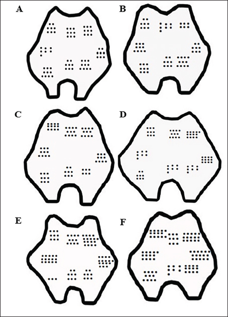
Fig. 7. Schematic distribution of taste buds on the palatal organ of C. rubrofuscus in different variants: kohaku (A), shiro (B), showa (C), sanke (D), platinum (E), and ogon (F). The diagrams illustrate the variation in taste bud density across different areas of the palatal organ for each koi fish variant. Understanding the distribution and density of taste buds is crucial for optimizing koi aquaculture feeding practices. Varieties with higher taste bud densities, like the ogon, might benefit from different feeding strategies compared with those with lower densities, such as the shiro. Tailoring diets to the sensory capabilities of each variety could improve feed efficiency and enhance growth rates. In addition, this knowledge could inform breeding practices to balance ornamental traits with functional attributes. Table 2. The proportion of taste buds in the same area across the six koi fish variants.
Future studies should explore the relationship between taste bud density and feeding behavior in koi fish to better understand how sensory capabilities influence food intake and growth. Investigating the effects of environmental factors and diet on taste bud development and function could provide further insights into optimizing aquaculture practices. Additionally, research into other sensory modalities, such as olfaction and vision, could complement the findings on taste buds and offer a comprehensive understanding of koi fish sensory biology. ConclusionThis study provides a detailed examination of the anatomical structure and distribution of taste buds in the palatal organs of six local koi fish varieties in Indonesia. The results highlight significant variations in taste bud density and distribution among the different varieties, with the ogon variety exhibiting the highest density. These findings have important implications for koi fish breeding, feeding management, and aquaculture practices, suggesting that sensory biology should be considered alongside traditional esthetic traits in the selective breeding process. The study also opens new avenues for research into the genetic and environmental factors that influence sensory organ development in koi fish. By expanding our understanding of how koi fish perceive and interact with their environment, we can develop more effective strategies for enhancing their health, growth, and overall quality in the aquaculture industry. AcknowledgmentsThe authors sincerely appreciate the support provided by the Faculty of Veterinary Medicine, Universitas Gadjah Mada. We are especially grateful for the research facilities and resources made available, which played a crucial role in the successful completion of this study. Conflict of interestThe authors declare that they have no conflict of interest. FundingThis study was funded by the Competitive Research Grant, under grant number 1846/UN.1/FKH/HK4/2022. Authors’ contributionAll authors contributed significantly to this research. SRNM conducted the study, performed the analysis, and drafted the manuscript. AH, KNA, and TWP contributed to data analysis, critically reviewed the draft, and provided valuable feedback to enhance the manuscript. All authors have thoroughly reviewed and approved the final version of the manuscript. Data availabilityAll data supporting the findings of this study are available in the manuscript. ReferencesAndrian, K.N., Aisy, N.R., Novindasari, B.B.M., Nurrahmi, I.A., Santi, M.D. and Haryanto, A. 2023. Random amplified polymorphic DNA-polymerase chain reaction analysis of four koi fish (Cyprinus carpio var. koi) variants from Yogyakarta, Indonesia. IOP Conf. Ser. Earth Environ. Sci. 1174(1), 012007. Andrian, K.N., Wihadmadyatami, H., Wijayanti, N., Karnati, S. and Haryanto, A. 2024. A comprehensive review of current practices, challenges, and future perspectives in koi fish (Cyprinus carpio var. koi) cultivation. Vet. World. 17(8), 1846–1854. Assan, D., Huang, Y., Mustapha, U.F., Addah, M.N., Li, G. and Chen, H. 2021. Fish feed intake, feeding behavior, and the physiological response of apelin to fasting and refeeding. Front. Endocrinol. 12, 798903. Atkinson, C.J.L., Martin, K.J., Fraser, G.J. and Collin, S.P. 2016. Morphology and distribution of taste papillae and oral denticles in the developing oropharyngeal cavity of the bamboo shark, Chiloscyllium punctatum. Biol. Open. 5(12), 1759–1769. Cohen, K.E. and Hernandez, L.P. 2018. The complex trophic anatomy of silver carp, highlighting a novel type of epibranchial organ. J. Morphol. 279(11), 1615–1628. de Kock, S. and Gomelsky, B. 2015. Japanese ornamental koi carp: origin, variation and genetics. In Biology and ecology of carp. Eds., Pietsch, C. and Hirsch, P. Boca Raton, FL: CRC Press, pp: 27–53. Doosey, M.H. and Bart Jr, H.L. 2011. Morphological variation of the palatal organ and chewing pad of catostomidae (Teleostei: Cypriniformes). J. Morphol. 272 (9), 1092–1108. Hansen, A., Ghosal, R., Caprio, J., Claus, A.W. and Sorensen, P.W. 2014. Anatomical and physiological studies of bigheaded carps demonstrate that the epibranchial organ functions as a pharyngeal taste organ. J. Exp. Biol. 217(21), 3945–3954. Kasumyan, A.O. 2019. The taste system in fishes and the effects of environmental variables. J. Fish Biol. 95(1), 155–178. Kasumyan, A.O. and Isaeva, O.M. 2023. Taste preferences of Cyprinid fishes (Cyprinidae): a comparative study. J. Ichthyol. 63(1), 119–146. Milla, S., Pasquet, A., Mohajer, L.E. and Fontaine, P. 2020. How domestication alters fish phenotypes. Rev. Aquac. 13(1), 388–405. Wang, J., Chen, G., Yu, X., Zhou, X., Zhang, Y. and Wu, Y. 2023. Transcriptome analyses reveal differentially expressed genes associated with development of the palatal organ in bighead carp (Hypophthalmichthys nobilis). Comp. Biochem. Physiol. Part D Genomics Proteomics. 46, 101072. | ||
| How to Cite this Article |
| Pubmed Style Mahesti SRN, Haryanto A, Andrian KN, Pangestiningsih TW. Anatomical study and distribution of taste buds in the palatal organs of various types of local koi fish (Cyprinus rubrofuscus) in Indonesia. Open Vet. J.. 2025; 15(3): 1157-1165. doi:10.5455/OVJ.2025.v15.i3.8 Web Style Mahesti SRN, Haryanto A, Andrian KN, Pangestiningsih TW. Anatomical study and distribution of taste buds in the palatal organs of various types of local koi fish (Cyprinus rubrofuscus) in Indonesia. https://www.openveterinaryjournal.com/?mno=228451 [Access: January 25, 2026]. doi:10.5455/OVJ.2025.v15.i3.8 AMA (American Medical Association) Style Mahesti SRN, Haryanto A, Andrian KN, Pangestiningsih TW. Anatomical study and distribution of taste buds in the palatal organs of various types of local koi fish (Cyprinus rubrofuscus) in Indonesia. Open Vet. J.. 2025; 15(3): 1157-1165. doi:10.5455/OVJ.2025.v15.i3.8 Vancouver/ICMJE Style Mahesti SRN, Haryanto A, Andrian KN, Pangestiningsih TW. Anatomical study and distribution of taste buds in the palatal organs of various types of local koi fish (Cyprinus rubrofuscus) in Indonesia. Open Vet. J.. (2025), [cited January 25, 2026]; 15(3): 1157-1165. doi:10.5455/OVJ.2025.v15.i3.8 Harvard Style Mahesti, S. R. N., Haryanto, . A., Andrian, . K. N. & Pangestiningsih, . T. W. (2025) Anatomical study and distribution of taste buds in the palatal organs of various types of local koi fish (Cyprinus rubrofuscus) in Indonesia. Open Vet. J., 15 (3), 1157-1165. doi:10.5455/OVJ.2025.v15.i3.8 Turabian Style Mahesti, Synthia Regita Noor, Aris Haryanto, Krisna Noli Andrian, and Tri Wahyu Pangestiningsih. 2025. Anatomical study and distribution of taste buds in the palatal organs of various types of local koi fish (Cyprinus rubrofuscus) in Indonesia. Open Veterinary Journal, 15 (3), 1157-1165. doi:10.5455/OVJ.2025.v15.i3.8 Chicago Style Mahesti, Synthia Regita Noor, Aris Haryanto, Krisna Noli Andrian, and Tri Wahyu Pangestiningsih. "Anatomical study and distribution of taste buds in the palatal organs of various types of local koi fish (Cyprinus rubrofuscus) in Indonesia." Open Veterinary Journal 15 (2025), 1157-1165. doi:10.5455/OVJ.2025.v15.i3.8 MLA (The Modern Language Association) Style Mahesti, Synthia Regita Noor, Aris Haryanto, Krisna Noli Andrian, and Tri Wahyu Pangestiningsih. "Anatomical study and distribution of taste buds in the palatal organs of various types of local koi fish (Cyprinus rubrofuscus) in Indonesia." Open Veterinary Journal 15.3 (2025), 1157-1165. Print. doi:10.5455/OVJ.2025.v15.i3.8 APA (American Psychological Association) Style Mahesti, S. R. N., Haryanto, . A., Andrian, . K. N. & Pangestiningsih, . T. W. (2025) Anatomical study and distribution of taste buds in the palatal organs of various types of local koi fish (Cyprinus rubrofuscus) in Indonesia. Open Veterinary Journal, 15 (3), 1157-1165. doi:10.5455/OVJ.2025.v15.i3.8 |

