


| Research Article | ||
Open Vet. J.. 2025; 15(3): 1226-1238 Open Veterinary Journal, (2025), Vol. 15(3): 1226-1238 Research Article Identification of tampering methods among 12,385 Arabian camels using different diagnostic imaging techniquesMohamed Tharwat1* and Abdulla Al-Hawas21Department of Clinical Sciences, College of Veterinary Medicine, Qassim University, Buraidah, Saudi Arabia 2Al-Hawas Comprehensive Veterinary Clinics, Al Mithnab, Saudi Arabia *Corresponding Author: Mohamed Tharwat. Department of Clinical Sciences, College of Veterinary Medicine, Qassim University, Buraidah, Saudi Arabia. Email: atieh [at] qu.edu.sa Submitted: 22/11/2024 Accepted: 13/02/2025 Published: 31/03/2025 © 2025 Open Veterinary Journal
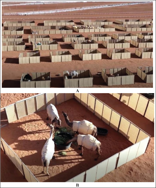
AbstractBackground: Tampering is an illegal manipulation of Arabian (dromedary) camels for cosmetic purposes and to increase their market value. Because of the tremendous prizes awarded to the owners of the winning camels, methods of illegal tampering were frequently developed in camel beauty festivals. The detection of such practices constitutes a significant challenge for veterinarians supervising these festivals. One of these challenges is the large number of examined camels at beauty festivals. The second is the disappearance of injected hormones in the blood from days to months after their preliminary injection. Such challenges may be faced by using well-trained and professional veterinarians as well as developing new recent and advanced laboratory techniques that can discover traces of injected hormones or even blood proteins released as a result of specific hormone injection. Aim: This study was conducted to document the most frequently used methods of illegal practices of tampering in dromedary camels and their detection by different methods, including (1) physical examination, (2) diagnostic imaging of ultrasonography, radiography, and thermography, and (3) hormonal analysis. Methods: A total number of 12,385 dromedary camels (12,080 females and 305 males) were examined for detection of different illegal manipulations of tampering. The camels were examined at two points: external and internal. This study was conducted in Saudi Arabia during the events of the 7th King Abdulaziz Camel Festival, Riyadh, Saudi Arabia, from December 28, 2022 to January 12, 2023. Results: The suspected camels were examined using different methods, including sonography, thermography, radiography, and blood examinations. Of the 12,385 examined camels, 943 (7.6%) (921 females and 22 males) were confirmed to have different forms of tampering. These forms included stretching of the lips in 499 camels (52.9%), binding of the lips in 139 camels (14.7%), injection of the nose by a filler material in 121 camels (12.8%), increased testosterone level in 74 camels (7.8%), injection of the lips by filler material in 60 camels (6.4%), fibrosis of the nostril in 33 camels (3.5%), and fibrosis of the lips in 17 camels (1.8%). Conclusion: A combination of clinical examination with sonography, radiography, thermography, and laboratory evaluation of testosterone and growth hormones is recommended for the detection of different forms of illegal manipulations of tampering in dromedary camels. There is no doubt that these illegal practices will negatively affect the welfare of camels and can lead to many physical and psychological drawbacks. Keywords: Camels, Diagnostic imaging, Radiography, Thermography, Ultrasonography. IntroductionArabian (dromedary) camels were a source of pride for their shepherds throughout the Arabian Peninsula. They are distinguished by characteristics over others, the most prominent of which is their pride, which is reflected in their character to a greater degree, and their trust in camels, as the animals that they share within many details of their daily lives. This indicates the importance of camels and their close association with civilization in the Arabian Peninsula throughout the ages, and this is proven by the finding of a group of inscriptions archeological sites in many historical sites, where camels formed a pillar for trade caravans in the past in transporting goods and fetching water, in addition to the multiple uses of its skins and tufts, including meat and milk production. The Arabs also mentioned camels extensively in their books, in prose and poetry, until their characteristics became proverbial in literary texts. Principally, the camel was and is still loved by every Bedouin and is considered the most valuable piece of ware. Bones of camels are entombed in the temples at Al-Fao in Saudi Arabia (Khan, 2022). Because of the importance that camels represent in the history of Saudi Arabia, and the distinguished cultural heritage they carry that is a source of pride for Saudis, the Camel Club has launched the King Abdulaziz Camel Festival (KACF) to educate the community and clarify its role in taking care of this sector and supporting related industries as an economic source for Saudi Arabia, and introducing the economic and developmental benefits of camels. During the activities of the KACF, medical committees are allocated to reveal the tampering that some camel owners resort to and to impose strict penalties against violators of tampering, represented by exclusion from participation, and depriving the violator and his camels from participating in the current and future versions, as well as referring the violator to the competent authorities to apply the legal penalties against him (Tharwat and Al-Hawas, 2021, 2023; Tharwat et al., 2021a,b). The KACF is held annually from the beginning of December to nearly the middle of January on the land of the southern deserts of Al-Dahna, Riyadh, Saudi Arabia. This festival contributes to introducing the relationship of camels to the people of the Arabian Peninsula, which is an authentic symbol of life in the desert and has a cultural, social, and economic connection that affects the history and life of Arab people throughout the ages. The people of the Arabian Peninsula relied on the gifts of nature in a greater way, as camels were present in all their details in their daily lives (Tharwat and Al-Hawas, 2021, 2023; Tharwat et al., 2021a,b). Camels from different Gulf countries, including Saudi Arabia, the United Arab Emirates, Qatar, Kuwait, and Oman, are usually enrolled in the KACF. The value of the prizes at the KACF in its seventh edition, which was held from December 2022 to January 2023, increased to a value of 300 million Saudi riyals ($80 million). Because of the increase in the festival’s prizes, some owners have resorted to unauthorized methods to beautifying camels, including, for example, but not limited to injections in the lips, stretching the lips, and the injection in the nose. Because these practices negatively affect the physical and psychological states of camels, they are prohibited by the medical committees supervising the KACF competitions. This study was conducted to document the most frequently used methods of illegal practices of tampering among 12,385 dromedary camels and their detection by different methods, including (1) physical examination, (2) diagnostic imaging of ultrasonography, radiography, and thermography, and (3) hormonal analysis. Material and MethodsCamelsA total of 12,385 dromedary camels (Camelus dromedarius) (12,080 females and 305 males) were examined to investigate different forms of tampering during the events of the 7th KACF in the southern Sayahed district of Al-Dahnaa, 90 km from Riyadh, the capital of Saudi Arabia, from December 28th, 2022 to January 12th, 2023. All the camels participating in this festival, on which this study was conducted, were healthy, and their ages ranged from one to 20 years. Different breeds of camels were examined, including black camels or Majaheem and colored camels or Maghateer. The camels that were colored included Waddah or white camels, Homor or camels with a red coat, Sofor or camels with a yellow-brown coat, Shaele camels with a gray coat that went sometimes to brown-red, Shageh camels with a luminous coat, Asail camels with a yellow to brown coat color, and Saheli camels that lived near coasts and had a red coat color. Camels were treated based on the Guidelines of Laboratory Animal Control Ethics of Qassim University, which were basically aligned to the Guide for the Use and Care and Laboratory Animals of the National Institutes of Health in the USA (NIH publications 86-23, revised 1996). Methods of festival participationThere are two methods of participating in the activities of the festival. The first step was to bring the camels to the Tatamman camel hotel. For check-in, a comprehensive medical examination is required to confirm that camels are free of any form of tampering. In this way, camels participated in the activities have obtained medical permission and were not examined by the medical committee once they had participated in the festival rounds. The second method to participate in the festival activities is direct participation in events without entering the Tatamman Hotel. The camels participating in this method will be subject to examination by the medical committee to prove that they are free of tampering. The Tatamman Hotel has 480 rooms to house its very unique guests, the camels. This desert hotel is designed to host camels near the KACF, and it is the first of its kind in the world. A full board stay including fodder and barley costs around $100 a night plus another $27 for milk consumption. The hotel offers services ranging from veterinary examinations, guarding camels, and security. The services also include transportation to the hotel, a tub, and camel cleaning. Camels must pass a medical examination for check-in. This hotel also has more than 200 professional workers who can do hairdressing and great care for camels. There are also veterinarians who specialize in caring for camels (Fig. 1). Examination of camel tamperingThe camels were examined at two points 500 m apart. The first point is the external examination point and is set up specifically to examine the camels before participating in the festival, before entering the Tatamman camel hotel and before the buying and selling operations. At this point, a total of 1,035 (980 females and 55 males) participants were examined. Examinations were conducted at 16:00–20:00 hours. As for the second point, it is the main or internal point, and it is the one set for examination of the actual camel that participated in the various activities of the festival. At this point, a total of 11,350 (1,100 females and 250 males) participants were examined. Examinations were conducted at 07:00–12:00 PM. The camels participating in this study were examined through a virtual examination in the first stage; then, the suspected camels were examined by several methods, including ultrasound examination, thermography, radiography, and biochemical determination of testosterone and growth hormone levels (Tharwat and Al-Hawas, 2021; Tharwat et al., 2021a, 2024a,b; Tharwat 2023, 2024). The testosterone and growth hormone results were compared with those of healthy camels in our recent reports (Tharwat et al., 2021b; Tharwat and Al-Hawas, 2024).
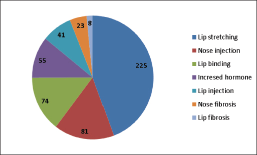
Fig. 1. The Tatamman Hotel. A hotel designed to host camels near the King Abdul Aziz Camel Festival. Camels must pass a medical examination for check-in. It has the largest capacity, with 480 rooms and integrated services (A). These services include transportation to the hotel, a bathtub, and camel cleaning. This hotel also has more than 200 professional workers who can do hairdressing and great care for camels. There are also veterinarians who specialize in caring for camels. Image (B) shows a close-up view of the image A. ResultsExternal examination pointCamels came to the external inspection point loaded on trucks, and a few of them came on foot from close distances (Fig. 2). The camels were then unloaded and examined at a recumbent position. On the other side, camels come to the internal inspection point on foot from a distance of approximately 700 m. Camels were first examined in the standing position, and any camel with any external abnormality was lying down and examined in the recumbent position (Fig. 3). At the first examination or external point, of the 1,035 examined camels, 507 (49%) camels were confirmed to have different forms of tampering. Of the 507 camels, 500 were female and seven were males. Figure 4 illustrates different types of tampering in camels examined at the external point. These forms included stretching of the lips in 225 camels (43.4%), injection of the nose by a filler material in 81 camels (15.6%), binding of the lips in 74 camels (14.3%), increased testosterone level in 55 camels (10.6%), injection of the lips by filler material in 41 camels (7.9%), fibrosis of the nostril in 23 camels (4.4%), and fibrosis of the lips in 8 camels (1.5%).
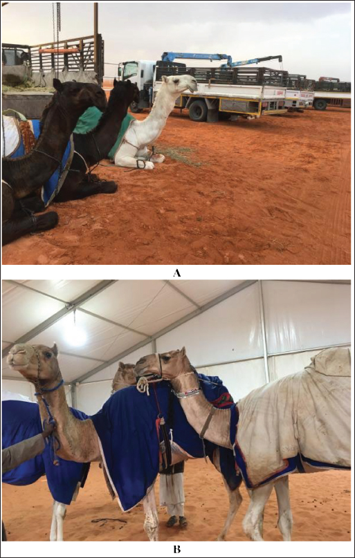
Fig. 2. External examination point where most camels are admitted when loading in trucks (A) and few are admitted when walking from a short distance (B).
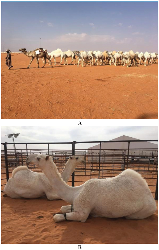
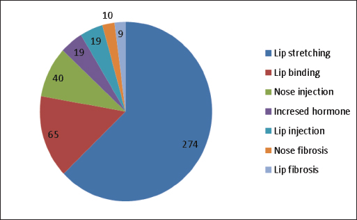
Fig. 3. Internal examination point at which most camels are admitted walking on foot (A). Camels suspected of having any form of tampering are examined in the sitting position (B). Internal examination pointsAt the second examination point, of the 11,350 examined camels, 436 (3.8%) camels were confirmed to have different forms of tampering. Of the 436 camels, 421 were female and 15 were males. Figure 5 shows various forms of tampering in camels examined at the inside of the passage. These forms include stretching of the lips in 274 camels (63.3%), binding of the lips in 65 camels (15%), injection of the nose by a filler material in 40 camels (9.2%), increased testosterone level in 19 camels (4.4%), injection of the lips by filler material in 19 camels (4.4%), fibrosis of the nostril in 10 camels (2.3%), and fibrosis of the lips in 9 camels (2.1%).
Fig. 4. Different forms of tampering among 1,035 camels examined at an external point. Different types of tampering were detected in 518 camels and included stretching of the lips, injection of the nose by a filler material, binding of the lips, increased testosterone level, injection of the lips by a filler material, fibrosis of the nostril, and fibrosis of the lip.
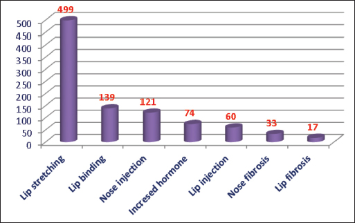
Fig. 5. Different forms of tampering among 11,350 camels examined at the internal point. The types of tampering detected in 433 camels included stretching of the lips, binding of the lips, injection of the nose by a filler material, increased testosterone level, injection of the lips by a filler material, fibrosis of the nostril, and fibrosis of the lips. Overall examination pointsCollectively, at two examination sites, 12,385 (12,080 females and 305 males) camels were examined. Of the 12,385 examined camels, 943 (7.6%) (921 females and 22 males) camels were confirmed to have different forms of tampering (Fig. 6). These forms included stretching of the lips in 499 camels (52.9%), binding of the lips in 139 camels (14.7%), injection of the nose by a filler material in 121 camels (12.8%), increased testosterone level in 74 camels (7.8%), injection of the lips by filler material in 60 camels (6.4%), fibrosis of the nostril in 33 camels (3.5%), and fibrosis of the lips in 17 camels (1.8%).
Fig. 6. Overall tampering methods among 12,385 camels examined at external and internal examination points. The types of tampering detected in 943 camels included stretching of the lips, binding of the lips, injection of the nose by a filler material, increased testosterone level, injection of the lips by a filler material, fibrosis of the nostril, and fibrosis of the lips. Methods of tamper detectionExtension and stretching of the lips in camels due to daily massage of the lips and sometimes binding with a rubber band resulted in blood congestion and lip enlargement (Fig. 7). Radiographic evaluation of filler materials in the perinasal region showed swelling of the soft tissues to the extent of variable degrees owing to the injected fillers; the injected substances appeared gray and had a soft tissue density (Fig. 8). In the injected lips, the injected filler was hypoechoic, and the scanning manner of the injected lips appeared heterogeneous (Fig. 9). In the perinasal area, the filler material appeared as either hypoechogenic or anechoic spots (Fig. 10). In infrared thermography (IRT), stretched lips appeared lighter and more heterogeneous than the darker and homogenous lips of healthy camels. In addition, the injected lip sites appeared darker than the surrounding tissue as their temperature was relatively higher when compared with healthy non-injected lip tissue (Fig. 11). Figure 12 shows a camel with enlargement of the lips due to filler injection, while Figure 13 shows enlargement of the soft palate or dulla and clitoris due to testosterone hormonal injection. DiscussionIn camel medicine, ultrasonography has been used extensively in dromedary camel medicine during the past decade, either in healthy (El-Tookhy and Tharwat, 2012; Tharwat et al., 2012a,b,c; Tharwat, 2013a) or diseased (Tharwat et al., 2012d,e, 2018a,b, 2023, 2024c, 2025a,b; Tharwat, 2013b, 2019, 2020a,b,c, 2021a,b, 2024; Tharwat and Al-Hawas, 2021; Tharwat and El-Tookhym 2021; Sadan et al., 2024) states. In addition, the effectiveness of thermography has been reported in camel beauty festivals through the detection of injected as well as extended lips (Tharwat et al., 2021a; Tharwat and Al-Hawas, 2023).
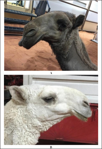
Fig. 7. Enlargement and protrusion of the lips in a female camel (A) due to stretching and extending of the lips and then pulling them outward compared with a control healthy camel (B).
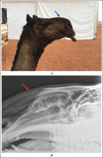
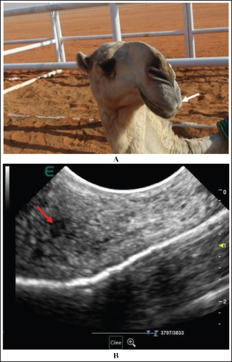
Fig. 8. Enlargement of the perinasal area in a female camel due to injection of filler material (blue arrow) (A). Image (B) shows a lateral radiographic view of the same camel, showing the gray-colored injected filler material in the perinasal region (red arrow). In humans, the application of filler materials, even when ratified by the US FDA, can imitate early or delayed, reversible, or irreversible drawbacks. These drawbacks may include ecchymosis, cellulite, edema, hypersensitivity reactions, granulomas, palpable or visible masses, abscesses, dermatopathy, necrosis, fistulas, and even blindness (Schutz et al., 2012; Shahrabi-Farahani et al., 2014; Kassir et al., 2020). In camels, some owners may inject testosterone or growth hormones into their camels, especially females at young ages, to change the general shape of the animal. Injecting these hormones has clear symptoms on camels that are well known, including, for example, but not limited to, the large head and neck, the length of the bones, the large size of muscles, the enlargement of the clitoris, and the high rate of female infertility. In camels, some drawbacks were also observed following different cosmetic interventions. Abscess formation can be detected in situ within the lips at the injection site. Others include granulomas, palpable nodules, or masses, and necrosis of the overlying tissue (Tharwat and Al-Hawas, 2021, 2024).
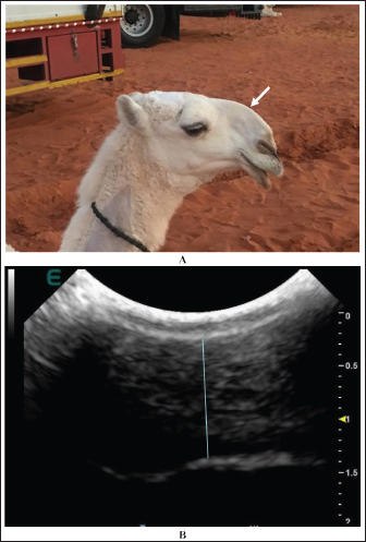
Fig. 9. A female camel with injected upper lip by a filler material. The upper lip is enlarged and pendulous (white arrow) (A). Sonographically, the injected substance appeared anechoic (red arrow), and the surrounding tissue appeared heterogeneous (B). In a bid to enhance their camel’s natural beauty, some owners have resorted to unnatural methods where inexperienced people or even unqualified persons perform these procedures (Tharwat and Al-Hawas, 2021, 2023; Tharwat et al., 2021a, 2024a,b; Tharwat, 2024). A total of 1,035 (980 females and 55 males) camels were examined at this point; of these, 507 (49%) camels were confirmed to have different forms of tampering. At the second point of examination, the internal one, a total of 11,350 (11,100 females and 250 males) participants in the festival events were examined. Of the 11,350 examined camels, 436 (3.8%) camels were confirmed to have different forms of tampering. Overall, at the two examination sites, 12,385 (12,080 females and 305 males) camels were examined. Of the 12,385 examined camels, 943 (7.6%) (921 females and 22 males) camels were confirmed to have different forms of tampering.
Fig. 10. Enlargement of the perinasal area in a female camel due to injection of filler material (white arrow) (A). Image (B) shows the ultrasonographic detection of the injected filler material (blue line) that appeared hypoechoic with anechoic spots. The detectable forms of illegal practices of tampering in this study included stretching of the lips in 499 camels, binding of the lips in 139 camels, injection of the nose by a filler material in 121 camels, increased testosterone level in 74 camels, injection of the lips by filler material in 60 camels, fibrosis of the nostril in 33 camels, and fibrosis of the lips in 17 camels. Stretching of the lips is usually performed through daily massage and pulling of the lips using an oil, which finally leads to drooping of the lips. Binding the lips is usually done with a rubber band, followed by pulling the lip outward. Compressing the lips using a piece of plastic sheet to stimulate inflammation, blood congestion, and pronounced enlargement of the lips is another methodology. Using a local anesthetic, either in the form of injections or ointments, to induce laxity in the lips is one of the methods of tampering in camels, as it leads to the drooping of the lower lip and changing the appearance of the camel. Enlargement of the nose, as well as the lips, is also performed via injection of a filler material subcutaneously in the nose region or in the upper or lower lips. Fibrosis of the nose skin or lips is usually noted when injecting an irritant material or in long-standing conditions (Tharwat and Al-Hawas, 2021).
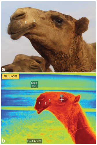
Fig. 11. Injection points (1, 2, 3) of the lips in a female camel using filler material (a). Image (b) shows the detection of the three injected points by thermography. Tampering is usually detected in camels either by close clinical examination, ultrasound examination, and thermography or by laboratory tests to estimate the level of hormones in the blood (Tharwat and Al-Hawas, 2021, 2023; Tharwat et al., 2021a, 2024a,b). In this study, ultrasonography was valuable for verifying the different cosmetic methods used by camels. The injected substance appeared of low echogenicity, and the scanning image of the injected lips showed heterogeneity with increasing overall lip thickness. In cases with lip binding, the tip of the lips appeared either isoechoic with a hypoechoic rim or hypoechoic with an echogenic center. This finding is in agreement with previously reported results (Tharwat and Al-Hawas, 2021). In the perinasal area, the filler material appeared as either hypoechoic or anechoic spots. These findings agree well with recently reported findings on human beings (Chai et al., 2022; Jiang et al., 2022). In this study, sonography was superior to radiography and thermography. Sonography was the easy, safe, and reliable methodology that was implemented in situ for a large number of examined camels to obtain quick and accurate results.
Fig. 12. Extension and stretching of the lips in two female camels (A and B) following filler injection. Radiography is also valuable for determining the injected filler materials in the perinasal region. Radiographic evaluation revealed swelling of the soft tissues to different extents owing to injected fillers; the injected substances appeared grayish with a variable soft tissue density. This finding was not correlated with those reported by Kwon et al. (2018), who found the radiopaque density of an unknown filler material in three cases, and Valiyaparambil et al. (2009) who reported a case of an unusual soft tissue radiopacity-radiographic appearance of dermal filler. The reason for the differences detected in tissue density may be due to the various types of injected fillers, whereas the most popular injected filler material in camels is hyaluronic acid (Tharwat and Al-Hawas, 2021). Our findings clearly demonstrate the usefulness of radiography as a diagnostic imaging technique for the precise detection of injected cosmetic fillers in the perinasal area and as a tool for detecting their expansion in the soft tissues. These findings agree well with recently reported findings in humans (Kroumpouzos et al., 2023) and camels (Tharwat et al., 2024b). The use of radiography in this study has some disadvantages. These drawbacks include not using the X-ray machine near the camels, as radiography can only be used in closed, protected rooms lined with lead. Therefore, it was necessary to move the suspected camel to safe places where the X-ray machine could be used, which would consequently waste time.
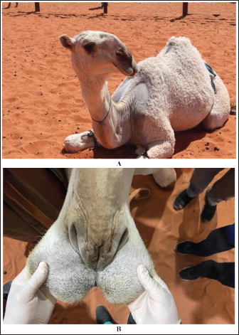
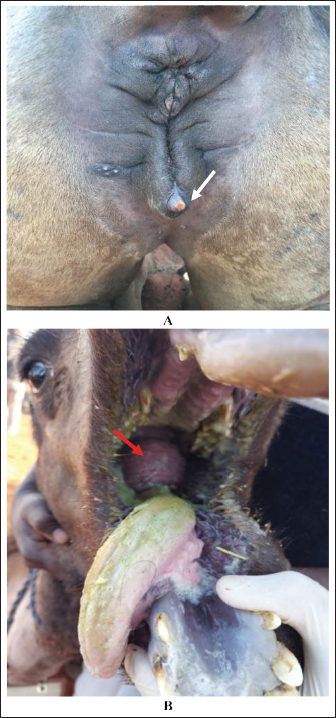
Fig. 13. Side effects of hormone injection in camels. Image (A) shows an enlarged clitoris (white arrow) in a female camel, and image (B) shows the development of the dulla (red arrow) in a yearling female camel. IRT is also valuable for detecting injected filler substances in the lisps and for verifying stretched or extended lips. According to IRT and similar to our published results (Tharwat et al., 2021a), the injected lip sites appeared darker than the surrounding tissue as their temperature was relatively higher than that of healthy non-injected lip tissue. The darker appearance of the injected lip sites may be explained by the presence of inflammation near the injection area. Stelletta et al. (2012) reported abnormal thermal images that can be monitored from the skin to superficial inflammations or alterations in blood flow. In addition, the stretched lips of IRT were lighter and more heterogeneous than the darker and homogenous lips of healthy camels. The light color of the image in this group could be explained by saliva retention within the salivary glands, as observed when examining the lips. This result is in agreement with our previously reported results (Tharwat et al., 2021b). Using a thermal camera to detect tampering also has some disadvantages. One disadvantage of this method is that it is sharply affected by the temperature of the atmosphere. For example, when examining a suspected case in the early morning, the temperature of the skin and the examined area differ greatly if the same animal is examined in the afternoon. Therefore, this process limits the efficiency of thermography when detecting tampering in camels. Conclusions, Challenges, and Future DirectionsBecause of the tremendous prizes awarded to the owners of the winning camels, methods of illegal tampering were frequently used in camel beauty shows. Therefore, the detection of these practices is a must; however, it constitutes a great challenge for veterinarians supervising camel festivals. A combination of close clinical examination with ultrasonography, radiography, thermography, and laboratory evaluation of testosterone and growth hormones is recommended to detect different forms of tampering in camels. There is no doubt that these unethical practices will negatively affect the welfare of camels and can lead to physical and psychological harm to these animals. Because of the great challenges faced by clinicians, the lack of time to examine large numbers of camels in beauty shows, and the disappearance of injected hormones from the blood days to months after their preliminary injection, the next research in this field should be directed to unconventional methods for precise detection of tampering. The integration of artificial intelligence to detect different types of tampering in camels is one of these approaches. In addition, developing recent and advanced laboratory techniques that can detect traces of injected hormones or even blood proteins released as a result of specific hormone injections is another approach that we are trying to implement in our future research. AcknowledgmentsNone. Conflict of interestThe authors declare no conflict of interest. FundingThis research received no specific grant. Author’s contributionMT: conceptualization, practical work, writing of manuscript draft, and preparation of figures and tables. AA: Practical work and revision of the manuscript draft. The authors have approved the final manuscript for publication. Data availabilityAll data supporting the findings of this study are available in the manuscript, and no additional data sources are required. ReferencesChai, H., Su, X., Yuan, L., Li, Z., Liu, Y., Dou, M. and Hu, J. 2022. Application of high-frequency ultrasound in detection and identification of nasal filling injection materials. J. Cosmet. Dermatol. 21, 4276–4287. Jiang, L., Yuan, L., Li, Z., Su, X., Hu, J. and Chai, H. 2022. High-frequency ultrasound of facial filler materials in the nasolabial groove. Aesthetic Plast. Surg. 46, 2972–2978. Kassir, M., Gupta, M., Galadari, H., Kroumpouzos, G., Katsambas, A., Lotti, T., Vojvodic, A., Grabbe, S., Juchems, E. and Goldust, M. 2020. Complications of botulinum toxin and fillers: a narrative review. J. Cosmet. Dermatol. 19, 570–573. Khan, M. 2022. Camel: the symbol of honor, pride and love of Arab Bedouins. Int. J. Archaeol. 10, 20–23. Kroumpouzos, G., Harris, S., Bhargava, S. and Wortsman, X. 2023. Complications of fillers in the lips and perioral area: prevention, assessment, and management focusing on ultrasound guidance. J. Plast. Reconstr. Aesthet. Surg. 84, 656–669. Kwon, Y.E., An, C.H., Choi, K.S., Lee, D.H. and An, S.Y. 2018. Radiographic study of dermal fillers in the facial area: a series of 3 cases. Imaging Sci. Dent. 48, 227–231. Sadan, M., Tharwat, M., Alkhedhairi, S., Refaai, W., Moghazy, H.M.E.L., Khodier, M.M., Alkhamiss, A.S. and Ghallab, A. 2024. Abdominal pedunculated leiomyoma in a male dromedary camel: clinical, hematobiochemical, ultrasonographic and pathologic findings. Int. J. Vet. Sci. 13, 458–462. Schutz, P., Ibrahim, H.H., Hussain, S.S., Ali, T.S., El-Bassuoni, K. and Thomas, J. 2012. Infected facial tissue fillers: case series and review of the literature. J. Oral Maxillofac. Surg. 70, 2403–2412. Shahrabi-Farahani, S., Lerman, M.A., Noonan, V., Kabani, S. and Woo, S.B. 2014. Granulomatous foreign body reaction to dermal cosmetic fillers with intraoral migration. Oral Surg. Oral Med. Oral Pathol. Oral Radiol. 117, 105–110. Tharwat, M. 2013a. Ultrasonography of the lungs and pleura in healthy camels (Camelus dromedarius). Acta Vet. Hung. 61, 309–318. Tharwat, M. 2013b. Ultrasonographic findings in camels (Camelus dromedarius) with trypanosomiasis. J. Camel Pract. Res. 20, 283–287. Tharwat, M. 2019. Chronic peritonitis in dromedary camels: clinical, ultrasonographic and pathologic findings. J. Camel Pract. Res. 26, 169–172. Tharwat, M. 2020a. Ultrasonography of the abdomen in healthy and diseased camels (Camelus dromedaries)–a review. J. Appl. Anim. Res. 48, 300–312. Tharwat, M. 2020b. Ultrasonography of the kidneys in healthy and diseased camels (Camelus dromedaries). Vet. Med. Intern. 2020, 7814927. Tharwat, M. 2020c. Ultrasonography of the liver in healthy and diseased camels (Camelus dromedaries). J. Vet. Med. Sci. 82, 399–407. Tharwat, M. 2021a. Ultrasonography of the thorax in healthy and diseased camels (Camelus dromedaries) – a mini-review. J. Camel Pract. Res. 28, 313–318. Tharwat, M. 2021b. Obstructive urolithiasis in dromedary camels: clinical, ultrasonographic and postmortem findings. J. Camel Pract. Res. 28, 85–93. Tharwat, M. 2023. Advanced biomarkers and its usage in Arabian camel medicine – a review. J. Appl. Anim. Res. 51, 350–357. Tharwat, M. 2024. Fundamentals of diagnostic ultrasound in dromedary camel medicine. Int. J. Vet. Sci. 13, 1–6. Tharwat, M. and Al-Hawas, A. 2021. Ultrasound detection of cosmic fillers injection of lips in camel beauty pageants: first report in Veterinary Medicine. Trop. Anim. Health Prod. 53, 53. Tharwat, M. and Al-Hawas, A. 2023. Infrared thermography in healthy Arabian camels (Camelus dromedarius). Int. J. Vet. Sci. 12, 762–767. Tharwat, M. and Al-Hawas, A. 2024. The emerging topic of cosmetic medicine in dromedary camels. Int. J. Vet. Sci. 13, 139–146. Tharwat, M., Al-Hawas, A. and Alkheraif, A.A. 2024a. Retrospective analysis of different cosmetic methods in 53090 dromedary camels. Int. J. Vet. Sci. 13, 545–549. Tharwat, M., Al-Hawas, A. and Albotti, Y. 2021a. Infrared thermography in healthy dromedary camels and its feasibility in injected and stretched lips in camel beauty pageants. J. Camel Pract. Res. 28, 355–359. Tharwat, M., Al-Hawas, A. and Aldhubayi, A. 2021b. Testosterone and growth hormone levels in female dromedary camels. J. Camel Pract. Res. 28, 373–376. Tharwat, M., Ali, H. and Alkheraif, A.A. 2025a. Clinical insights on paratuberculosis in Arabian camels (Camelus dromedarius): a review. Open Vet. J. 15, 8–17. Tharwat, M., Alkheraif, A.A., Elmoghazy, H.M.M., Haridy, M. and Marzok, M. 2025b. Disseminated pyogranulomas in a female dromedary camel: hematobiochemical, sonographic and pathologic investigations. Int. J. Vet. Sci. 14, 120–124. Tharwat, M., Al-Sobayil, F., Ali, A. and Buczinski, S. 2012a. Echocardiography of the normal camel (Camelus dromedarius) heart: technique and cardiac dimensions. BMC Vet. Res. 8, 130. Tharwat, M., Al-Sobayil, F., Ali, A. and Buczinski, S. 2012b. Transabdominal ultrasonographic appearance of the gastrointestinal viscera of healthy camels (Camelus dromedarius). Res. Vet. Sci. 93, 1015–1020. Tharwat, M., Al-Sobayil, F., Ali, A. and Buczinski, S. 2012c. Ultrasonography of the liver and kidneys of healthy camels (Camelus dromedarius). Can. Vet. J. 53, 1273–1278. Tharwat, M., Al-Sobayil, F., Ali, A. and Buczinski, S. 2012d. Ultrasonographic evaluation of abdominal distension in 52 camels (Camelus dromedarius). Res. Vet. Sci. 93, 448–456. Tharwat, M., Al-Sobayil, F., Ali, A., Hashad, M. and Buczinski, S. 2012e. Clinical, ultrasonographic, and pathologic findings in 70 camels (Camelus dromedarius) with Johne’s disease. Can. Vet. J. 53, 543–548. Tharwat, M., EL Moghazy, H.M. and Oikawa, S. 2023. Ultrasonographic verification of hepatic hydatidosis in a female dromedary camel: a case report. J. Vet. Med. Sci. 85, 1286–1290. Tharwat, M., El-Ghareeb, W.R. and Almundarij, T.I. 2024b. Depraved appetite in dromedary camels: clinical, ultrasonographic, and postmortem findings. Open Vet. J. 14, 652–663. Tharwat, M. and El-Tookhy, O. 2021. Ocular ultrasonography in camels (Camelus dromedarius): a review. J. Camel Pract. Res. 28, 185–190. Tharwat, M., Sadan, M. and Al-Hawas, A. 2024c. The emerging topic of injected cosmetic fillers in the perinasal region of dromedary camels: ultrasonographic and radiographic verification. Open Vet. J. 14, 840–845. Tharwat, M., Sadan, M., El-Shafaey, E., Al-Hawas, A. and Saeed, E.M.A. 2018a. Unilateral nephrectomy in a female dromedary camel with pyelonephritis caused by Staphylococcus lugdunensis. Pakistan Vet. J. 38, 116–118. Tharwat, M., Sadan, M., El-Shafaey, E., Saeed, E. and Al-Hawas, A. 2018b. Bilateral renal abscessation and chronic active pyelonephritis in a male camel (Camelus dromedarius) caused by Escherichia coli. J. Vet. Med. Sci. 80, 778–783. Valiyaparambil, J., Rengasamy, K. and Mallya, S.M. 2009. An unusual soft tissue radiopacity--radiographic appearance of a dermal filler. Br. Dent. J. 207, 211–212. | ||
| How to Cite this Article |
| Pubmed Style Tharwat M, Al-hawas A. Identification of tampering methods among 12,385 Arabian camels using different diagnostic imaging techniques. Open Vet. J.. 2025; 15(3): 1226-1238. doi:10.5455/OVJ.2025.v15.i3.14 Web Style Tharwat M, Al-hawas A. Identification of tampering methods among 12,385 Arabian camels using different diagnostic imaging techniques. https://www.openveterinaryjournal.com/?mno=229874 [Access: January 25, 2026]. doi:10.5455/OVJ.2025.v15.i3.14 AMA (American Medical Association) Style Tharwat M, Al-hawas A. Identification of tampering methods among 12,385 Arabian camels using different diagnostic imaging techniques. Open Vet. J.. 2025; 15(3): 1226-1238. doi:10.5455/OVJ.2025.v15.i3.14 Vancouver/ICMJE Style Tharwat M, Al-hawas A. Identification of tampering methods among 12,385 Arabian camels using different diagnostic imaging techniques. Open Vet. J.. (2025), [cited January 25, 2026]; 15(3): 1226-1238. doi:10.5455/OVJ.2025.v15.i3.14 Harvard Style Tharwat, M. & Al-hawas, . A. (2025) Identification of tampering methods among 12,385 Arabian camels using different diagnostic imaging techniques. Open Vet. J., 15 (3), 1226-1238. doi:10.5455/OVJ.2025.v15.i3.14 Turabian Style Tharwat, Mohamed, and Abdulla Al-hawas. 2025. Identification of tampering methods among 12,385 Arabian camels using different diagnostic imaging techniques. Open Veterinary Journal, 15 (3), 1226-1238. doi:10.5455/OVJ.2025.v15.i3.14 Chicago Style Tharwat, Mohamed, and Abdulla Al-hawas. "Identification of tampering methods among 12,385 Arabian camels using different diagnostic imaging techniques." Open Veterinary Journal 15 (2025), 1226-1238. doi:10.5455/OVJ.2025.v15.i3.14 MLA (The Modern Language Association) Style Tharwat, Mohamed, and Abdulla Al-hawas. "Identification of tampering methods among 12,385 Arabian camels using different diagnostic imaging techniques." Open Veterinary Journal 15.3 (2025), 1226-1238. Print. doi:10.5455/OVJ.2025.v15.i3.14 APA (American Psychological Association) Style Tharwat, M. & Al-hawas, . A. (2025) Identification of tampering methods among 12,385 Arabian camels using different diagnostic imaging techniques. Open Veterinary Journal, 15 (3), 1226-1238. doi:10.5455/OVJ.2025.v15.i3.14 |

