


| Research Article | ||
Open Vet. J.. 2025; 15(3): 1310-1321 Open Veterinary Journal, (2025), Vol. 15(3): 1310-1321 Research Article Therapeutic effects of kecemcem leaves extract (Spondias pinnata (L.f.) Kurz) on LDL levels, cytokines, and microscopic liver changes in hyperlipidemic rat modelLia Cahya Sari1, Nunuk Dyah Retno Lastuti2*, Theresia Indah Budhy1, Yuliasih Yuliasih3, Ni Luh Ayu Megasari1 and Indra Prastiwi Jamaluddin11Immunology Program, Postgraduate School, Universitas Airlangga, Surabaya, Indonesia 2Postgraduate School, Universitas Airlangga, Surabaya, Indonesia 3Departement of Internal Medicine, Faculty of Medicine, Universitas Airlangga, Surabaya, Indonesia *Corresponding Author: Nunuk Dyah Retno Lastuti. Postgraduate School, Universitas Airlangga, Surabaya, Indonesia. Email: nunukdyah53 [at] gmail.com Submitted: 06/12/2024 Accepted: 12/02/2025 Published: 31/03/2025 © 2025 Open Veterinary Journal
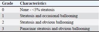
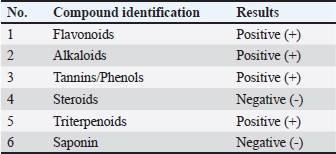
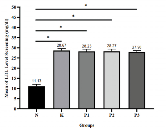
AbstractBackground: Modern lifestyle, which is characterized by a high-fat diet, increases the risk of hyperlipidemia, a primary trigger for cardiovascular diseases and metabolic disorders. Excessive fat consumption can trigger hepatic steatosis and oxidative stress, leading to inflammation and liver damage, which contributes to the development of non-alcoholic fatty liver disease. Conventional cholesterol-lowering therapies are often expensive and come with side effects, making herbal alternatives such as kecemcem leaves a promising option. Kecemcem leaves extract contains flavonoids such as quercetin and catechin, which exhibit antioxidant properties, regulate lipid metabolism, and reduce inflammation. Aim: This study aimed to evaluate the therapeutic effects of kecemcem leaves extract [Spondias pinnata (L.f.) Kurz] in reducing low-density lipoprotein (LDL) levels and its impact on pro-inflammatory cytokines [Interleukin-1 beta (IL-1β), tumor necrosis factor-alpha (TNF-α) and anti-inflammatory cytokines Interleukin-10 (IL-10)], as well as liver histology in hyperlipidemic Wistar rats. Methods: Hyperlipidemia was induced through a high-fat diet for 16 weeks, after which the rats were administered kecemcem leaves extract at doses of 250, 500, and 1,000 mg/kg body weight for 1 week. One week after the administration of kecemcem leaves extract, blood and liver samples were collected from all rats to measure LDL, IL-1β, TNF-α, and IL-10 levels, as well as to perform liver histological analysis. Results: Kecemcem leaves extract significantly reduced LDL, IL-1β, and TNF-α levels while increasing IL-10 levels in hyperlipidemic rats, particularly at the 250 mg/kg body weight dose. In addition, there was an improvement in liver histology, as evidenced by decreased steatosis and hepatocyte ballooning. These effects are attributed to the flavonoid content in kecemcem, such as quercetin and catechins, which inhibit cholesterol synthesis, reduce oxidative stress, and decrease liver fat accumulation. Conclusion: Kecemcem leaves extract potentially reduced LDL, IL-1β, and TNF-α levels, increased IL-10 levels, and improved liver histology. This effect is supported by the flavonoid content of quercetin and catechin. Thus, kecemcem leaves extract has shown promising therapeutic effects in managing conditions related to hyperlipidemia and inflammation. Keywords: Cytokines, Hyperlipidemia, Immunology, LDL, Spondias pinnata (L.f.) Kurz. IntroductionModern lifestyle, which often promotes the consumption of high-cholesterol foods, complicates efforts to control blood cholesterol levels, leading to hyperlipidemia. Hyperlipidemia, a condition where blood cholesterol levels rise, has been identified as a major trigger for various cardiovascular diseases and metabolic disorders, such as coronary heart disease, atherosclerosis, metabolic syndrome, and stroke (Karr, 2017; Hong et al., 2022). Low-density lipoprotein (LDL) is considered the primary atherogenic lipoprotein and is frequently used as an indicator for diagnosing hyperlipidemia (Aman et al., 2019). According to Indonesia’s Basic Health Research in 2018, the prevalence of hyperlipidemia is very high, with 72.8% of individuals aged 15 years and older having LDL levels above 100 mg/dl, and 28.8% having total cholesterol levels exceeding 200 mg/dl (Rahmawaty et al., 2022). If left untreated, hyperlipidemia can increase the risk of death from cardiovascular diseases, which are currently the leading cause of death worldwide (Hayudanti et al., 2016). Excessive fat consumption not only leads to elevated blood cholesterol levels but also causes fat accumulation in liver cells, which is known as hepatic steatosis. The accumulated fat undergoes oxidation, producing free radicals, which in turn trigger oxidative stress that can damage liver cells and cause inflammation. This process is followed by the activation of Kupffer cells, which release pro-inflammatory cytokines such as interleukin-1 beta (IL-1β) and tumor necrosis factor-alpha (TNF-α) while simultaneously reducing the production of the anti-inflammatory cytokine interleukin-10 (IL-10), thereby exacerbating the body’s inflammatory response (Sarwanti et al., 2020). Chronic inflammation, driven by increased pro-inflammatory cytokine production and reduced anti-inflammatory cytokine levels, contributes to the progression of hepatic steatosis, which may evolve into non-alcoholic fatty liver disease (NAFLD), a leading cause of chronic liver disease globally (Anjani, 2023). Pharmacological cholesterol-lowering therapies often come with high costs, various side effects, and long-term consumption (Alloubani et al., 2021). Safer and more affordable alternatives, particularly herbal remedies with natural antioxidant properties and minimal side effects, are now gaining interest. A promising plant is kecemcem [Spondias pinnata (L.f.) Kurz], which has been traditionally used in folk medicine. In Balinese ethnobotanical traditions, kecemcem leaves are processed into an herbal drink called “Loloh Cemcem”, which is used to treat illnesses and promote overall health (Aryasa et al., 2021). Research shows that methanol extract from kecemcem fruit exhibits significant antihyperlipidemic activity, demonstrated by reducing total cholesterol, LDL, and triglyceride levels while increasing high-density lipoprotein levels in hyperlipidemic-induced rats (Raju et al., 2017). Additionally, Loloh Cemcem has been shown to lower total cholesterol levels in mice fed a high-fat diet (Dewi et al., 2022). The phytochemical content of kecemcem leaves, especially flavonoids such as quercetin and catechin, plays a significant role in antioxidant activity and lipid metabolism regulation (Azizah et al., 2019; Laksemi, 2019). These flavonoids not only prevent oxidative stress by reducing reactive oxygen species (ROS) production but also reduce inflammation by suppressing the expression of pro-inflammatory proteins such as TNF-α and IL-1β while promoting the production of the anti-inflammatory cytokine IL-10. Given the crucial role of flavonoids in lowering cholesterol levels and preventing inflammation, the utilization of kecemcem leaves as a natural flavonoid source has the potential to be an effective solution for managing hyperlipidemia and its complications, including hepatic steatosis. Therefore, examining LDL levels, cytokines, and liver histology in this study on hyperlipidemic Wistar rats treated with kecemcem leaves ethanol extract is essential to further understand the therapeutic effects of kecemcem leaves, ensuring more targeted and effective management of hyperlipidemia, with a focus on preventing disease progression and reducing the risk of complications. Materials and MethodsSpecimen collectionWe obtained kecemcem leaves from kecemcem plants grown in Penglipuran Village, Bangli Regency, Bali Province, Indonesia. Taxonomic identification of kecemcem leaves was performed by the UPT Laboratorium Herbal Materia Medica Batu, Malang Regency, East Java Province, Indonesia. Kecemcem leaves preparation and extractionA total of 5,000 g of fresh, green, and pest-free leaves were wet-sorted to remove unnecessary parts. The leaves were washed with running water and drained for 24 hours. After draining, the leaves were sliced and dried in a shaded room to prevent direct sunlight exposure. The dried leaves were then dry-sorted, and the simplicia was ground into powder. The kecemcem leaves extract was prepared using a maceration method. A total of 2,500 g of kecemcem leaves powder was placed in a macerator and mixed with 96% ethanol in a 1:15 ratio. The mixture was stirred and macerated for 24 hours and filtered to obtain the filtrate, followed by re-maceration using the same procedure. The re-macerated filtrate was evaporated and further concentrated over a water bath to yield 270 g of thick kecemcem leaves ethanol extract. Phytochemical analysis of kecemcem leaves ethanol extractQualitative and quantitative analyses were performed to determine the phytochemical content of the kecemcem ethanol extract. The qualitative determination of flavonoids and tannins was performed using an alkaline reagent test and a ferric chloride test (Kancherla et al., 2019). The Dragendorff and Mayer reagents were used to determine the alkaloids content, while the Bouchardat reagent was used to determine the presence of both alkaloids and terpenoids content (Nurjannah et al., 2022; Khoirunnisa and Zakia, 2024). The qualitative determination of saponins was performed as previously described (Khoirunnisa and Zakia, 2024). The total flavonoid content was determined using ultraviolet-visible spectrophotometry at 435 nm wavelength (Evayana and Aminah, 2022). Development of hyperlipidemic animal modelThirty male Wistar rats with an average body weight of 200 g were acclimatized for 7 days and fed standard feed. Following acclimatization, the rats were randomly divided into five groups: normal group (N), positive control group (K), and treatment group (P1, P2, and P3). All groups continued to receive standard feed ad-libitum, whereas the control group (K) and treatment groups 1–3 (P1, P2, and P3) were given an additional high-fat diet (approximately 52.8% of fat content per feed) consisting of a 1:1:1 mixture of cow brain, duck egg yolk, and used cooking oil. The high-fat diet was administered every morning for 16 weeks via gastric gavage at 1% of the body weight. Administration of kecemcem leaves extractKecemcem leaves extract was administered at varying doses. Treatment groups P1, P2, and P3 received 250, 500, and 1,000 mg/kgBW, respectively. The extract was dissolved in 1% CMC-Na solution and administered via gastric gavage every morning for one week following the development of the animal model. Blood and liver collectionBlood collection was first performed following the 16-week development of the animal model. Rats were fasted for 8–10 hours and anesthetized with ketamine. Approximately 1 ml of blood was taken from the lateral tail vein for serum LDL analysis. Following the 1-week administration of kecemcem leaves extract, rats were fasted for 8–10 hours, then anesthetized using ketamine. Approximately 5 ml of blood was drawn via cardiac puncture and collected using a red top tube, and liver samples were collected for microscopic examination. The serum was then isolated from blood samples as previously described (Megasari et al., 2021). LDL levels measurementThe LDL levels in serum samples were measured using an Erba Mannheim Biochemical Analyzer XL-640 (Erba Lachema, Czech Republic). The normal LDL-C level in rats was determined by a measured value of 27.20 mg/dl (Zaidan et al., 2024). Cytokine levels examinationCytokine levels, including IL-1β, TNF-α, and IL-10, in serum samples were measured using the rat IL-1β enzyme-linked immunosorbent assay (ELISA) kit (BT LAB, China), rat TNF-α ELISA kit (BT LAB, China), and rat IL-10ELISA Kit (BT LAB, China). Preparation of liver samples, hematoxylin & eosin (HE) staining, and microscopic examinationFollowing liver collection, samples were immersed in 10% neutral-buffered formalin for 24 hours for fixation. The fixed organs were then dehydrated and cleared using graded alcohols (70%, 80%, 90%, and absolute) and toluene. The samples were embedded in liquid paraffin and allowed to cool. Once cooled, blocks were sectioned using a microtome to a thickness of approximately 4-5 microns. For HE staining, the slides were deparaffinized by immersing them in xylene twice for 2 minutes each, followed by 95% alcohol for 30 seconds, and rinsing in water. The slides were stained with Gill’s hematoxylin for five minutes, rinsed with distilled water, and then dipped in ammonia solution until the tissue turned blue, followed by rinsing with distilled water and 70% alcohol. A counterstain was applied using 1% alcoholic eosin for four minutes. Dehydration was performed by immersing the samples in 95% alcohol twice for 30 seconds each, followed by drying the slides to remove excess water. The samples were then cleared with xylene twice for 5 minutes each. Finally, coverslips were placed on the slides and sealed with Entellan to preserve the samples. For microscopic examination, each liver preparation was examined across five microscopic fields at 100× magnification. The liver samples evaluated in this research were assessed for histopathological features of NAFLD, focusing on two criteria: steatosis and ballooning. The assessment used a semi-quantitative method consisting of four stages (Table 1). In Grade 0, there is no steatosis or <5% steatosis in hepatocytes, and ballooning is not observed. Grade 1 hepatocytes are characterized by 5%–33% steatosis and a few cases of ballooning. Grade 2 indicates 34%–66% steatosis with numerous ballooning events in the hepatocytes. Grade 3 is defined by >66% steatosis and extensive ballooning in the hepatocytes (Nassir et al., 2015). Data analysisData were analyzed using SPSS version 21 for Windows (IBM, United States of America) and GraphPad Prism Version 8 for Windows (GraphPad Software, Inc., United States of America). Statistical analysis was performed using one-way ANOVA or the paired t-test and considered significant when p < 0.05. Ethical approvalThis research was approved by the Health Research Ethical Clearance Commission, Faculty of Dental Medicine, Airlangga University, Surabaya, Indonesia, under ethical clearance number: 0074/HRECC.FODM/II/2024. ResultsPhytochemical content of the kecemcem leaves extractThe qualitative analysis indicated that the ethanol extract of kecemcem leaves contains flavonoids, alkaloids, tannins/phenols, and triterpenoids (Table 2). Quantitative analysis revealed that the total flavonoid content in the ethanol extract of kecemcem leaves was approximately 13.0804 mg QE/g. LDL levelsThe success of creating hyperlipidemic rat models was determined by screening LDL-C levels that exceeded the normal value of 27.2 mg/dl after being fed a high-fat diet. The screening results showed that all rats in the high-fat diet groups had LDL levels above 27.2 mg/dl, whereas the normal group (N), which was not fed the high-fat diet, had the lowest LDL levels (Fig. 1). Therefore, it was concluded that all samples in groups K, P1, P2, and P3 were successfully established as hyperlipidemic rat models. Table 1. Histopathological criteria for NAFLD (Brunt’s et al., 1999).
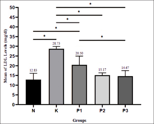
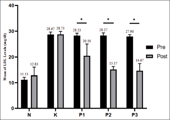
LDL levels were reexamined after the administration of kecemcem leaves extract. The extract was administered at doses of 250 mg/kgBW to group P1, 500 mg/kgBW to group P2, and 1,000 mg/kgBW to group P3. The normal (N) and control (K) groups did not receive kecemcem leaves extract. Group K had the highest LDL-C level at 28.73 mg/dl, while groups P1, P2, and P3 showed lower LDL-C levels than group K (Fig. 2). Administration of kecemcem leaves extract at doses of 250, 500, and 1,000 mg/kgBW resulted in differences in LDL levels between hyperlipidemic Wistar rats and control rats that did not receive the extract. The 250 mg/kgBW dose also showed a significant difference in LDL levels compared with the normal and 1,000 mg/kgBW treatment groups, but not with the 500 mg/kgBW group. The 500 mg/kgBW dose did not show a significant difference in LDL levels compared with the 1,000 mg/kgBW group. Additionally, the 500 and 1,000 mg/kgBW doses showed no difference in LDL levels compared with the normal group. Table 2. Qualitative analysis of the kecemcem leaves ethanol extract.
A comparison of pre-test and post-test LDL levels revealed that groups N and K, which were not given kecemcem leaves extract, showed a slight increase in LDL levels from pre-test to post-test, while groups P1, P2, and P3, which received the extract, showed a decrease in LDL levels (Fig. 3). Significant differences between pre- and post-test data were observed in groups P1, P2, and P3, whereas groups N and K did not show significant differences. These results indicate that administration of kecemcem leaves extract at doses of 250, 500, and 1,000 mg/kgBW resulted in significant changes in LDL levels in hyperlipidemic Wistar rats before and after treatment.
Fig. 1. LDL levels following 16-week administration of a high-fat diet to develop a hyperlipidemic animal model (* denotes p < 0.05). N: normal group; K: positive control group given high-fat diet; P1: treatment group 1 given high-fat diet; P2: treatment group 2 given high-fat diet; and P3: treatment group 3 given high-fat diet.
Fig. 2. LDL-C levels following 1-week administration of kecemcem leaves ethanol extract (* denotes p < 0.05). N: normal group; K: positive control group given high-fat diet; P1: treatment group 1 given (250 mg/kgBW extract); P2: treatment group 2 (500 mg/kgBW extract); and P3: treatment group 3 (1,000 mg/kgBW extract).
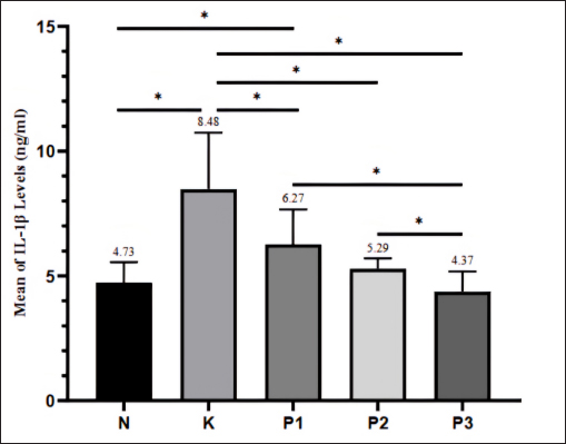
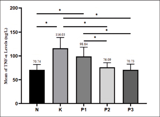
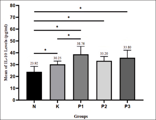
Fig. 3. LDL levels before and after the 1-week administration of kecemcem leaves ethanol extract (* denotes p < 0.05). Pre: prior to extract administration; Post: following extract administration. N: normal group; K: positive control group; P1: treatment group 1 (250 mg/kgBW extract); P2: treatment group 2 (500 mg/kgBW extract); and P3: treatment group 3 (1,000 mg/kgBW extract). Cytokines levelsIL-1β levels The results of IL-1β level examination showed that the control group (K), which did not receive kecemcem leaves extract, had the highest IL-1β level at 8.48 ng/ml. Groups P1, P2, and P3, which were given the extract at various doses, showed lower IL-1β levels compared to the control group (K) (Fig. 4). The results indicate that the administration of kecemcem leaves extract at doses of 250, 500, and 1,000 mg/kgBW led to differences in IL-1β levels in hyperlipidemic Wistar rats compared with the control group that did not receive the extract. The 250 mg/kgBW dose also showed a significant difference in IL-1β levels compared with the normal and 1,000 mg/kgBW groups, but not with the 500 mg/kgBW group. The 500 mg/kgBW dose resulted in a difference in IL-1β levels compared with the 1,000 mg/kgBW group, whereas both the 500 and 1,000 mg/kgBW doses did not show differences compared with the normal group. TNF-α levels The results of the TNF-α level examination showed that the control group (K) had the highest TNF-α level at 116.03 ng/l. Groups P1, P2, and P3, which were given the extract at various doses, showed lower TNF-α levels compared to the control group (K) (Fig. 5). The results indicate that the administration of kecemcem leaves extract at doses of 500 and 1,000 mg/kgBW led to differences in TNF-α levels in hyperlipidemic Wistar rats compared with the control group that did not receive the extract. The 250 mg/kgBW dose showed a difference in TNF-α levels compared with the normal group and the 500 and 1,000 mg/kgBW groups, but not with the control group. The 500 mg/kgBW dose did not show the difference in TNF-α levels compared with the 1,000 mg/kgBW group, whereas both the 500 mg/kgBW and 1,000 mg/kgBW doses did not show differences compared with the normal group. IL-10 levels The results of the IL-10 level examination showed that group P1, which received kecemcem leaves extract at a dose of 250 mg/kgBW, had the highest IL-10 level of 38.76 pg/ml. Groups P1, P2, and P3, which received the extract at different doses, exhibited higher IL-10 levels compared with the control group (K) (Fig. 6). The results indicate that administration of kecemcem leaves extract at doses of 250, 500, and 1,000 mg/kgBW, as well as no extract treatment, caused differences in IL-10 levels in Wistar rats with hyperlipidemia compared with the control group. However, only the 250 mg/kg BW dose showed a significant difference in IL-10 levels compared with the control group. The 500 and 1,000 mg/kgBW doses did not result in differences in IL-10 levels compared with the control or 250 mg/kgBW treatment groups. Additionally, no significant difference in IL-10 levels was observed between the 500 and 10,00-mg/kgBW doses.
Fig. 4. IL-1β levels after 1 week of administration of kecemcem leaves ethanol extract (* denotes p < 0.05) N: normal group; K: positive control group; P1: treatment group 1 (250 mg/kgBW extract); P2: treatment group 2 (500 mg/kgBW extract); and P3: treatment group 3 (1,000 mg/kgBW extract).
Fig. 5. TNF-α levels after 1 week of administration of kecemcem leaves ethanol extract (* denotes p < 0.05). N: normal group; K: positive control group; P1: treatment group 1 (250 mg/kgBW extract); P2: treatment group 2 (500 mg/kgBW extract); and P3: treatment group 3 (1,000 mg/kgBW extract).
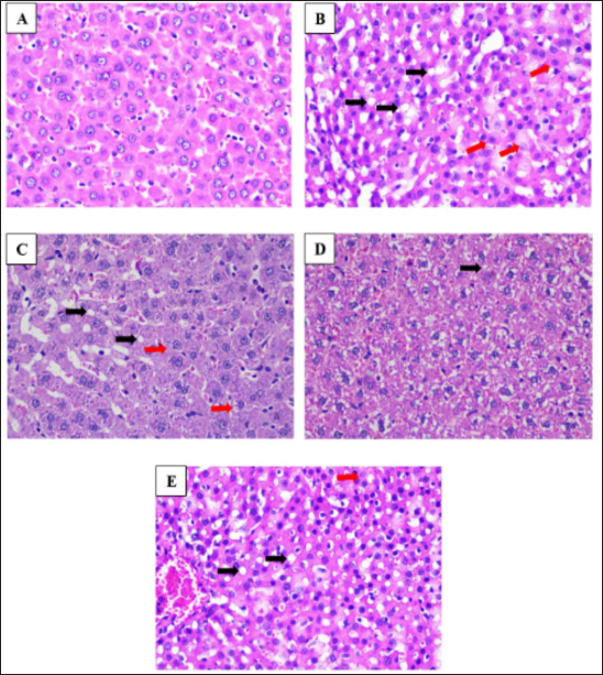
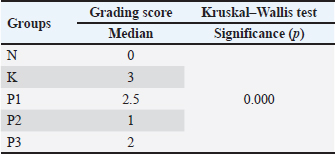
Fig. 6. IL-10 levels after 1-week of administration of kecemcem leaves ethanol extract (* denotes p < 0.05) N: normal group n; K: positive control group; P1: treatment group 1 (250 mg/kgBW extract); P2: treatment group 2 (500 mg/kgBW extract); and P3: treatment group 3 (1,000 mg/kgBW extract). Liver microscopic examinationThe microscopic observation of liver tissue preparations from rats treated with kecemcem leaves extract was performed. The preparations, stained with HE and examined under a light microscope at 100× magnification, are shown in Figure 7. According to Brunt et al. (1999) grading score for liver histopathological changes, the normal group had the lowest grading score compared with all other groups, indicating minimal liver damage (Table 3). The control group had the highest grading score, indicating the most severe liver damage. Among the treatment groups, the P2 group had the lowest grading score, closely approaching that of the normal group, suggesting that the 500 mg/kgBB dosage had the most protective effect against liver damage induced by hyperlipidemia. DiscussionThe study results demonstrated that the administration of kecemcem leaves extract at doses of 250, 500, and 1,000 mg/kgBW effectively reduced LDL levels in hyperlipidemic Wistar rats, whereas the normal and control groups, which did not receive the extract, showed no significant changes in LDL levels. The reduced LDL levels observed in the groups receiving kecemcem leaves extract may be attributed to the flavonoid compounds present in the leaves. Flavonoids play a crucial role in lipid metabolism by inhibiting the enzyme 3-hydroxy-3-methylglutaryl coenzyme A (HMG-CoA) reductase, which is a key enzyme in cholesterol biosynthesis, thereby helping to lower LDL levels. This enzyme converts HMG-CoA into mevalonate. Inhibiting this enzyme decreases mevalonate production, which in turn reduces cholesterol production in the liver. This reduction in cholesterol activates SREBP, a process involving the translocation of SREBP from the endoplasmic reticulum to the Golgi, where it is processed by protease enzymes into its active form. The active SREBP then moves to the nucleus and binds to sterol regulatory elements on the DNA, enhancing the transcription of genes encoding LDL receptors (LDLR). This increased transcription results in the production of more LDLR by cells. These newly produced LDLRs are then transported to the surface of liver cells, where they capture more LDL particles from the bloodstream, thereby lowering blood LDL cholesterol levels. Flavonoids also inhibit the acyl-CoA cholesterol acyl transferase in HepG2 cells, reducing cholesterol esterification in the liver and decreasing cholesterol ester levels. Cholesterol esters are essential components of very low density lipoprotein (VLDL) formation, thereby reducing their production. Additionally, flavonoids decrease Apo B100 secretion, a component of VLDL, and reduce the activity of microsomal triglyceride transfer protein. Consequently, this reduction in VLDL synthesis leads to lower LDL levels (Zeka et al., 2017; Hijriani et al., 2023).
Fig. 7. Microscopic examination of liver tissue at 100× magnification. Black arrows denote steatosis, and red arrows denote ballooning. A: N (normal group); B: K (positive control group); C: P1 treatment group 1 (250 mg/kgBW extract); D: P2 treatment group 2 (500 mg/kgBW extract); and E: P3 treatment group 3 (1,000 mg/kgBW extract). Table 3. Grading score results of liver microscopic features after 1-week administration of kecemcem leaves extract.
The active flavonoid compounds found in kecemcem include quercetin and catechins. Catechins have been shown to reduce lipid droplet accumulation in the liver, prevent fat buildup, and significantly decrease total cholesterol and serum LDL levels. Catechins lower cholesterol by inhibiting HMG-CoA synthesis (Wan et al., 2024). Other studies have also indicated that catechins can reduce lipid peroxidation, inhibit pro-inflammatory cytokines, lower aspartate aminotransferase, and alanine aminotransferase levels, and protect the liver from excessive fat accumulation. The antioxidant properties of quercetin and catechins help prevent cholesterol elevation by inhibiting LDL oxidation, thereby reducing oxidized LDL (oxLDL) levels. Inhibiting LDL oxidation protects blood vessel walls by preventing oxLDL formation, reducing endothelial cell damage, decreasing monocyte/macrophage recruitment and activation, lowering foam cell formation, reducing pro-inflammatory cytokines secretion, minimizing oxidative stress, and enhancing atherosclerotic plaque stability. Collectively, these mechanisms contribute to reducing the risk of atherosclerosis and cardiovascular complications (Agusmansyah, 2021; Hijriani et al., 2023). Hyperlipidemia caused by a high-fat diet can trigger inflammation, as indicated by increased IL-1β and TNF-α levels. A stronger inflammatory response is correlated with higher IL-1β and TNF-α levels. Hyperlipidemia leads to cholesterol accumulation in the endothelial cells of arterial walls, initiating an inflammatory response and increasing ROS. ROS oxidizes LDL, leading to lipid peroxidation and the formation of oxLDL. OxLDL is recognized by LOX-1 and toll-like receptor (TLR) 4 (TLR4) receptors, activating intracellular signaling pathways like MAPK and PI3K/Akt, which in turn activates nuclear factor-kappa B (NF-κB). NF-κB induces the transcription of pro-inflammatory genes like IL-1β and TNF-α. ROS also directly activates MAPK pathways, further contributing to NF-κB activation (Hermawan and Hastuti, 2019). The results of this study showed that administration of kecemcem leaves extract at doses of 250, 500, and 1,000 mg/kgBW significantly affected IL-1β levels compared with the control group that did not receive the extract. However, only the 500 and 1,000 mg/kgBW doses significantly affected TNF-α levels compared with the control. The IL-1β and TNF-α levels in the groups receiving kecemcem leaves extract than in the control group. These findings are attributed to the anti-inflammatory effects of the flavonoid compounds quercetin and catechin present in the kecemcem leaves extract. Quercetin is a dietary polyphenol and bioactive flavonoid known to inhibit oxidative stress, inflammation, hypercholesterolemia, and atherosclerosis. Quercetin has been shown to strongly inhibit IL-1β and TNF-α secretion triggered by cholesterol crystals in macrophages (Vahdat-Lasemi et al., 2021). The anti-inflammatory effects of flavonoids mainly occur through the inhibition of the NF-κB pathway. Quercetin exerts its anti-inflammatory activity by inhibiting extracellular signal-regulated JNK, which subsequently blocks MAPK, AP-1, and NF-κB activity. Catechin, another type of flavonoid, also inhibits MAPK, AP-1, and NF-κB by blocking c-Jun N-terminal kinase and p38 kinase. These findings suggest that flavonoids target the MAPK pathway and AP-1 transcription factor to reduce inflammatory responses. Regarding inflammation, quercetin reduces NF-κB protein expression, leading to decreased secretion of pro-inflammatory cytokines, such as IL-1β and TNF-α (Ribeiro et al., 2015; Zheng et al. 2016). Flavonoids also exhibit anti-inflammatory activity because of their strong antioxidant properties, which reduce free radicals in the body. They inhibit the expression of inducible nitric oxide (NO) synthase and the production of NO. NO contributes to the formation of peroxynitrite, a highly damaging compound, under oxidative stress conditions. LDL oxidation is often induced by peroxynitrite, formed through the interaction of NO with superoxide ions produced by activated immune cells, such as macrophages. Therefore, flavonoids can inhibit LDL oxidation, reducing the formation of oxLDL, which acts as a danger-associated molecular pattern to activate TLR-4 (Al-Khayri et al., 2022). A high-fat diet elevates free fatty acid (FFA) levels, which can increase pro-inflammatory cytokine levels and reduce anti-inflammatory cytokines. High FFA levels also enhance TLR expression in macrophages, activating M1 macrophages that release TNF-α, IL-1, and MCP-1 while reducing IL-10 levels. However, this study found higher IL-10 levels in hyperlipidemic rats than in normal controls, which is consistent with the findings of Malik et al. (2020), which suggests a compensatory effect of adipose tissue. During acute inflammation, the release of pro-inflammatory cytokines is followed by anti-inflammatory responses, such as IL-10, to limit TNF-α effects. Although the body has natural mechanisms to increase IL-10 levels in response to inflammation, therapeutic interventions could provide additional benefits by reducing excessive inflammation and mitigating hyperlipidemia-related complications. The study analyzed the effect of kecemcem leaves extract on IL-10 levels in hyperlipidemic Wistar rats. The 250 mg/kgBW dose significantly increased IL-10 levels compared with the control group, whereas the 500 and 1,000 mg/kgBW doses did not show significant differences. The increase in IL-10 levels can be explained by two mechanisms. First, the flavonoid compounds in kecemcem leaves extract directly increase IL-10 production through modulation of immune signaling pathways, inhibition of pro-inflammatory cytokines, reduction of oxidative stress, and macrophage polarization. Quercetin, a flavonoid in kecemcem leaves extract, improves hyperlipidemia, inflammatory cytokines, and oxidative stress through the AMP-activated protein kinase (AMPK)/Sirtuin 1 (SIRT1)/NF-κB pathway. AMPK reduces inflammation and endoplasmic reticulum stress caused by oxLDL, improving endothelial dysfunction and atherosclerosis. Quercetin enhances AMPK phosphorylation and increases SIRT1 expression and activity, a key protein in anti-atherosclerosis effects that regulates endothelial and vascular function. AMPK activation also suppresses NF-κB, IL-1β, and TNF-α expression while increasing IL-10 levels. Thus, the modulation of the AMPK/SIRT1/NF-κB pathway by quercetin underlies its anti-hyperlipidemia, anti-inflammatory, and antioxidant effects against atherosclerosis (Zhang et al., 2020). This explains the study’s finding that the IL-10 levels were higher in the groups receiving kecemcem leaves extract than in the control group. Second, the increase in pro-inflammatory cytokine levels stimulates IL-10 production as part of a feedback regulation mechanism to control inflammation and prevent tissue damage in the context of hyperlipidemia-induced inflammation. In inflammatory conditions such as hyperlipidemia, the body naturally tries to balance the inflammatory response by increasing anti-inflammatory cytokine production, such as IL-10, which can be triggered by elevated pro-inflammatory cytokines. Pro-inflammatory cytokines like IL-1β and TNF-α can stimulate IL-10 production as a protective mechanism to prevent tissue damage caused by uncontrolled inflammation. This may explain the study’s finding that the IL-10 levels in the 250 mg/kgBW group were higher than those in the 500 and 1,000 mg/kgBW groups, as the pro-inflammatory cytokine IL-1β levels were higher in the 250 mg/kgBW group than in the 500 and 1,000 mg/kgBW groups. Both mechanisms synergistically increase IL-10 levels, thereby balancing the immune response and reducing inflammation in hyperlipidemia. The administration of kecemcem leaves extract at doses of 250, 500, and 1,000 mg/kgBW demonstrated that the 250 mg/kgBW dose was more effective in increasing IL-10 levels compared to the control group. A high-fat diet affects liver lipid metabolism by increasing triglyceride lipolysis in adipocytes, releasing more FFA into circulation. Excess FFAs are delivered to the liver, where they are either oxidized by mitochondria or stored as triglycerides, leading to fat accumulation and fatty liver (Zhang et al., 2020). After treatment with kecemcem leaves extract at different doses, there was a noticeable reduction in hepatocyte steatosis and ballooning in the 250 mg/kgBW group (P1), compared with the control group, although it was still significantly different from the normal group. The 500 mg/kgBW group (P2) showed fewer hepatocytes with fat accumulation and ballooning, with results similar to those of the normal group, indicating better recovery. The 1,000 mg/kgBW group (P3) also showed improvement, but it was not significantly better than the 500 mg/kgBW group. Kecemcem leaves extract exerts its effects through flavonoids like quercetin and catechins, which reduce liver fat accumulation by inhibiting lipogenesis (Wardhani, 2020). Quercetin influences enzymes like acetyl-CoA carboxylase and fatty acid synthase, which are involved in fat synthesis, while enhancing beta-oxidation of fatty acids and reducing triglyceride buildup. Additionally, quercetin boosts LPL enzyme activity, converting VLDL to IDL, reducing VLDL accumulation in the liver, and preventing steatosis (Hastuti et al., 2021). Both quercetin and catechins have anti-inflammatory and antioxidant properties, reducing oxidative stress and blocking NF-κB transcription factors, thereby lowering proinflammatory cytokines involved in NAFLD pathogenesis. Based on the results of this study, it can be concluded that the administration of kecemcem leaves extract at doses of 250, 500, and 1,000 mg/kgBW effectively reduced LDL levels, pro-inflammatory cytokines (IL-1β and TNF-α), and improved liver histology in hyperlipidemic rats. These effects are attributed to the flavonoid compounds in the extract, particularly quercetin, and catechins, which inhibit cholesterol synthesis, reduce oxidative stress, and decrease liver fat accumulation. Additionally, the extract promoted an anti-inflammatory response by increasing IL-10 levels, especially at the 250 mg/kg BW dose, which contributed to improved liver health and reduced inflammation. This study supports the potential of kecemcem as a natural therapy for managing hyperlipidemia and its associated complications. ConclusionKecemcem leaves extract reduced LDL, IL-1β, and TNF-α levels, increased IL-10 levels, and improved liver histology. These effects were supposedly due to the flavonoid content of quercetin and catechin. Kecemcem leaves extract exhibits promising therapeutic effects for managing conditions related to hyperlipidemia and inflammation. AcknowledgmentsThe authors would like to express their gratitude to the Lembaga Penelitian dan Pengabdian Masyarakat Airlangga University for the financial support in this research through the Airlangga Research Fund 2024, as well as to the laboratory staff at the Experimental Animal Unit, Department of Physiology and Medical Biochemistry, Faculty of Medicine, Airlangga University who have assisted during the research process. Conflicts of interestThe authors declare that they have no conflicts of interest. FundingThis research received financial support from Lembaga Penelitian dan Pengabdian Masyarakat Airlangga University through the Airlangga Research Fund 2024, under grant number: 747/UN3.SPS/PT.01.03/2024. Author’s contributionLCS, NDRL, TIB, Y, NLAM, and IPJ determined the concept and method of the research. LCS and IPJ prepared reagents, materials, and extracts until the extract testing. LCS, IPJ, and NDRL provided treatment to the animal model. LCS and Y checked LDL levels. CSF and NLAM checked cytokine levels. IPJ and TIB conducted a microscopic examination of the liver. LCS, TIB, and IPJ performed data analysis and interpretation. LCS, NDRL, TIB, Y, NLAM, and IPJ contributed to drafting, revising, and finalizing the manuscript. All authors have read and approved the manuscript for publication. Data availabilityAll research data are available in the manuscript. ReferencesAgusmansyah, S. 2021. Syzygium polyanthum extract as a therapy for dyslipidemia. OAIJMR 1(5), 111–115. Al-Khayri, J.M., Sahana, G.R., Nagella, P., Joseph, B.V., Alessa, F.M. and Al-Mssallem, M.Q. 2022. Flavonoids as potential anti-inflammatory molecules: a review. Molecules 27(9), 2901. Alloubani, A., Nimer, R. and Samara, R. 2021. Relationship between hyperlipidemia, cardiovascular disease and stroke: a systematic review. Curr. Cardiol. Rev. 17(6), e051121189015. Aman, A.M., Soewondo, P., Soelistijo, S.A., Arsana, P.M., Wismandari, Zufry, H. and Rosandi, R. 2019. Pedoman Pengelolaan Dislipidemia Di Indonesia 2019. Jakarta: PB PPERKENI. Anjani, D.A.V.N. 2023. Non-alcoholic fatty liver disease: diagnosis and treatment. J. Biol. Tropis. 23(3), 213–224. Aryasa, I.W.T., Artini, N.P.R. and Juliari, P.G.A.E. 2021. Uji Nilai Gizi Dan Kapasitas Antioksidan Pada Loloh Tanaman Cemcem (Spondias Pinnata (L.F) Kurz.) Daerah Desa Bebalang, Kecamatan Bangli, Kabupaten Bangli, Bali. Stannum: Jurnal Sains dan Terapan Kimia. 15(2), 133–142. Azizah, S. and Nursamsiar, Nur, S. 2019. Uji Aktivitas Antioksidan Ekstrak Etanol Daun Kedondong Hutan (Spondias pinnata (L.F.) Kurz.) Dengan Berbagai Metode Uji. JIM 5(1), 91–96. Brunt, E.M., Janney, C.G., Di Bisceglie, A.M., Neuschwander-Tetri, B.A. and Bacon, B.R. 1999. Nonalcoholic steatohepatitis: a proposal for grading and staging the histological lesions. Am J Gastroenterol 94, 2467–2474. Dewi, A.C., Wiryanthini, I.A.D. and Surudarma, I.W. 2022. Pengaruh Konsumsi Loloh Cemcem Terhadap Kadar Kolesterol Total Pada Mencit (Mus Musculus L.) Dengan Diet Tinggi Lemak. E-J. Medika Udayana. 11(2), 98–102. Evayana, E. and Aminah, S. 2022. Determination of total levels of white turmeric flavonoids (Curcuma zedoria Rosc.) with variation of solvent types. Media Eksakta. 18, 1–5. Hastuti, P., Martantiningtyas, D.C. and Beandrade, M.U. 2021. Lipoprotein, apolipoprotein, dan Sindrom Metabolik. Depok, Indonesia: UGM Press. Hayudanti, D., Kusumastuty, I. and Tritisari, K.P. 2016. Pengaruh Pemberian Jus Jambu Biji Merah (Psidium guajava) dan Jeruk Siam (Citrus nobilis) terhadap Kadar High Density Lipoprotein (HDL) pada Pasien Dislipidemia (The Effect of Guava Extract (Psidium guajava) and Siam Citrus Fruit (Citrus nobilis) on HDL level in dyslipidemic patients). IJHN 3(1), 41–48. Hermawan, D. and Hastuti, P. 2019. High fiber diet decreases the level of interleukin 1. KnE Life Sci. 4(12), 30–35. Hijriani, B.I., Atfal, B., Kodariah, L., Hadiatun, N. and Ismatullah, N.K. 2023. Efektivitas Ekstrak Daun Salam (Syzygium polyanthum) Dalam Mencegah Kenaikan Kadar Kolesterol LDLTikus Putih (Rattus norvegicus) Diinduksi Kuning Telur Puyuh. Jurnal Kesehatan Rajawali. 13(2), 1–4. Hong, N., Lin, Y., Ye, Z., Yang, C., Huang, Y., Duan, Q. and Xie, S. 2022. The relationship between dyslipidemia and inflammation among adults in east coast China: a cross-sectional study. Front. Immunol. 13, 937201. Kancherla, N., Dhakshinamoothi, A., Chitra, K. and Komaram, R.B. 2019. Preliminary analysis of phytoconstituents and evaluation of anthelminthic property of Cayratia auriculata (In vitro). Maedica (Bucur) 14(4), 350–356. Karr, S. 2017. Epidemiology and management of hyperlipidemia. Am. J. Manag. Care. 23, S139–S148. Khoirunnisa, L. and Zakia, N. 2024. Analisis flavonoid batang brotowali sebagai antioksidan dan uji aktivitasnya dengan metode DPPH. Ar-Razi J. Ilm. 12(2), 73–83. Laksemi, D.A.A.S. 2019. Biological activity of Spondias pinnata: a review. Indones. J. Biomed. Sci. 13(2), 88–93. Malik, R., Sadewa, A.H. and Sunarti, S. 2020. High fiber diets enhance gene expression and Il-10 level on hyperlipidemic rats model. Curr. Res. Nutr. Food Sci. 8(2), 471–478. Megasari, N.L.A., Utsumi, T., Yamani, L.N., Juniastuti, Gunawan, E., Furukawa, K., Nishimura, M., Lusida, M.I. and Mori, Y. 2021. Seroepidemiological study of SARS-CoV-2 infection in East Java, Indonesia. PLoS One 16(5), e0251234. Nassir, F., Rector, R.S., Hammoud, G.M. and Ibdah, J.A. 2015. Pathogenesis and prevention of hepatic steatosis. Gastroenterol. Hepatol. (N Y). 11(3), 167–175. Nurjannah, I., Mustariani, B.A.A. and Suryani, N. 2022. Skrining Fitokimia dan Uji Antibakteri Ekstrak Kombinasi Daun Jeruk Purut (Citrus hystrix) dan Kelor (Moringa oleifera L.) Sebagai Zat Aktif pada Sabun Antibakteri. SPIN-J Kimia Pendidikan Kimia. 4(1), 23–36. Rahmawaty, A., Cahyani, F.R., Safitri, N., Sitepu, A.A.N.C., Hapitria, E.N. and Megantara, S. 2022. Uji In Silico Kandungan Senyawa Tanaman Anggur (Vitis vinifera L.) Untuk Kandidat Obat Anti Hiperlipidemia. Majalah Farmasi Dan Farmakologi. 26(2), 57–62. Raju, N.J., Pawar, V.S. and Vishala T.C. 2017. Anti hyperlipidemic activity of Spondias pinnata fruit extracts. IJPSDR 9(4), 178–181. Ribeiro, D., Freitas, M., Tomé, S.M., Silva, A.M.S., Laufer, S., Lima, J.L.F.C. and Fernandes. E. 2015. Flavonoids inhibit COX-1 and COX-2 enzymes and cytokine/Chemokine production in human whole blood. Inflammation 38(2), 858–870. Sarwanti, Stephanie, M. and Kodariah, R. 2020. Peran CD44 pada progresivitas non alcoholic steatohepatitis (NASH). MPI 29(2), 71–81. Vahdat-Lasemi, F., Aghaee-Bakhtiari, S.H., Tasbandi, A., Jaafari, M.R. and Sahebkar, A. 2021. Targeting interleukin-β by plant-derived natural products: implications for the treatment of atherosclerotic cardiovascular disease. PTR 35(10), 5596–5622. Wan, Y., Ma, D., Yu, L., Tian, W., Wang, T., Chen, X., Shang, Q. and Xu, H. 2024. The associations between dietary flavonoid intake and hyperlipidemia: data from the national health and nutrition examination survey 2007–2010 and 2017–2018. Front. Nutr. 11, 1374970. Wardhani, T.M. 2020. Pemanfaatan Tanaman Kelor (Moringa oleifera Lam.) sebagai Sumber Terapi Preventif dan Kuratif pada Pasien Perlemakan Hepar dengan Sindrom Metabolik. Dentika Dental J. 1(2), 12. Zaidan, S., Abdillah, S., Arfian, N. and Arozal, W. 2024. Hypolipidemic effect of brown seaweed (Sargassum crassifolium) extract in vivo (Study of histopathology, mRNA expression, and immunohistochemistry (IHC) with VCAM-1, ICAM-1, and MCP-1 parameters). ArXiv [Preprint] Feb 12, arXiv:2402.07497v1. Zeka, K., Ruparelia, K., Arroo, R.R.J., Budriesi, R. and Micucci, M. 2017. Flavonoids and their metabolites: prevention in cardiovascular diseases and diabetes. Diseases 5(3), 19. Zhang, F., Feng, J., Zhang, J., Kang, X. and Qian, D. 2020. Quercetin modulates AMPK/SIRT1/NF-κB signaling to inhibit inflammatory/oxidative stress responses in diabetic high fat diet-induced atherosclerosis in the rat carotid artery. Exp. Ther. Med. 20(6), 280. Zheng, J., Wu, J., Chen, J., Liu, J., Lu, Y., Huang, C., Hu, G., Wang, X. and Zeng, Y. 2016. Therapeutic effects of quercetin on early inflammation in hypertriglyceridemia-related acute pancreatitis and its mechanism. Pancreatology 16(2), 200–210. | ||
| How to Cite this Article |
| Pubmed Style Sari LC, Lastuti NDR, Budhy TI, Yuliasih Y, Megasari NLA, Jamaluddin IP. Therapeutic effects of kecemcem leaves extract (Spondias pinnata (L.f.) Kurz) on LDL levels, cytokines, and microscopic liver changes in hyperlipidemic rat model. Open Vet. J.. 2025; 15(3): 1310-1321. doi:10.5455/OVJ.2025.v15.i3.22 Web Style Sari LC, Lastuti NDR, Budhy TI, Yuliasih Y, Megasari NLA, Jamaluddin IP. Therapeutic effects of kecemcem leaves extract (Spondias pinnata (L.f.) Kurz) on LDL levels, cytokines, and microscopic liver changes in hyperlipidemic rat model. https://www.openveterinaryjournal.com/?mno=231802 [Access: January 25, 2026]. doi:10.5455/OVJ.2025.v15.i3.22 AMA (American Medical Association) Style Sari LC, Lastuti NDR, Budhy TI, Yuliasih Y, Megasari NLA, Jamaluddin IP. Therapeutic effects of kecemcem leaves extract (Spondias pinnata (L.f.) Kurz) on LDL levels, cytokines, and microscopic liver changes in hyperlipidemic rat model. Open Vet. J.. 2025; 15(3): 1310-1321. doi:10.5455/OVJ.2025.v15.i3.22 Vancouver/ICMJE Style Sari LC, Lastuti NDR, Budhy TI, Yuliasih Y, Megasari NLA, Jamaluddin IP. Therapeutic effects of kecemcem leaves extract (Spondias pinnata (L.f.) Kurz) on LDL levels, cytokines, and microscopic liver changes in hyperlipidemic rat model. Open Vet. J.. (2025), [cited January 25, 2026]; 15(3): 1310-1321. doi:10.5455/OVJ.2025.v15.i3.22 Harvard Style Sari, L. C., Lastuti, . N. D. R., Budhy, . T. I., Yuliasih, . Y., Megasari, . N. L. A. & Jamaluddin, . I. P. (2025) Therapeutic effects of kecemcem leaves extract (Spondias pinnata (L.f.) Kurz) on LDL levels, cytokines, and microscopic liver changes in hyperlipidemic rat model. Open Vet. J., 15 (3), 1310-1321. doi:10.5455/OVJ.2025.v15.i3.22 Turabian Style Sari, Lia Cahya, Nunuk Dyah Retno Lastuti, Theresia Indah Budhy, Yuliasih Yuliasih, Ni Luh Ayu Megasari, and Indra Prastiwi Jamaluddin. 2025. Therapeutic effects of kecemcem leaves extract (Spondias pinnata (L.f.) Kurz) on LDL levels, cytokines, and microscopic liver changes in hyperlipidemic rat model. Open Veterinary Journal, 15 (3), 1310-1321. doi:10.5455/OVJ.2025.v15.i3.22 Chicago Style Sari, Lia Cahya, Nunuk Dyah Retno Lastuti, Theresia Indah Budhy, Yuliasih Yuliasih, Ni Luh Ayu Megasari, and Indra Prastiwi Jamaluddin. "Therapeutic effects of kecemcem leaves extract (Spondias pinnata (L.f.) Kurz) on LDL levels, cytokines, and microscopic liver changes in hyperlipidemic rat model." Open Veterinary Journal 15 (2025), 1310-1321. doi:10.5455/OVJ.2025.v15.i3.22 MLA (The Modern Language Association) Style Sari, Lia Cahya, Nunuk Dyah Retno Lastuti, Theresia Indah Budhy, Yuliasih Yuliasih, Ni Luh Ayu Megasari, and Indra Prastiwi Jamaluddin. "Therapeutic effects of kecemcem leaves extract (Spondias pinnata (L.f.) Kurz) on LDL levels, cytokines, and microscopic liver changes in hyperlipidemic rat model." Open Veterinary Journal 15.3 (2025), 1310-1321. Print. doi:10.5455/OVJ.2025.v15.i3.22 APA (American Psychological Association) Style Sari, L. C., Lastuti, . N. D. R., Budhy, . T. I., Yuliasih, . Y., Megasari, . N. L. A. & Jamaluddin, . I. P. (2025) Therapeutic effects of kecemcem leaves extract (Spondias pinnata (L.f.) Kurz) on LDL levels, cytokines, and microscopic liver changes in hyperlipidemic rat model. Open Veterinary Journal, 15 (3), 1310-1321. doi:10.5455/OVJ.2025.v15.i3.22 |

