


| Review Article | ||
Open Vet. J.. 2025; 15(6): 2286-2297 Open Veterinary Journal, (2025), Vol. 15(6): 2286-2297 Review Article Pathogen-driven pregnancy loss in dairy cattle: An overview of diagnostic advances and innovationsNina Herlina1,2, Safika3, Ekowati Handharyani4 and Agus Setiyono4*1Animal Biomedical Study Program, IPB Postgraduate School, School of Veterinary Medicine and Biomedicine, IPB University, Bogor, Indonesia 2Center for Applied Zoology, Research Organization for Life Sciences, National Research and Innovation Agency (BRIN), Cibinong Science Center, Bogor, Indonesia 3Division of Microbiology, School of Veterinary Medicine and Biomedicine, IPB University, Bogor, Indonesia 4Division of Pathology, School of Veterinary Medicine and Biomedicine, IPB University, Bogor, Indonesia *Corresponding Author: Agus Setiyono. Division of Pathology, School of Veterinary Medicine and Biomedical Sciences, IPB University, Bogor, Indonesia. Email: agusse [at] apps.ipb.ac.id Submitted: 06/01/2025 Revised: 10/05/2025 Accepted: 20/05/2025 Published: 30/06/2025 © 2025 Open Veterinary Journal
AbstractPregnancy failure or abortion in dairy cattle is a major reproductive concern that affects animal welfare and the dairy industry’s economic stability. Pathogens are a key factor contributing to this issue. Accurate diagnosis is essential for effective management and preventive measures. This review discusses various pathogens responsible for abortion in dairy cattle, including bacteria, viruses, and protozoa. Conventional diagnostic methods, which are important, may not always comprehensively detect all pathogens involved in abortion. This study focuses on current advances in diagnostic technology, particularly molecular approaches such as polymerase chain reaction and enzyme-linked immunosorbent assays, as well as the promise of metagenomic techniques for increased accuracy. By integrating recent research findings, this review emphasizes the significance of these advancements in enhancing disease management, prevention measures, and overall herd health. Keywords: Abortion, Dairy cattle, Diagnostic, Metagenomic, Molecular. IntroductionPregnancy loss in dairy cattle, which occurs as abortions between 42 and 260 days of gestation, poses a major challenge for the dairy industry (Mee, 2020). Up to 20% of pregnancies are affected by this problem, which causes significant financial losses for the dairy industry (Albuja et al., 2019). Abortions in dairy cows result in higher expenses for reproduction, veterinary services, and feed while adversely impacting milk yield, fertility, and overall health, leading to increased culling and turnover rates (Keshavarzi et al., 2020). Abortion rates typically range from 1% to 2%. Finding the root causes requires careful consideration if this rate rises above 2%–5%. To investigate causes, potential remedies, and preventive actions, more assessment is necessary (Van Loo et al., 2024). According to Serrano-Martínez et al. (2019), abortions may be caused by infectious agents, factors of unknown origin, or noninfectious causes. Abortion in cattle is caused by noninfectious conditions, such as hormonal imbalances, metabolic abnormalities, heat stress, physical damage, and malnutrition. These factors may interfere with a healthy pregnancy and result in fetal death (Khan et al., 2023). Other factors linked to abortion are the consumption of toxic plants, exposure to mycotoxins, and hormonal fluctuations due to progesterone deficiencies or the use of glucocorticoids, oxytocin, estrogen, and prostaglandins. Nutritional insufficiencies, genetic factors, chromosomal disorders, umbilical torsion, and twin pregnancies can also contribute (Yadav et al., 2021). Toxins from certain plants, such as Astragalus species and Ponderosa pine needles, along with estrogenic mycotoxins can further lead to abortion incidents (Buroni et al., 2020). Various infectious agents, including bacteria, viruses, protozoa, and fungi, have been implicated in bovine abortion. Many factors, particularly infectious agents, contribute to pregnancy loss and perinatal deaths in cattle. Girmay et al. (2024) found that the seroprevalence of bovine viral diarrhea virus (BVDV) and BHV1 was higher in dairy cows with reproductive problems in Ethiopia than in Neospora caninum and Chlamydia burnetii. A number of agents associated with cattle reproductive failure, including Salmonella enterica, Chlamydia abortus, Listeria monocytogenes, Brucella species, Leptospira species, C. burnetii, C. abortus, S. enterica, Campylobacter species, and Yersinia enterocolitica, pose zoonotic risks (Ebani, 2022). These pathogens are therefore crucial to public health and animal husbandry. Using the One Health concept, cooperation between veterinary and medical specialists is crucial for successfully reducing illnesses linked to fertility (Sevik, 2025). However, despite improvements in diagnostic methods, the identification of the causes of reproductive losses has not surpassed 50% accuracy and remains stagnant over time. The dairy industry faces economic losses due to expected calf deaths and resulting delays in milk production, which are vital to farm profitability. Identifying these pathogens quickly and accurately is essential for effective management and control. Laboratories performing abortion diagnostic tests should include standard assessments for the main infectious diseases linked to abortion. Prioritizing the most prevalent abortifacient drugs is crucial for efficient disease management and control, even when financial limitations may restrict the range of tests. Traditional diagnostic techniques are crucial, but they usually overlook different causal agents, emphasizing the need for improvements in diagnostic technologies. Pathogens Involved in Bovine AbortionWorldwide studies indicate that N. caninum (22.2%) is the most common pathogen. Meanwhile, C. burnetii was found at the lowest percentage, at 9.5%. The percentages of opportunistic bacteria and the Chlamydiaceae family were in the middle, with 10.9% and 21.4%, respectively (Hecker et al., 2023). Vidal et al. (2017a,b) investigated the prevalence of zoonotic infections in cases of bovine abortion in Switzerland. According to the findings, Coxiella burnetii was found in 15.9% of cases, C. abortus in 38.5%, and Leptospira species in 21.4%. The study revealed that diagnostic tools, including real-time polymerase chain reaction (PCR) and histological examinations, should be utilized to enhance the specific identification of bovine abortion instances and accurately determine the prevalence of these zoonotic infections. Pathogens such as bovine herpesvirus type 1 (BoHV-1) and bovine viral diarrhea (BVD) occur at rates of approximately 5%. Intermittently isolated or opportunistic pathogens, such as Escherichia coli, Salmonella spp., Staphylococcus spp., Pasteurella spp., Pseudomonas spp., Trueperella pyogenes, Streptococcus spp., Bacillus licheniformis, Enterobacter spp., Pajaroellobacter abortibovis, Klebsiella spp., Acinetobacter spp., Histophilus somni, Bordetella spp., Actinomyces spp., Yersinia paratuberculosis, Aeromonas spp., and Cardiobacterium spp., were observed in 21.4% of cases (Hecker et al., 2023). Table 1 shows the list of pathogenic microorganisms causing abortion in dairy cattle in the past 5 years (2019–2024). Neospora caninumDubey et al. (1988) reported that N. caninum is a parasitic organism from the Apicomplexa family that primarily affects dogs but also a variety of animals, especially livestock. The parasite has a life cycle involving definitive hosts, mainly dogs and other canids, and intermediate hosts such as cattle. Dog waste can contain oocysts and can contaminate food, water, and the surroundings. Cattle become infected by consuming these oocysts. Vertical transmission from mother to fetus is also crucial, as this facilitates the spread of infection within herds (Dubey, 2003). The protozoan parasite N. caninum plays an important role in abortion in cattle. Infection can occur through horizontal transmission via the ingestion of oocysts expelled by canids or vertically, from an infected mother to her fetus, potentially leading to abortion, stillbirth, or the birth of offspring that are subclinically infected (Gliga et al., 2025). In dairy cattle, abortion is the main symptom, often occurring in the second trimester. Infected cows may also experience stillbirths, premature births, and weak calves (Buxton et al., 2002). N. caninum is the leading abortifacient agent in both dairy and beef cattle. A 2023 study in Egypt highlighted the substantial role of N. caninum in causing abortions among dairy cattle, emphasizing the parasite’s contribution to economic losses in the livestock industry (Selim et al., 2023). Infected cows may not show symptoms, but their fetuses can be infected and may experience paralysis or sensory deficits. Diagnosing N. caninum in dairy cattle employs various methods. PCR as a molecular approach in identified DNA of N. caninum in various tissues and fluids provides fast and precise identification (Buxton et al., 2002). Moreover, histopathological analysis of aborted fetuses can detect specific lesions, including nonsuppurative encephalitis and myocarditis, and help identify the parasite in tissues (Dubey and Lindsay, 1996). Various serological techniques, such as the indirect fluorescent antibody test (IFAT), enzyme-linked immunosorbent assays (ELISA), immunoblotting, and direct agglutination tests, are available for detecting specific antibodies against N. caninum in cattle (Paré et al., 1995; Dubey and Schares, 2006; Selim et al., 2023). Udonsom et al. (2024) evaluated the effectiveness of three N. caninum proteins as biomarkers for immune-based diagnosis for detecting IgG antibodies against N. caninum compared with the IFAT as a reference method for identifying N. caninum antibodies. The present study emphasizes NcSAG1 as a valuable antigen marker, whereas NcPrx2 and NcMIC4 show promise for immunodiagnostic applications in detecting N. caninum infections in field collections. Chlamydia abortusChlamydia abortus is an intracellular bacterium that leads to significant abortion rates in dairy cattle, resulting in major economic losses for the dairy sector (Longbottom and Coulter, 2003). This pathogen can also infect humans, posing health risks. It is associated with pregnancy losses in cattle, particularly in Argentina. Chlamydia abortus is primarily transmitted through the ingesting or inhalation of contaminated materials such as aborted fetuses, placental tissues, and vaginal secretions. It may also be transferred vertically from the mother to the fetus (Wheelhouse and Longbottom, 2015). Dairy cattle, along with other ruminants such as sheep and goats, are key reservoirs for this bacterium. Infected animals shed the bacteria, contaminating their surroundings and spreading the infection among herds (Borel et al., 2018). The most common clinical symptom in dairy cattle is abortion, which usually occurs in the latter trimester. Other reproductive issues include stillbirth, weak calves, and infertility (Longbottom and Coulter, 2003). Infected cows might show symptoms of systemic illness such as fever and lethargy, although many cases can be asymptomatic, complicating detection and management (Wheelhouse and Longbottom, 2015). Diagnosis of C. abortus in dairy cattle uses various methods. Serological tests, such as the complement fixation test (CFT) and ELISA, help identify antibodies against bacteria, revealing both acute and chronic infections (Sachse et al., 2009). Molecular techniques such as PCR frequently help to identify C. abortus DNA in samples such as placental tissues, vaginal swabs, and aborted fetuses, providing a quick and precise diagnostic method (Pantchev et al., 2009; Sachse et al., 2009). Although it is feasible to isolate C. abortus from clinical samples, this is difficult because of the bacterium’s intracellular nature and the need for specialized lab facilities (Borel et al., 2018). Pinto et al. (2025) identified 2.2% positivity for the Chlamydiaceae family in pregnancy loss of ruminants using real-time PCR (qPCR). Table 1. List of pathogens causing abortion in dairy cattle in the past 5 years (2019–2024).
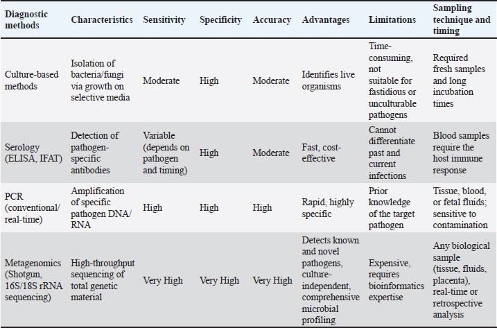
Brucella abortusBrucella abortus is a highly contagious bacteria classified as a Gram-negative coccobacillus that results in a zoonotic disease, brucellosis, which affects animals and public health (Wang et al., 2024). The disease spreads substantially through direct contact with infected creatures or their concealment, including abandoned fetuses, placental accouterments, and uterine fluids. Contaminated feed, water, and equipment can also transmit the disease (Khurana et al., 2021). Wild animals such as deer and bison act as reservoirs, making their control challenging in cattle herds (Moreno, 2014). Common signs of dairy cattle include late-term abortions, retained placenta, and metritis (Narnaware et al., 2017). Herds may experience abortion storms (Zhou et al., 2021). Infected cows often exhibit diminished milk production, whereas bulls may show signs of orchitis and arthritis (Radostits et al., 2007). The diagnosis of brucellosis involves various methods. Serological tests, such as the Rose Bengal test, CFT, and ELISA, are frequently used to identify antibodies against Brucella (Rahman et al., 2024). The gold standard for diagnosis remains bacteriological methods, which isolate Brucella from milk, blood, or tissues (OIE, 2009). Additionally, molecular techniques such as PCR allow for quick and precise detection of Brucella DNA in clinical samples (Islam et al., 2025). Rahimoon et al. (2024) highlighted that molecular diagnostics outperform traditional methods because they pose no risk to laboratory personnel since DNA is noninfectious. In addition, they offer greater reliability and adaptability. Coxiella burnetiiCoxiella burnetii is a bacteria that causes Q fever. It is a Gram-negative intracellular organism that affects various animals, especially dairy cattle. This pathogen poses serious health threats to livestock and humans, significantly affecting the dairy industry (Celina and Cerny, 2022). Coxiella burnetii is tough, able to survive in extreme conditions and survive in the environment for a long time. The main ways it spreads are through inhaling contaminated aerosols, direct contact with infected animals or their fluids (such as placental tissues, birth fluids, and milk), and consumption of contaminated food or water (Clark and Magalhaes, 2018). Dairy cattle and other ruminants, including sheep and goats, act as major reservoirs. Infected animals can discharge bacteria in their milk, feces, urine, and birthing materials, thereby contributing to environmental contamination and further transmission (Celina and Cerny, 2022). In dairy cattle, C. burnetii often leads to reproductive issues such as abortions, stillbirths, weak calves, and infertility, primarily in the last trimester of pregnancy. Although many infected cattle show no symptoms, some may present with fever, lethargy, and mastitis, which can reduce milk production (Gisbert et al., 2024). Several methods have been used to diagnose Coxiella burnetii infection in dairy cattle (Sahu et al., 2020). Antibody detection for C. burnetii is usually performed using serological tests, such as the CFT and ELISA, in which acute and chronic infections are identified. Molecular techniques, such as PCR, which detects C. burnetii DNA in milk, placental tissue, and feces, offer a rapid and specific diagnostic approach (Lyoo et al., 2017; Saegerman et al., 2022). Although C. burnetii can be isolated from clinical samples, isolation is rarely accomplished due to the bacterium’s intracellular characteristics and the need for specialized labs to grow this fastidious organism (Francis et al., 2020). Trueperella pyogenesTrueperella pyogenes is a Gram-positive, facultatively anaerobic bacteria that commonly infects livestock, especially cattle. It is responsible for various pus-producing illnesses. This bacteria produces multiple virulence factors, including pyolysin (a hemolysin), neuraminidase, and various proteases, which increase its ability to cause disease (Wen et al., 2025). It adheres to and invades host tissues, releasing toxins and enzymes that harm host cells and tissues, causing inflammation and pus production (Rzewuska et al., 2019). Trueperella pyogenes can induce sporadic abortions at any stage of pregnancy and is often isolated from the placenta and abomasal contents. The fetus frequently lacks identifiable abnormalities, but the placenta may exhibit diffuse inflammation. Currently, there is no effective vaccine for therapy. Trueperella pyogenes can be diagnosed using a variety of ways. Blood agar is used in traditional culture procedures to isolate and identify bacteria in clinical samples, such as pus or tissue biopsies. Molecular technologies such as PCR enable speedy and exact detection (Zhang et al., 2015). Furthermore, histopathology examination of tissue samples may reveal the characteristic shape of bacteria as well as any related tissue damage. Recent findings by Wente et al. (2024) used MALDI-TOF to identify 151 T. pyogenes isolates from 16 herds and described them through RAPD-PCR typing. Bovine herpesvirus type 1BoHV-1 is a prominent virus that causes bovine rhinotracheitis (IBR), a contagious disease that has a considerable impact on the cattle industry (Biswas et al., 2013). BoHV-1, an encapsulated double-stranded DNA virus, belongs to the Herpesviridae and Alphaherpesvirinae subfamily. There are several subtypes, including BoHV-1.1, which is linked to respiratory and reproductive issues, and BoHV-1.2, which is associated with genital infections (Weiqiang et al., 2022). The virus spreads mostly through respiratory secretions, direct contact, and contaminated objects, and can also be transmitted through sexual contact (Brock et al., 2020). BoHV-1 can be latent in the sensory ganglia before reactivating and shedding during stress or immune suppression (Dai et al., 2022). The infection can take many forms, depending on the affected organ systems, age, and immunity of the cattle. Symptoms of the respiratory variant known as IBR include high fever, nasal discharge, conjunctivitis, trouble breathing, and substantial upper respiratory inflammation, which can be worsened by subsequent bacterial infections. In cows, the reproductive form can cause abortion, infertility, and vulvovaginitis, whereas bulls can develop balanoposthitis. The ocular variant manifests as conjunctivitis and keratitis, frequently with respiratory symptoms (Brock et al., 2020). BoHV-1 infection in young calves can result in severe disease with significant mortality rates (Lemaire et al., 2000). Antibodies against BoHV-1 can be detected using procedures like ELISA and virus neutralization tests (VNT), indicating a previous or present infection. PCR is widely used to detect BoHV-1 DNA in nasal swabs, blood, and tissue samples, enabling a rapid and accurate diagnostic alternative. Although virus culture using clinical samples can prove BoHV-1, it is time-consuming and requires specialized equipment. Furthermore, identifying viral antigens in tissue samples may be helpful in the diagnosis of abortion cases (Brock et al., 2020). Bovine viral diarrheaBovine viral diarrhea (BVD) is a serious viral disease affecting cattle worldwide. The disease is caused by a virus from the genus Pestivirus belonging to the Flaviviridae family. This disease has significant economic consequences for the dairy and meat industries because it affects animal health, reproduction, and productivity (Tura et al., 2025). BVDV has two biotypes: cytopathic (cp) and non-cp, as well as two primary genotypes: BVDV-1 and BVDV-2, each with many subgenotypes (Ridpath et al., 2010). The virus spreads through direct contact with infected animals or contaminated objects (such as feed, water, and equipment). Vertical transmission from cow to fetus is also prevalent, resulting in persistently infected (PI) calves if the fetus becomes infected during early gestation (Smith et al., 2020). PI animals are the main reservoirs of the virus, shedding enormous amounts of it throughout their lifetimes and spreading the infection within herds (Newcomer and Givens, 2016). BVD can manifest in a variety of clinical forms, ranging from asymptomatic to severe disease, depending on the virus strain, host immunological response, and presence of additional infections. Acute BVD symptoms include fever, diarrhea, respiratory difficulties, decreased milk production, and immunosuppression, which can develop into secondary infections (Baker, 2021). Mucosal illness occurs in PI animals when they are superinfected with a cp strain, resulting in severe gastrointestinal ulcerations, chronic diarrhea, weight loss, and often death (Givens, 2020). BVD also causes reproductive problems, such as abortion, congenital abnormalities, stillbirths, and weak calves, and infected fetuses may suffer deformities, such as cerebellar hypoplasia (Newcomer et al., 2017). Calves infected in utero between 45 and 125 days of gestation become PI animals, acting as lifelong carriers who shed the virus (Grooms, 2018). There are numerous ways for diagnosing BVD. Serological techniques, such as ELISA and VNT, are used to detect antibodies against BVDV, indicating past or current infection (Lanyon et al., 2014). Molecular techniques such as PCR and RT-PCR are commonly employed to detect BVDV RNA in blood, tissue, or milk samples, allowing for a quick and accurate diagnosis (Meyers and Thiel, 2020). Virus isolation, which involves growing the virus from clinical samples in cell culture, can confirm BVDV infection, but this method is time-consuming and requires special equipment (Walz et al., 2020). Furthermore, ear notch testing is a standard method for identifying PI animals by examining ear notch samples using PCR or antigen-capture ELISA (Ridpath et al., 2012). Diagnostic Advances and InnovationsTraditional diagnostic methodsTraditional approaches include culture-based procedures and serological assays. Although these methods are useful for detecting specific diseases, they frequently lack the sensitivity and speed required for proper diagnosis. This deficiency can lead to disease underreporting and mismanagement, compromising herd health and productivity. Molecular diagnostic techniquesThe development of molecular diagnostics has transformed pathogen detection. Prior to 2010, most investigations used culture-based methods. Since then, molecular techniques have been used to identify previously unknown bacteria associated with the condition (Galvão et al., 2019). Molecular diagnostics uses molecular biology techniques, especially those involving DNA, RNA, or proteins, to identify specific genes, mutations, or infections in samples. Molecular diagnostics have transformed the detection of infectious agents in dairy cattle (Modise-Tlotleng et al., 2024). These approaches detect low-abundance pathogens with high sensitivity and specificity and provide rapid results, allowing for early intervention. PCR-based assays have been developed to detect a variety of bacterial and viral infections, including E. coli, Salmonella spp., T. pyogenes, bovine herpesvirus 1, and BVDV (Goecke et al., 2021). Wolf-Jäckel et al. (2021) studied abortifacients from 162 aborted or stillborn bovine fetuses and placentas in Denmark using real-time quantitative PCR to detect Chlamydia spp. and chlamydia-like organisms. All 162 cases were negative for Chlamydiaceae using PCR. Nine instances were positive for Chlamydiales by PCR, with DNA found solely in the placenta. Metagenomic approachesOver the last two decades, high-throughput DNA sequencing (next-generation sequencing) has rapidly advanced, with new technologies being marketed (Slatko et al., 2018). Metagenomic next-generation sequencing is a revolutionary method for diagnosing infectious diseases. The method uses unbiased high-throughput sequencing to identify and analyze microbial genomes (Zhao et al., 2024). Metagenomics represents a tremendous advancement in diagnostic ability. Bazzazan et al. (2025) used metagenomics as an effective technique for studying the uterine microbiome in dairy cattle. This technique enables the simultaneous detection of many pathogens in a single sample, resulting in a thorough understanding of the microbial population. Metagenomic research has identified novel pathogens related to abortion in dairy cattle, including Fusobacterium necrophorum and Porphyromonas levii (Çömlekcioğlu et al., 2024). Metagenomics provides a complete overview of pathogen diversity and abundance by sequencing the DNA of microbial communities in the reproductive tract. This method can distinguish between culturable and nonculturable species, providing a more detailed understanding of the reproductive microbiome and its role in abortion. Metagenomic techniques provide a thorough examination of the microbial community in the reproductive tract, allowing the identification of possible infections that may lead to abortion (Ong et al., 2021). These sophisticated approaches help us comprehend the complicated interactions between the microbiome and the host, offering light on the etiology of abortifacient agents (Juli et al., 2024). Furthermore, the capacity to detect the virus at an early stage allows for timely intervention, thus minimizing the spread of infection across the herd (Periyasamy et al., 2024). Metagenomic approaches to abortion in dairy cattle can be used using a variety of biological samples, including fetal tissues, placental tissues, and maternal blood (Vidal et al., 2017a,b). By analyzing these samples, researchers can identify potential pathogens and gain a better understanding of the dysbiosis (microbial imbalance) associated with abortion (Appiah et al., 2020). Metagenomics, which includes whole-genome sequencing or 16S/18S/ITS rRNA gene sequencing, has been shown to be effective in detecting bovine abortion cases. Recent research has shown that metagenomic 16S rDNA sequencing techniques can effectively detect bacterial communities in aborted fetal tissues and placental samples (Wolf-Jäckel et al., 2021). For example, a study evaluating placental tissues from aborted fetuses using shotgun metagenomics revealed a varied spectrum of bacterial species, including the existences of viral, bacterial, and fungal species that were previously undetectable using conventional approaches (Vidal et al., 2017a,b). Metagenomics has revealed previously unknown pathogens, such as T. pyogenes, C. abortus, and Coxiella burnetii, that focused on PCR approaches had been missed. Another study used long-read sequencing technologies (PacBio, Oxford Nanopore) to enhance genome assembly and taxonomy classification of complicated microbial populations. This indicates the presence of several coinfecting organisms in abortion cases, emphasizing the complexity of microbial interactions (Akter et al., 2021). More precise diagnosis, more focused therapies, and more successful biosecurity measures are all possible with improved pathogen detection (Periyasamy et al., 2024). Table 2 shows a comparison of the features of different diagnostic techniques for identified pathogens of aborted cattle. Metagenomic techniques can be applied to management procedures, such as reproductive health monitoring and breeding plans. Many studies have indicated that microbial dysbiosis in the reproductive tract may be associated with decreased fertility (Gupta et al., 2024). Adnane and Chapwanya (2024) highlighted the potential of metagenomics to improve our understanding of microbial communities that regulate cattle reproductive health and to guide breeding strategies targeting increasing fertility and decreasing abortion rates. Genomic approachesGenome discovery has been one of the most significant developments in the improvement of tools for the genetic enhancement of cattle in recent decades (Gutierrez-Reinoso et al., 2021). Productivity and genetic scanning of genes related to abortion as an effort to prevent pregnancy loss in dairy cattle was reported by Sigdel et al. (2021). The study presented the findings of genetic factors linked with abortion in 58,000 Holstein cows in the United States, which included nulliparous, primiparous, and multiparous animals. Seven genomic areas were identified as being implicated in abortion in both heifers and lactating cows: BTA2, BTA10, BTA14, BTA16, BTA21, BTA24, and BTA29. These areas regulate genes involved in pregnancy maintenance and fetal growth, including CHST14, IGF1R, IGF2, PSEN2, SLC2A5, and WNT4. Key scientific terminology and mechanisms related to pregnancy loss include calcium signaling, cell-cell adhesion, cellular proliferation, fetal development, immunity, membrane permeability, and steroid metabolism. The work highlights unique ways to enhance pregnancy maintenance in dairy cows through marker-assisted breeding, leading to a better knowledge of the genetic and biochemical causes of pregnancy loss. Suarez et al. (2024) used a genome-wide association analysis to identify spontaneous abortion in artificially inseminated heifers and discovered that 216 loci and 413 positional candidate genes were linked to spontaneous abortion. Identification of loci associated with spontaneous abortion in artificial insemination-bred heifers may be exploited to reduce fetal loss via genomic selection. Improving the reproductive health of dairy cattle can be significantly improved by incorporating cutting-edge diagnostic methods into standard herd management procedures (Gupta et al., 2024). Table 2. Comparison of the features of different diagnostic techniques for identifying pathogens in aborted cattle.
Rapid searches for parent-progeny links and identical genotypes among all possible genotypes can significantly reduce processing time. Sequence data are used to identify more informative single-nucleotide polymorphisms and incorporate existing ones. Sequencing-based genotyping can enhance marker selection flexibility. The genetic selection program for dairy cattle has grown significantly both domestically and internationally. More than 50 qualities were evaluated. Recently added metrics included feed efficiency, heifer and cow livability, age at first calving, six health characteristics, and gestation length. To better depict the economic value of selecting candidates, work is being done to establish hoof health evaluations (Wiggans and Carrillo, 2022). Future research should focus on improving these diagnostic tools, lowering costs, and making them more accessible for regular diagnostic and widespread use. ConclusionAbortion of dairy cattle presents serious challenges to the livestock industry, affecting both economics and animal welfare. This complex problem necessitates novel diagnostic approaches. Although traditional approaches have advantages, they frequently fail to properly capture complex microbial interactions at work. Recent advances in molecular and metagenomic approaches have significantly improved the detection and identification of abortion-causing agents. Metagenomic techniques provide crucial information about the microbiome associated with abortion. Using these cutting-edge methods, researchers and veterinarians can develop more effective approaches to diagnosing, preventing, and managing abortion in dairy cattle, thereby boosting herd health and productivity. AcknowledgmentsThe authors thank Indonesia’s Endowment Fund for Education Agency for funding this study. Conflict of interestThe authors declare no conflicts of interest. FundingFunding for this study was provided by the Endowment Fund for Education Agency Indonesia. Authors’ contributionsNH, SS, EH, and AS developed the concepts and drafted the manuscript and the final approval. Data availabilityAll data required to substantiate the findings of this research are included in the manuscript. ReferencesAçici, M., Bölükbaş, C., Pekmezci, G., Gürler, H., Genç, O., Gürler, A., Kaya, S. and Umur, Ş. 2019. A diagnostic survey of Neospora caninum infection in aborted fetuses in the middle Black Sea region and Sivas Province, Turkey. Turk. J. Vet. Anim. Sci. 43, 761–766; doi:10.3906/vet-1908-16 Adnane, M. and Chapwanya, A. 2024. Microbial gatekeepers of fertility in the female reproductive microbiome of cattle. Int. J. Mol. Sci. 25, 10923; doi:10.3390/ijms252010923 Akter, R., El-Hage, C.M., Sansom, F.M., Carrick, J., Devlin, J.M. and Legione, A.R. 2021. Metagenomic investigation of potential abortigenic pathogens in foetal tissues from Australian horses. BMC Genomics 22(1), 713; doi:10.1186/s12864-021-08010-5 Albuja, C., Ortiz, O., López, C. and Hernández-Cerón, J. 2019. Economic impact of pregnancy loss in an intensive dairy farming system. Vet. Méx. OA. 6(1), 1–8; doi:10.22201/fmvz.24486760e.2019.1.572 Appiah, M.O., Wang, J. and Lu, W. 2020. Microflora in the reproductive tract of cattle: a review. Agriculture 10, 232; doi:10.3390/agriculture10060232 Baker, J.C. 2021. The clinical manifestations of bovine viral diarrhea infection. Vet. Clin. North Am. Food Anim. Pract. 37(1), 25–42. Bazzazan, A., Costa, M., Segura, M. and Lefebvre, R. 2025. Prepartum vaginal microbiota and postpartum uterine microbiota in cows. Clin. Theriogenol. 17, 1–9; doi:10.58292/CT.v17.10988 Biswas, S., Bandyopadhyay, S., Dimri, U. and Patra, P.H. 2013. Bovine herpesvirus-1 (BHV-1) – a re-emerging concern in livestock: a revisit to its biology, epidemiology, diagnosis, and prophylaxis. Vet. Q. 33(2), 68–81; doi:10.1080/01652176.2013.799301 Borel, N., Polkinghorne, A. and Pospischil, A. 2018. A review on Chlamydial diseases in animals: still a challenge for pathologists? Vet. Pathol. 55(3), 374–390. Brock, J., Lange, M., Guelbenzu-Gonzalo, M., Meunier, N., Vaz, A.M., Tratalos, J.A., Dittrich, P., Gunn, M., More, S.J., Graham, D. and Thulke, H.H. 2020. Epidemiology of age-dependent prevalence of Bovine Herpes Virus Type 1 (BoHV-1) in dairy herds with and without vaccination. Vet Res. 51, 124; doi:10.1186/s13567-020-00842-5 Buroni, F., Gardner, D.L., Boabaid, F.M., Oliveira, L.G.S., de Nava, L., Lopez, F. and Riet-Correa, F. 2020. Spontaneous abortion in cattle after consumtion of Hesperocyparis (Cupressus) macrocarpa (Hartw), Bartel and Cupresus arizonica (Greene) needles in Uruguay. Toxicon 181, 53–56. Buxton, D., McAllister, M.M. and Dubey, J.P. 2002. The comparative pathogenesis of neosporosis. Trends Parasitol. 18(12), 546–552; doi:10.1016/s1471-4922(02)02414-5 Celina, S.S. and Cerný, J. 2022. Coxiella burnetii in ticks, livestock, pets and wildlife: a mini-review. Front. Vet. Sci. 11(9), 1068129; doi:10.3389/fvets.2022.1068129 Changoluisa, D., Rivera-Olivero, I.A., Echeverria, G., Garcia-Bereguiain, M.A. and de Waard, J.H. 2019. Serology for Neosporosis, Q fever and Brucellosis to assess the cause of abortion in two dairy cattle herds in Ecuador. BMC Vet. Res. 15, 194; doi:10.1186/s12917-019-1924-7 Clark, N.J. and Soares Magalhães, R.J. 2018. Airborne geographical dispersal of Q fever from livestock holdings to human communities: a systematic review and critical appraisal of evidence. BMC Infect. Dis. 18, 218; doi:10.1186/s12879-018-3135-4 Dai, H., Wu, J., Yang, H., Guo, Y., Di, H., Gao, M. and Wang, J. 2022. Construction of BHV-1 UL41 defective virus using the CRISPR/Cas9 system and analysis of viral replication properties. Front. Cell Infect. Microbiol. 12, 942987; doi:10.3389/fcimb.2022.942987 Dubey, J.P., Carpenter, J.L., Speer, C.A., Topper, M.J. and Uggla, A. 1988. Newly recognized fatal protozoan disease of dogs. J. Am. Vet. Med. Assoc. 192(9), 1269–1285. Dubey, J.P. and Lindsay, D.S. 1996. A review of Neospora caninum and neosporosis. Vet. Parasitol. 67(1–2), 1–59. Dubey, J.P. 2003. Review of Neospora caninum and neosporosis in animals. Korean J. Parasitol. 41(1), 1–16. Dubey, J.P. and Schares, G. 2006. Diagnosis of bovine neosporosis. Vet. Parasitol. 140, 1–34. Ebani, V.V. 2022. Reproductive disorders in domestic ruminants: a one health concern. Pathogens 11(10), 1139; doi:10.3390/pathogens11101139 Francis, R., Mioulane, M., Le Bideau, M., Mati, M.C., Fournier, P.E., Raoult, D., Bou Khalil, J.Y. and La Scola, B. 2020. High-content screening, a reliable system for Coxiella burnetii isolation from clinical samples. J. Clin. Microbiol. 58(5), e02081-19; doi:10.1128/JCM.02081-19 Galvão, K.N, Bicalho, R.C. and Jeon, S.J. 2019. Symposium review: the uterine microbiome associated with the development of uterine disease in dairy cows. J. Dairy Sci. 102(12), 11786–11797; doi:10.3168/jds.2019-17106 Çömlekcioğlu, U., Jezierska, S., Opsomer, G. and Pascottini, O.B. 2024. Uterine microbial ecology and disease in cattle: a review. Theriogenology 213, 66–78; doi:10.1016/j.theriogenology.2023.09.016 Girmay, G., Emeru, B.A., Tegegne, D.T., Bora, S.K., Gudeta, W.F., Dersso, B.S., Hurrisa, B.U., Werid, G.M., Yalew, S.T. and Messele, Y.E. 2024. Seroprevalence of bovine Herpesvirus-1, bovine viral diarrhoea virus, Neospora caninum and Coxiella burnetii in dairy cows in Ethiopia. BMC Res. Notes. 17(1), 394; doi:10.1186/s13104-024-07059-1 Gisbert, P., Garcia-Ispierto, I., Quintela, L.A. and Guatteo, R. 2024. Coxiella burnetii and reproductive disorders in cattle: a systematic review. Animals 14(9), 1313; doi:10.3390/ani14091313 Givens, M.D. 2020. Review of pathogenesis and diagnosis of bovine viral diarrhea virus infection. Vet. Clin. North Am. Food Anim. Pract. 36(1), 31–47. Gliga, D.S., Zumthor, J.P., Frey, C.F. and Basso, W. 2025. Enhancing farmer awareness: vertical transmission of Neospora caninum in aborting cattle and the value of diagnostics tools. J. Vet. Par. 334, 110403. Goecke, N.B., Nielsen, B.H., Petersen, M.B. and Larsen, L.E. 2021. Design of a high-throughput real-time PCR system for detection of bovine respiratory and enteric pathogens. Front. Vet. Sci. 8, 677993; doi:10.3389/fvets.2021.677993 Grégoire, F., Bakinahe, R., Petitjean, T., Boarbi, S., Delooz, L., Fretin, D., Saulmont, M. and Mori, M. 2020. Laboratory diagnosis of bovine abortions caused by non-maintenance pathogenic Leptospira spp.: necropsy, serology and molecular study out of a Belgian experience. Pathogens 9(6), 413; doi:10.3390/pathogens9060413 Grooms, D.L. 2018. Reproductive consequences of infection with bovine viral diarrhea virus. Vet. Clin. North Am. Food Anim. Pract. 34(2), 279–291. Gutierrez-Reinoso, M.A., Aponte, P.M. and Garcia- Herreros, M. 2021. Genomic analysis, progress and future perspectives in dairy cattle selection: a review. Animals 11(3), 599; doi:10.3390/ani11030599 Gupta, D., Sarkar, A., Pal, Y., Suthar, V., Chawade, A. and Kushwaha, S.K. 2024. Bovine reproductive tract and microbiome dynamics: current knowledge, challenges, and its potential to enhance fertility in dairy cows. Front. Microbiomes 3, 1473076; doi:10.3389/frmbi.2024.1473076 Hecker, Y.P., González-Ortega, S., Cano, S., Ortega- Mora, L.M. and Horcajo, P. 2023. Bovine infectious abortion: a systematic review and meta- analysis. Front. Vet. Sci. 10, 1249410; doi:10.3389/fvets.2023.1249410 Islam, M.S., Habib, M.A., Tonu, N.S., Haque, M.S. and Rahman, M.M. 2025. Beyond serology: a meta- analysis of advancements in molecular detection of Brucella spp. in seronegative animals and biological samples. Vet. Med. Sci. 11(1), e70200; doi:10.1002/vms3.70200 Juli, M.S.B., Raza, A., Forutan, M., Siddle, H.V., Fordyce, G., Muller, J., Boe-Hansen, G.B. and Tabor, A.E. 2024. Characterisation of reproductive tract microbiome and immune biomarkers for bovine genital campylobacteriosis in vaccinated and unvaccinated heifers. Front. Microbiol. 15, 1–16; doi:10.3389/fmicb.2024.1404525 Keshavarzi, H., Sadeghi-Sefidmazgi, A., Ghorbani, G.R., Kowsar, R., Razmkabir, M. and Amer, P. 2020. Effect of abortion on milk production, health, and reproductive performance of Holstein dairy cattle. Anim. Reprod. Sci. 217, 106458; doi:10.1016/j.anireprosci.2020.106458 Khan, I., Mesalam, A., Heo, Y.S., Lee, S.H., Nabi, G. and Kong, I.K. 2023. Heat stress as a barrier to successful reproduction and potential alleviation strategies in cattle. Animals (Basel) 13(14), 2359; doi:10.3390/ani13142359 Khurana, S.K., Sehrawat, A., Tiwari, R., Prasad, M., Gulati, B., Shabbir, M.Z., Chhabra, R., Karthik, K., Patel, S.K., Pathak, M., Iqbal Yatoo, M., Gupta, V.K., Dhama, K., Sah, R. and Chaicumpa, W. 2021. Bovine brucellosis - a comprehensive review. Vet. Q. 41(1), 61–88; doi:10.1080/01652176.2020.1868616 Kim, J., Kim, J.W., Lee, K.K., Lee, K., Ku, B.K. and Kim, H.Y. 2024. Laboratory investigation of causes of bovine abortion and stillbirth in the Republic of Korea, 2014–2020. J. Vet. Diagn. Invest. 36(3), 428–437; doi:10.1177/10406387241239919 Lanyon, S.R., Hill, F.I., Reichel, M.P. and Brownlie, J. 2014. Bovine viral diarrhoea: pathogenesis and diagnosis. Vet. J. 199(2), 201–209. Lemaire, M., Weynants, V., Godfroid, J., Schynts, F., Meyer, G., Letesson, J.J. and Thiry, E. 2000. Effects of bovine herpesvirus type 1 infection in calves with maternal antibodies on immune response and virus latency. J. Clin. Microbiol. 38(5), 1885–1894; doi:10.1128/JCM.38.5.1885-1894.2000 Liu, P., Jinyan, W., Weimin, M., Yamin, Y., Lv, L., Jiang, C., Zhijie, L., Jijun, H., Youjun, S., Zhaocai, L. and Xiaoan, C. 2024. Molecular detection and characterization of Coxiella burnetii in aborted samples of livestock in China. Acta Tropica. 254, 107163; doi:10.1016/j.actatropica.2024.107163 Longbottom, D. and Coulter, L.J. 2003. Animal chlamydioses and zoonotic implications. J. Comp. Pathol. 128(4), 217–244. Lyoo, K.S., Kim, D., Jang, H.G., Lee, S.J., Park, M.Y. and Hahn, T.W. 2017. Prevalence of antibodies against Coxiella burnetii in Korean native cattle, dairy cattle, and dogs in South Korea. Vector Borne Zoonotic Dis. 17(3), 213–216; doi:10.1089/vbz.2016.1977 Mee, J.F. 2020. Investigation of bovine abortion and stillbirth/perinatal mortality - similar diagnostic challenges, different approaches. Ir. Vet. J. 73, 20; doi:10.1186/s13620-020-00172-0 Meyers, G. and Thiel, H.J. 2020. Molecular characterization of pestiviruses. Adv. Virus Res. 58(1), 123–162. Mioni, M.D.S.R., Henker, L.C., Teixeira, W.S.R., Lorenzett, M.P., Labruna, M.B., Pavarini, S.P., Driemeier, D., Rousset, É., Sidi-Boumedine, K., Thiéry, R. and Megid, J. 2022. Molecular detection of Coxiella burnetii in aborted bovine fetuses in Brazil. Acta Trop. 227, 106258; doi:10.1016/j.actatropica.2021.106258 Modise, B.M., Mpoloka, S.W., Settypalli, T.B., Hyera, J., Natale, A., Ceglie, L., Gcebe, N., Marobela- Raborokgwe, C., Viljoen, G.J., Cattoli, G. and Lamien, C.E. 2023. A novel multiplex qPCR-HRM assay for the simultaneous detection of four abortive zoonotic agents in cattle, sheep, and goats. Sci. Rep. 13, 12282; doi:10.1038/s41598-023-39447-1 Modise-Tlotleng, B.M., Mpoloka, S.W., Settypalli, T.B.K., Hyera, J., Kgotlele, T., Kumile, K., Sechele, M.E., Raboloko, O.O., Marobela- Raborokgwe, C., Viljoen, G.J., Cattoli, G. and Lamien, C.E. 2024. Molecular testing of zoonotic bacteria in cattle, sheep, and goat abortion cases in Botswana. Microorganisms 12, 2644; doi:10.3390/microorganisms12122644 Moreno, E. 2014. Retrospective and prospective perspectives on zoonotic brucellosis. Front. Microbiol. 5, 213. Morrell, E.L., Campero, C.M., Cantón, G.J., Odeón, A.C., Moore, D.P., Odriozola, E., Paolicchi, F. and Fiorentino, M.A. 2019. Current trends in bovine abortion in Argentina. Pesq. Vet. Bras. 39, 12–19; doi:10.1590/1678-5150-PVB-5668 Narnaware, S.D., Dahiya, S.S., Kumar, S., Tuteja, C.F., Nath, K. and Patil, V.N. 2017. Pathological and diagnostic investigations of abortions and neonatal mortality associated with natural infection of Brucella abortus in dromedary camels. Comp. Clin. Pathol. 26, 79–85. Newcomer, B.W. and Givens, M.D. 2016. Prevention of bovine viral diarrhea virus infection in cattle using a combination vaccine containing a modified-live virus. Clin. North Am. Food Anim. Pract. 32(2), 267–283. Newcomer, B.W., Chamorro, M.F. and Walz, P.H. 2017. Vaccination of cattle against bovine viral diarrhea virus. Vet. Microbiol. 206(1), 78–83. OIE. 2009. Bovine brucellosis: manual of diagnostic tests and vaccines for terrestrial animals. Paris, France: OIE, pp: 409–435. Available via http://www.oie.int/international-standard-setting/terrestrial-manual/access-online Ong, C.T., Turni, C., Blackall, P.J., Boe-Hansen, G., Hayes, B.J. and Tabor, A.E. 2021. Interrogating the bovine reproductive tract metagenomes using culture-independent approaches: a systematic review. Anim. Microbiome 3, 41; doi:10.1186/s42523-021-00106-3 Pantchev, A., Sting, R., Bauerfeind, R., Tyczka, J. and Sachse, K. 2009. New real-time PCR tests for species-specific detection of Chlamydophila psittaci and Chlamydophila abortus from tissue samples. Vet. J. 181(2), 145–150. Paré, J., Hietala, S.K. and Thurmond, M.C. 1995. Interpretation of an indirect fluorescent antibody test for diagnosis of Neospora sp. infection in cattle. J. Vet. Diagn. Invest. 7(2), 273–275. Periyasamy, D., Huang, Y. and Hill, J.E. 2024. Targeted syndromic next-generation sequencing panel for simultaneous detection of pathogens associated with bovine reproductive failure. J. Clin. Microbiol. 63(1), e01433-24; doi:10.1128/jcm.01433-24 Pinto, A.P.V., Teixeira, W.S.R., Malossi, C.D., Possebon, F.S., Henker, L.C., Lorenzett, M.P., Pavarini, S.P., Driemeier, D., Casagrande, R.A., Araújo Júnior, J.P. and Megid, J. 2025. Molecular detection of Chlamydiales organisms in ruminant abortions in Brazil. Braz. J. Microbiol. 56, 643–650; doi:10.1007/s42770-024-01574-w Radostits, O.M., Gay, C.C., Hinchcliff, K.W. and Vonstable, P.D. 2007. Veterinary medicine. In A text book of disease of cattle, sheep, pigs, goats and horses. Eds., Constable, P.D., Hinchcliff, K.W., Done, S.H. and Grünberg, W. London, UK: W.B. Saunders, pp: 963–985. Rahimoon, M., Mirani, A., Sahito, J., Bhutto, A., Khoso, P., Laghari, R., Kaka, A., Aqeel, M. and Rahimmon, G. 2024. Brucellosis and its diagnostic techniques in animals: a comprehensive review. J. Biores. Manag. 11(3), 15. Rahman, M.Z., Bose, P., Ahamed, T., Sultana, P., Zaman, M.M., Sobur, K.A., Khatun, M.M. and Islam, M.A. 2024. Exploring bovine brucellosis in Bangladesh: current scenario and future outlook. J. Adv. Vet. Anim. Res. 11(4), 888–913. Ridpath, J.F., Fulton, R.W., Kirkland, P.D. and Neill, J.D. 2010. Prevalence and antigenic differences observed between bovine viral diarrhea virus subgenotypes isolated from cattle in Australia and feedlots in the Southwestern United States. J. Vet. Diagn. Invest. 2(2), 184–191; doi:10.1177/104063871002200203 Ridpath, J.F., Fulton, R.W., Kirkland, P.D., Neill, J.D. and Ince, T. 2012. Diagnosis of bovine viral diarrhea virus in persistently infected animals: comparison of testing of ear notches, serum, and buffy coats. J. Vet. Diagn. Invest. 24(3), 548–551. Robi, D.T., Ararsa, B., Beksisa, U., Melkam, A. and Shiferaw, T. 2023. Neglected zoonotic bacteria causes and associated risk factors of cattle abortion in different agro-ecological zones of southwest Ethiopia. Vet. Immunol. Immunopathol. 259, 110592; doi:10.1016/j.vetimm.2023.110592 Rzewuska, M., Kwiecień, E., Chrobak-Chmiel, D., Kizerwetter-Świda, M., Stefańska, I. and Gieryńska, M. 2019. Pathogenicity and virulence of Trueperella pyogenes: a review. Int. J. Mol. Sci. 20(11), 2737. Sachse, K., Vretou, E., Livingstone, M., Borel, N., Pospischil, A. and Longbottom, D. 2009. Recent developments in the laboratory diagnosis of chlamydial infections. Vet. Microbiol. 135(1–2), 2–21. Saegerman, C., Grégoire, F. and Delooz, L. 2022. Diagnosis of Coxiella burnetii cattle abortion: a one-year observational study. Pathogens 11(4), 429; doi:10.3390/pathogens11040429 Sahu, R., Rawool, D.B., Vinod, V.K., Malik, S.V.S. and Barbuddhe, S.B. 2020. Current approaches for the detection of Coxiella burnetii infection in humans and animals. J. Microbiol. Methods. 179, 106087; doi:10.1016/j.mimet.2020.106087 Salehi, B., Amouei, A, Dodangeh, S., Daryani, A., Sarvi, S., Safari-Kharyeki, M.R., Salehi, S., Hosseini, S.A. and Hosseininejad, Z. 2021. Molecular identification of Neospora caninum infection in aborted fetuses of sheep, cattle, and goats in Mazandaran Province, Northern Iran. Iran. J. Parasitol. 16(3), 483–489. Santori, D., Fanelli, R., Donato, F.D., Dottarelli, S., Barone, A. and Sezzi, E. 2024. Biomolecular survey on the main infectious causes of abortion in sheep in the Italian regions of Latium and Tuscany. Open Vet. J. 14(6), 1447–1452; doi:10.5455/OVJ.2024.v14.i6.12 Sarangi, L.N, Tharani, N., Polapally, S., Rana, S.K., Thodangala, N., Bahekar, V.S., Prasad, A., Reddy, R.V.C., Surendra, K.S.N.L., Gonuguntla, H.N., Ponnanna, N.M. and Sharma, G.K. 2021. Infectious bovine abortions: observations from an organized dairy herd. Braz. J. Microbiol. 52, 439–448; doi:10.1007/s42770-020-00414-x Selim, A., Alshammari, A., Gattan, H.S., Mohamed, M., Mohamed, S. and Omar, A.A. 2023. Neospora caninum infection in dairy cattle in Egypt: a serosurvey and associated risk factors. Sci. Rep. 13, 15489; doi:10.1038/s41598-023-42538-8 Serrano-Martínez, M.E., Cisterna, C.A., Romero, R.C., Huacho, M.A., Bermabé, A.M. and Albornoz, L.A. 2019. Evaluation of abortions spontaneously induced by Neospora caninum and risk factors in dairy cattle from Lima, Peru. Rev. Bras. Parasitol. Vet. 28, 215–220. Sevik, M. 2025. Zoonotic abortifacient agents in bovine abortion: diagnostic assessment of 125 cases (2015- 2017). Vet. Med. Sci. 11(3), e70354; doi:10.1002/vms3.70354 Sigdel, A., Bisinotto, R.S. and Peñagaricano, F. 2021. Genes and pathways associated with pregnancy loss in dairy cattle. Sci. Rep. 11, 13329. Silva da Costa, L., Jéssica, A.W., João, V.B., Isadora, C.M., Alessandra, S., Gabriella, B.N., Luiz, C.M., Anderson, B.M. and Renata, A.C. 2022. Neospora caninum-associated abortions in cattle from Southern Brazil: anatomopathological and molecular characterization. Vet. Parasitol. Reg. Stud. Rep. 36, 100802; doi:10.1016/j.vprsr.2022.100802 Slatko, B.E., Gardner, A.F. and Ausubel, F.M. 2018. Overview of next-generation sequencing technologies. Curr. Protoc. Mol. Bio. 122, e59; doi:10.1002/cpmb.59 Smith, D.R., Anderson, R.R. and Sneed, L. 2020. Epidemiology of bovine viral diarrhea virus infection. Vet. Clin. North Am. Food Anim. Pract. 36(1), 1–15. Suarez, V.H., Martínez, G.M. and Wirsh, S. 2024. Abortion effects on production, reproductive performance, and health of dairy cattle. Acta Vet. Eurasia. 50(2), 87–93. Subekti, D.T., Fatmawati, M., Khoiriyah, A., Pramesthi, A., Fong, S., Desem, M.I, Azmi, Z., Kusumaningtyas, E., Endrawati, D. and Purwanto, E.S. 2021. Seroprevalence of seven reproductive diseases in beef and dairy cows from three provinces in Indonesia. Hindawi Vet. Med. Int. 2021(6492289), 1–9; doi:10.1155/2021/6492289 Tura, T., Tamiru, Y., Dima, C., Garoma, A., Kebede, A. and Abdeta, D. 2025. Seroprevalence of bovine viral diarrhea virus infection and its associated risk factors in dairy cattle in and around Sebeta sub city, Ethiopia. Sci. Rep. 15, 812; doi:10.1038/s41598-024-80602-z Udonsom, R., Adisakwattana, P., Popruk, S., Reamtong, O., Jirapattharasate, C., Thiangtrongjit, T., Rerkyusuke, S., Chanlun, A., Hasan, T., Kotepui, M. and Siri, S. 2024. Evaluation of immunodiagnostic performances of Neospora caninum peroxiredoxin 2 (NcPrx2), microneme 4 (NcMIC4), and surface antigen 1 (NcSAG1) recombinant proteins for bovine neosporosis. Animals 14, 531; doi:10.3390/ani14040531 Van Loo, H., Pascottini, O.B., Ribbens, S., Hooyberghs, J., Opsomer, G. and Pardon, B. 2024. Enhancing bovine abortion surveillance: a learning experience. J. Dairy Sci. 107(3), 1766–1777. Vidal, S., Kegler, K., Greub, G., Aeby, S., Borel, N., Dagleish, M.P., Posthaus, H., Perreten, V. and Rodriguez-Campos, S. 2017a. Neglected zoonotic agents in cattle abortion: tackling the difficult to grow bacteria. BMC Vet. Res. 13(1), 373; doi:10.1186/s12917-017-1294-y Vidal, S., Kegler, K., Posthaus, H., Perreten, V. and Rodriguez-Campos, S. 2017b. Amplicon sequencing of bacterial microbiota in abortion material from cattle. Vet. Res. 48, 64; doi:10.1186/s13567-017-0470-1 Walz, P.H., Bell, T.G., Wells, J.L. and Givens, M.D. 2020. Understanding the importance of BVDV vaccine performance. Vet. Clin. North Am. Food Anim. Pract. 36(1), 121–138. Wang, Y., Vallée, E., Heuer, C., Wang, Y., Guo, A., Zhang, Z. and Compton, C. 2024. A scoping review on the epidemiology and public significance of Brucella abortus in Chinese dairy cattle and humans. One Health 18, 100683; doi:10.1016/j.onehlt.2024.100683 Weiqiang, G., Jia, X., Jingyi, L., Hongjun, C. and Yong- Sam, J. 2022. The full-genome characterization and phylogenetic analysis of bovine herpesvirus type 1.2 isolated in China. Front. Microbiol. 13, 1033008; doi:10.3389/fmicb.2022.1033008 Wen, X., Cheng, J. and Liu, M. 2025. Virulence factors and therapeutic methods of Trueperella pyogenes: a review. Virulence 16(1), 2467161; doi:10.1080/21505594.2025.2467161 Wente, N., Leimbach, S., Woudstra, S. and Krömker, V. 2024 Trueperella Pyogenes-strain diversity and occurrence in dairy herds. Pathogens 13(7), 534; doi:10.3390/pathogens13070534 Wheelhouse, N. and Longbottom, D. 2015. Chlamydia- related organisms: infection in ruminants and potential for zoonotic transmission. Curr. Clin. Micro. Rpt. 2, 1–9; doi:10.1007/s40588-014-0011-x Wiggans, G.R. and Carrillo, J.A. 2022. Genomic selection in United States dairy cattle. Front. Genet. 13, 994466; doi:10.3389/fgene.2022.994466 Wolf-Jäckel, G.A., Strube, M.L., Schou, K.K., Schnee, C., Agerholm, J.S. and Jensen, T.K. 2021 Bovine abortions revisited—enhancing abortion diagnostics by 16S rDNA amplicon sequencing and fluorescence in situ hybridization. Front. Vet. Sci. 8, 623666; doi:10.3389/fvets.2021.623666 Yadav, R., Yadav, P., Singh, G., Kumar, S., Dutt, R. and Pandey, A.K. 2021. Non-infectious causes of abortion in livestock animals - a review. Int. J. Livestock Res. 11(2), 1–13; doi:10.5455/ijlr.20201031 Zhang, Y., Lin, X., Wang, Q., Zhang, Y., Xu, G. and Ma, Z. 2015. Development of a loop-mediated isothermal amplification assay for rapid detection of Trueperella pyogenes in bovine mastitis. J. Vet. Sci. 16(2), 195–201. Zhang, H., Deng, X., Cui, B., Shao, Z., Zhao, X., Yang, Q., Song, S., Wang, Z., Wang, Y., Wang, Y. and Liu, Z. 2020. Abortion and various associated risk factors in dairy cow and sheep in Ili, China. PLoS One 15, e0232568; doi:10.1371/journal.pone.0232568 Zhao, Y., Zhang, W. and Zhang, X. 2024. Application of metagenomic nextgeneration sequencing in the diagnosis of infectious diseases. Front. Cell Infect. Microbiol. 14, 1458316; doi:10.3389/fcimb.2024.1458316 Zhou, Y., Meng, Y., Ren, Y., Liu, Z. and Li, Z. 2021. A retrospective survey of the abortion outbreak event caused by brucellosis at a blue fox breeding farm in Heilongjiang Province, China. Front. Vet. Sci. 8, 666254; doi:10.3389/fvets.2021.666254 | ||

| How to Cite this Article |
| Pubmed Style Herlina N, Safika S, Handharyani E, Setiyono A. Pathogen-driven pregnancy loss in dairy cattle: An overview of diagnostic advances and innovations. Open Vet. J.. 2025; 15(6): 2286-2297. doi:10.5455/OVJ.2025.v15.i6.3 Web Style Herlina N, Safika S, Handharyani E, Setiyono A. Pathogen-driven pregnancy loss in dairy cattle: An overview of diagnostic advances and innovations. https://www.openveterinaryjournal.com/?mno=236255 [Access: January 24, 2026]. doi:10.5455/OVJ.2025.v15.i6.3 AMA (American Medical Association) Style Herlina N, Safika S, Handharyani E, Setiyono A. Pathogen-driven pregnancy loss in dairy cattle: An overview of diagnostic advances and innovations. Open Vet. J.. 2025; 15(6): 2286-2297. doi:10.5455/OVJ.2025.v15.i6.3 Vancouver/ICMJE Style Herlina N, Safika S, Handharyani E, Setiyono A. Pathogen-driven pregnancy loss in dairy cattle: An overview of diagnostic advances and innovations. Open Vet. J.. (2025), [cited January 24, 2026]; 15(6): 2286-2297. doi:10.5455/OVJ.2025.v15.i6.3 Harvard Style Herlina, N., Safika, . S., Handharyani, . E. & Setiyono, . A. (2025) Pathogen-driven pregnancy loss in dairy cattle: An overview of diagnostic advances and innovations. Open Vet. J., 15 (6), 2286-2297. doi:10.5455/OVJ.2025.v15.i6.3 Turabian Style Herlina, Nina, Safika Safika, Ekowati Handharyani, and Agus Setiyono. 2025. Pathogen-driven pregnancy loss in dairy cattle: An overview of diagnostic advances and innovations. Open Veterinary Journal, 15 (6), 2286-2297. doi:10.5455/OVJ.2025.v15.i6.3 Chicago Style Herlina, Nina, Safika Safika, Ekowati Handharyani, and Agus Setiyono. "Pathogen-driven pregnancy loss in dairy cattle: An overview of diagnostic advances and innovations." Open Veterinary Journal 15 (2025), 2286-2297. doi:10.5455/OVJ.2025.v15.i6.3 MLA (The Modern Language Association) Style Herlina, Nina, Safika Safika, Ekowati Handharyani, and Agus Setiyono. "Pathogen-driven pregnancy loss in dairy cattle: An overview of diagnostic advances and innovations." Open Veterinary Journal 15.6 (2025), 2286-2297. Print. doi:10.5455/OVJ.2025.v15.i6.3 APA (American Psychological Association) Style Herlina, N., Safika, . S., Handharyani, . E. & Setiyono, . A. (2025) Pathogen-driven pregnancy loss in dairy cattle: An overview of diagnostic advances and innovations. Open Veterinary Journal, 15 (6), 2286-2297. doi:10.5455/OVJ.2025.v15.i6.3 |

