


| Research Article | ||
Open Vet. J.. 2025; 15(6): 2471-2477 Open Veterinary Journal, (2025), Vol. 15(6): 2471-2477 Research Article The efficacy of mung bean sprouts suspension in improving reproductive organs of female albino ratsAndriyanto Andriyanto1*, Aulia Andi Mustika1, Hamdika Yendri Putra2, Amaq Fadholly1, Mawar Subangkit3, Elpita Tarigan2, Yusa Irarang2 and Wasmen Manalu41Division of Pharmacology and Toxicology, School of Veterinary Medicine, IPB University, Bogor, Indonesia 2IRatCo Laboratory, Bogor, Indonesia 3Divison of Pathology, School of Veterinary Medicine, IPB University, Bogor, Indonesia 4Division of Physiology, School of Veterinary Medicine, IPB University, Bogor, Indonesia *Corresponding Author: Andriyanto Andriyanto. Division of Pharmacology and Toxicology, School of Veterinary Medicine, IPB University, Bogor, Indonesia. Email: andriyanto [at] apps.ipb.ac.id Submitted: 07/01/2025 Revised: 15/05/2025 Accepted: 21/05/2025 Published: 30/06/2025 © 2025 Open Veterinary Journal
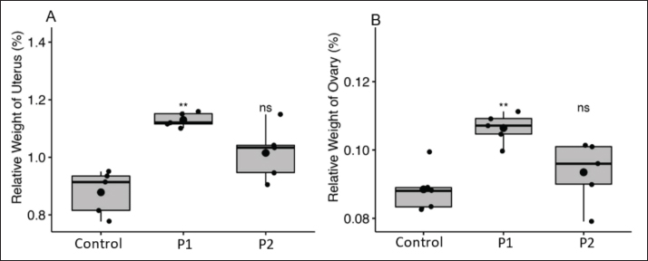
AbstractBackground: Mung bean sprouts (Vigna radiata) (MBS) are recognized for their antioxidant and anti-inflammatory properties. Aim: The objective of this study was to determine the impact of MBS suspension on the reproductive organs of female rats. Methods: A total of 15 eight-week-old female Sprague–Dawley rats in their diestrus phases were allocated into three distinct groups: one control group receiving aquadest (P0) and two experimental groups receiving 1% MBS suspension (P1) and 5% MBS suspension (P2) for 20 days. The assessment of reproductive organs was conducted using measured serum levels of follicle-stimulating hormone (FSH), estradiol, and progesterone. The measurements of ovarian and uterine relative weights, uterine cornual diameter, uterine cornual length, uterine vascularization, histological assessment of uterine gland diameter, and the quantification of primary, secondary, tertiary, de Graaf follicles, corpus luteum, and Vascular Endothelial Growth Factor (VEGF) expression in the ovary was also evaluated. Results: The P1 group exhibited a substantial rise in relative weights of the uterus and ovaries, whereas the P2 group had an increase in uterine cornual width, length, and vascularization. Neither the P1 nor P2 groups exhibited an increase in uterine gland diameter or the quantity of ovarian follicles and corpus luteum, despite the observation of larger mature superficial follicles. Serum concentrations of FSH in both P1 and P2 groups were significantly lower than those in the P0 group. The suspension treatment of MBS showed a nonsignificant influence on serum estradiol concentrations across all groups. A notable rise in serum progesterone levels and VEGF expression was detected in both P1 and P2 groups. Conclusion: The 20-day administration of MBS enhanced the reproductive system and function in female rats. Keywords: Female rat, Mung bean sprouts, Hormones Concentration. IntroductionThe mung bean (Vigna radiata) is a significant edible legume crop, widely consumed by households across Asia. It was extensively utilized as a dietary supplement for human health (Hou et al., 2019). The mung bean comprises a balanced array of nutrients, encompassing protein, dietary fiber, minerals, vitamins, and substantial quantities of bioactive substances. Antioxidant substances, including vitamin E, vitamin C, and polyphenols, are present in the mung bean (Ali et al., 2013; Mehta et al., 2021). Vitamin E present in mung beans functions as a natural antioxidant, safeguarding cells from free radical damage (Rizvi et al., 2014). Excessive reactive oxygen species resulting from oxidative stress might impact early embryonic development by altering critical transcription factors that regulate gene expression (Siauciunaite et al., 2019). Concerning the effect of oxidative stress upon female reproduction, the elevated levels of reactive oxygen species in the female reproductive system may adversely impact oocyte fertilization and hinder embryonic implantation (Mauchart et al., 2023). Recent studies have illustrated that elevated levels of reactive oxygen species were found in different animals that suffered from anestrus (Amin et al., 2019, Amin et al., 2017b), pyometra (Amin et al., 2021c), and abortion (Amin et al., 2023d), indicating that elevated levels of reactive oxygen species have deleterious effects on reproductive health. In addition, several studies have indicated that materials with antioxidant properties help alleviate these deleterious effects of reactive oxygen species (Amin et al., 2022e). The other research indicates that mung bean sprouts (MBSs) exhibit enhanced biological activity and increased levels of secondary metabolites post-germination, since relevant biosynthetic enzymes are active during the early stages of this process. Consequently, germination is believed to enhance the nutritional and therapeutic properties of mung beans (Tang et al., 2014; Cheng et al., 2023). Research on mung beans and their sprouts in animal models has demonstrated significant health benefits, including hypoglycemic and hypolipidemic effects, antihypertensive properties, anticancer activity, hepatoprotective effects, and immunomodulatory functions (Ma et al., 2022; Matemu et al., 2021). The estrogenic activity of mung beans has been documented; however, no research has examined the impact of MBS on the reproductive organs and functions of female animals. This study aimed to assess the advantageous effects of Indonesian MBS on enhancing the female reproductive organs of albino rats prior to mating, with the objective of optimizing the growth and development of reproductive systems to facilitate successful reproduction and yield quality offspring. Materials and MethodsEthical approvalThe Animal Ethics Committee of the School of Veterinary Medicine and Biomedical Science, IPB University approved this study (007/KEH/SKE/I/2020) dated: January 2, 2020. Study period and locationThis research was performed from August 2021 to December 2021 at the Laboratory Animal Management Unit, School of Veterinary Medicine and Biomedicine, Bogor Agricultural University. Preparation of mung bean sprouts suspensionThe MBS were collected from local farmers in Dramaga, Bogor, Indonesia. Identification of the plant was done by Herbarium Bogoriense Research Centre and National Innovation Bogor with the number of identification was B-746/V/DI.05.07/3/2022. Preparation of MBS suspension starts with 100 grams of fresh MBS collected from local farmers located in Dramaga, Bogor, Indonesia were pulverized and thereafter introduced into 100 ml of boiling water. The suspension was maintained at room temperature for 1 hour and subsequently filtered through a 100 μm wire mesh to get a stock solution. One percent and 5% drinking solutions were made by diluting the stock solution with mineral water. The stock solutions were stored in a 4°C refrigerator and produced every 3 days. Experimental animalsFifteen female Sprague–Dawley rats, aged 8 weeks and weighing around 200–230 g, were acquired from iRatco Veterinary Laboratory Services in Bogor, West Java, Indonesia. The chosen experimental rats were in the diestrus phase of their estrous cycles. Evaluation of the estrous cycle was performed through the preparation of vaginal smears, with the estrous stage determined based on the types of cells observed microscopically. The experimental rats were categorized into three treatment groups: a control group (P0) and two experimental groups receiving MBS suspension combined with mineral water at dosages of 1% (P1) and 5% (P2) in their drinking water. The experimental rats underwent MBS suspension treatment in their drinking water for 20 days, with the water being replaced every 12 hours. Upon completion of the treatment, Euthanasia was performed using cervical dislocation following anesthesia induced by intraperitoneal administration of ketamine (80 mg/kg BW) and xylazine (5 mg/kg BW). After ensuring complete anesthesia, terminal blood collection was carried out via intracardiac puncture. Approximately 2 ml of blood was collected in a plain tube. Then, the serum was separated using the centrifuge at 3,000 rpm for 5 minutes. The collected serum will serve as the sample for subsequent laboratory assay FSH, estradiol, and progesterone. Determinations of serum FSH, estradiol, and progesterone concentrationsConcentrations of serum FSH, estradiol, and progesterone were quantified utilizing Rat FSH (Follicle Stimulating Hormone) ELISA, Rat/porcine E2 (Estradiol) ELISA, and P4 (Progesterone) ELISA (Elabscience, US), in accordance with the manufacturer’s guidelines. Macroscopic analysisImages of the uterus were captured for vascular examination after euthanasia. The dimensions of the left and right uterine cornua, the diameter of the uterine cornua, and the ratio of uterine blood vessels were quantified by image analysis (ImageJ NIH, US), while the relative weight of the uterus was determined by weighing the uterus in relation to body weight. The total count of superficial follicles in the right and left ovaries of each animal was manually determined during the macroscopic exams using a light stereo microscope. The quantities of primary, secondary, tertiary, and de Graaf follicles in the ovary were quantified by image analysis techniques. The ovaries’ relative weight was assessed using a procedure analogous to that employed for determining the relative weight of the uterus. Relative organ weight is preferred over absolute (individual) organ weight because it provides a more accurate and standardized measure of treatment effects by accounting for variations in body weight among the animals. In experimental animal studies, individual body weights may vary naturally or due to treatment, and assessing organ weight relative to body weight (e.g., mg/g body weight) allows for more objective and meaningful comparisons across groups. Moreover, relative organ weight better reflects physiological or pathological changes in the organ itself, minimizing the confounding influence of overall body mass fluctuations. Histology of uterus and ovaryAfter euthanasia, the ovaries and uterus of the experimental animals identified, harvested, and were then preserved in 10% formalin. Fixed ovarian and uterine tissues fixed in paraffin blocks were sectioned into 4 μm thick slices using a Leica RM2135 Microtome (Leica, DE) and subsequently mounted on slides. The tissue was subsequently deparaffinized in xylene, rehydrated in graded alcohols, stained with hematoxylin and eosin, and finally sealed with Entellan® (Merck, DE). The organ slides were subsequently analyzed using an optical microscope. Immunohistochemical stainingFor immunohistochemistry, paraffin slices of the ovary were dewaxed, rehydrated, and washed in phosphate- buffered saline (PBS), followed by a 15-minute incubation with methanol containing 0%–3% H2O2 to inhibit endogenous peroxidase activity. For improved antigen retrieval, we employed HIER solution epitope retrieval (Cat. no. CPL500, ScyTek, US) for 5 minutes at 121°C. The slices were rinsed with 10 mM PBS at pH 7.4 for 5 minutes, and incubated with a particular Vascular Endothelial Growth Factor (VEGF) antibody. The sections were subsequently treated with a mouse monoclonal antibody to VEGF at a dose of 4 μg/ml (sc-7269; Santa Cruz Biotechnology, US), followed by a 5-minute buffer immersion and amplification using polymer EnVision Flex™ (Dako, US). Data analysisData analyses were conducted utilizing R software (version 3.5.1). The results, presented as mean ± SD, were subjected to the one-way analysis of variance followed by the Duncan post hoc test. Statistical significance was determined at p < 0.05. ResultsEffect of MBS suspension administration on the organs’ relative weightThe results of the study showed that the relative uterine weight in group P1 was significantly higher compared to the control group. Although group P2 also exhibited an increase in uterine weight compared to the control group, the difference was not statistically significant A consistent pattern was observed in the ovaries, where the P1 group showed a significant increase in relative ovarian weight compared to the control group (p < 0.05). The P2 group also demonstrated a higher ovarian weight relative to body weight than the control, although statistical significance was not specified. Figure 1 illustrates the comparative weight of the uterine and ovary in experimental rats administered MBS. Effect of MBS suspension administration on the uterine profileThe diameters of rat uterine cornua and the extent of uterine vascularization exhibited an increase in both the P1 and P2 groups relative to the P0 (control) group, suggesting a positive effect of MBS therapy on uterine tissue development. Although the length of the uterine cornua also showed an upward trend, the difference was not statistically significant when compared to the control group. These findings imply that the treatment may influence certain aspects of uterine morphometry more selectively as illustrated in Figure 2. Effect of MBS suspension administration on the ovarian follicle numberMacroscopically, the quantity of bigger mature follicles was greater in the P1 and P2 groups than in the P0 group (Fig. 3). Histological assessment revealed no significant changes in the histological architecture of ovary follicles between the control and experimental rats (Fig. 4).
Fig. 1. Relative weight of rat uterine (A) and ovary (B) of the experimental rats after 20 days of treatment with MBS maceration at group Control, P1 and P2. The dot inside each bar represents the mean value, Stripes inside each bar represent the median value. The dots outside the bar refers to outliers data. ns refers the no significant difference between each data compared to control while (**) imply significance value with p < 0.01.
Fig. 2. Diameter (A), length (B), and vascularization (C) of cornua uteri in the experimental rats after 20 days of treatment with MBS maceration at group Control, P1 and P2. The dot inside each bar represents the mean value, Stripes inside each bar represent the median value. The dots outside the bar refers to outliers data. ns refers the no significant difference between each data compared to control while (*) imply significance value with p < 0.05.
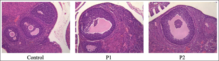
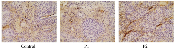
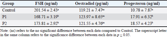
Fig. 3. Macroscopic evaluation of the ovary of experimental rats after 20 days of treatment with MBS suspension with 10x magnification. Effect of MBS suspension administration on the VEGF protein expressionsThe immunohistochemistry labeling revealed that VEGF expression was localized in the ovarian interstitial tissue, indicating encouragement of growth and neovascularization development. The expression of VEGF in both the P1 and P2 groups was elevated relative to the P0 group. The P1 group has the highest VEGF expression among all groups (Fig. 5). Effect of MBS suspension administration on the FSH, estradiol, and progesterone serum concentrationsSerum concentrations of FSH in both P1 and P2 groups were significantly lower than in the P0 group. The suspension treatment of MBS had a nonsignificant influence on serum estradiol concentrations across all groups. A notable elevation in serum progesterone levels was detected in both P1 and P2 groups relative to the P0 group (Table 1). DiscussionThe administration of MBS suspension tin, this experiment demonstrated significant potential for enhancing the health and gonadal growth and development of female animals. Treatment with MBS suspension considerably elevated serum progesterone levels while reducing serum FSH and estrogen concentrations at the conclusion of the treatment. Nonetheless, there was no augmentation in the quantity of follicles (primary, secondary, tertiary, and de Graaf follicles) and corpus luteum. This suggests that the elevation in serum progesterone levels is not associated with the augmented quantity of follicles and corpus luteum (Andriyanto et al., 2022; Cáceres et al., 2024). The lack of increase in the number of corpus luteum in the experimental rats treated with MBS suspension, compared to the control, indicates that the elevated progesterone concentrations in these rats are associated with the augmented number and activity of cells responsible for synthesizing and producing progesterone within the existing corpus luteum. The notion was proven by the enlarged diameters of mature follicles observed both macroscopically and microscopically, which were significantly greater in rats subjected to MBS suspension treatment. Unlike our prior studies, the majority of the elevations in serum estrogen and progesterone levels were ascribed to the augmented quantity of developing follicles and corpus luteum (Ricciotti and FitzGerald, 2011; Coughlan et al., 2022).
Fig. 4. Panel of histopathology comparisons. All groups exhibited normal microstructural conditions, and there were no significant changes observed throughout the experiments. Haematoxylin and eosin staining. Magnification 200x.
Fig. 5. Expression of VEGF with immunohistochemical staining in the ovary of experimental rats after 20 days of treatment with MBS maceration group control, P1 and P2. Magnification 200x. Table 1. FSH, Estradiol, and Progesterone serum concentration in each group during this research.
This study demonstrated that the administration of MBS suspension in albino rats resulted in an increase in the relative weight of both the ovary and uterus. The augmented mass of these two organs correlates with the physiological processes involved in follicular growth and development in the ovary, facilitating the synthesis and secretion of estrogen during estrus and the production of progesterone post-ovulation. The active uterus producing Prostaglandin E2 hormones promotes its growth and development, as evidenced by increased vascularization and local edema (Siriparu et al., 2022). The prior investigation indicated that MBS are useful in enhancing ovarian function (Cao et al., 2011). Mung bean possesses a significant concentration of flavonoid and phenolic chemicals, which contribute to its elevated antioxidant activity. The supplementation of antioxidant substances is significant in the treatment of reproductive diseases and fertility by regulating oxidative stress (Haldar et al., 2022; Supasatyankul et al., 2022). The decreased serum FSH levels, along with a significant inverse correlation between serum FSH concentration and VEGF expression, indicate that active ovarian tissue effectively utilized FSH to promote follicular growth and development. FSH binds to its receptors on ovarian cell membranes, activating signaling pathways essential for folliculogenesis. MBS significantly elevated serum FSH levels in experimental rats as shown in this study. This discrepancy may be associated with the timing of serum FSH concentration measurements during follicular ovulation, as both serum FSH and estrogen levels were reduced at this period (Arslan et al., 2003; Bachelot et al., 2009). The reduced serum levels of estrogen and FSH in the experimental groups were hypothesized to correlate with the negligible rise in the quantity of follicles and corpus luteum. The elevated serum progesterone levels in the experimental rats administered MBS suspension are likely associated with the augmented quantity and activity of cells in the developed corpus luteum during follicular ovulation. Post-ovulation, the ovulated follicles transform into the corpus luteum, which secretes progesterone to promote the growth and development of the uterine glands in anticipation of pregnancy, despite the experimental rats not being mated (Perry et al., 2023; Guo et al., 2021). The elevation of serum progesterone levels in the experimental groups will enhance the expression of VEGF to promote uterine vascularization (Massri et al., 2023). This experiment demonstrated that elevated serum progesterone levels in the experimental group enhance uterine vascularization intensity by upregulating VEGF expression, as evidenced by the strong positive correlations between progesterone concentrations and uterine vascularization. Increased vascularization in the uterus enhances the movement of nutrients, oxygen, and growth factors, hence supporting the growth and development of embryos and fetuses following mating. In this observation, although the experimental rats were not mated, the uterine preparations for implantation to facilitate the creation of components necessary for the implanted zygote nonetheless transpired (Kumar et al., 2012; Mandala et al., 2020). Consequently, the enhanced prenatal environment, achieved by augmenting the synthesis and secretion of gestational hormones through MBS suspension, substantiates that MBS holds promise for enhancing reproductive success and child quality. The enhanced uterine environment and the characteristics of oocytes will facilitate the formation and development of embryos and fetuses throughout gestation, resulting in superior-quality progeny (Cao et al., 2022). ConclusionThe suspension of MBS applied to albino rats over 4–5 estrus cycles prior to mating significantly enhances the growth and development of reproductive organs, as evidenced by improvements in the morphology and functions of gonads. The suspension of MBS shows significant potential for enhancing the reproductive systems of female animals, thereby improving reproductive performance and the quality of offspring produced. AcknowledgmentsThe authors like to acknowledge the Ministry of Education, Culture, Research, and Technology of Indonesia and IPB University for their financial support of this research. Conflict of interestThe authors assert that there are no conflicts of interest pertaining to any financial, personal, or other affiliations with individuals or organizations relevant to the content addressed in the work. Author’s contributionsAA: Conception, Data curation, Formal analysis, Writing original draft, Supervision. AAU: Data acquisition, Data curation. HYP: Data acquisition, Data curation AF: Writing, review the manuscript. MS: Data acquisition. ET: Data acquisition, Data curation. YI: Supervision, Conception, Validation. WM: Supervision, Conception, Validation. Data availabilityAll data supporting the findings of this study are available within the manuscript. ReferencesAli, N.M., Yusof, H.M., Long, K., Yeap, S.K., Ho, W.Y., Beh, B.K., Koh, S.P., Abdullah, M.P. and Alitheen, N.B. 2013. Antioxidant and hepatoprotective effect of aqueous extract of germinated and fermented mung bean on ethanol-mediated liver damage. BioMed. Res. Int. 13, 693–613. Amin, Y.A., Ali, R.A., Fouad, S.S. and Ibrahim, R.M. 2021c. The deleterious effect of postpartum pyometra on the reproductive indices, the metabolic profile, and oxidant/antioxidant parameters of dairy cows. Vet. World. 14(2), 329. Amin, Y.A., El-Naga, E.M.A., Noseer, E.A., Fouad, S.S. and Ali, R.A. 2019. Synchronization with controlled internal drug release (CIDR) and prostaglandin F2α (PGF2α) influences oxidant/antioxidant biomarkers and mineral profile in summer-stressed anoestrous buffalo (Bubalus bubalis). Theriogenology 134, 34–41. Amin, Y.A., Mahmoudand, H.Y. and Megahed, G.A. 2017b. Relation between oxidant/antioxidant status and postpartum anestrous conditions. World J. Res. Rev. 9(10). Amin, Y.A., Omran, G.A., Fouad, S.S., Fawy, M.A., Ibrahim, R.M., Khalifa, F.A. and Ali, R.A. 2023d. Abortion associated with postpartum opportunistic bacterial invasion reduces fertility and induces disturbances of reproductive hormones, hematological profile, and oxidant/antioxidant profiles in dairy cows. J. Adv. Vet. Anim. Res. 10(4), 654. Amin, Y.A., Youssef, N.A.M., Mahmoud, A.E.Z.E., Salah, M., Khalil, A.M., Shanab, O. and Hassaneen, A.S.A. 2022e. Impact of polyherbal formulation oral administration on the estrus response, luteal activity, and oxidative stress in postpartum dairy cows with ovarian subfunction. Vet. World. 15(2), 360. Andriyanto, A., Widi, L.N., Subangkit, M., Tarigan, E., Irarang, Y., Nengsih, R.F. and Manalu, W. 2022. Potential use of Indonesian basil (Ocimum basilicum) maceration to increase estradiol and progesterone synthesis and secretion to improve prenatal growth of offspring using female albino rats as an animal model. Vet. World. 15(5), 1197–1207. Arslan, A.A., Zeleniuch-Jacquotte, A., Lukanova, A., Rinaldi, S., Kaaks, R. and Toniolo, P. 2003. Reliability of follicle-stimulating hormone measurements in serum. Reprod. Biol. Endocrinol. 1, 49. Bachelot, A., Beaufaron, J., Servel, N., Kedzia, C., Monget, P., Kelly, P.A., Gibori, G. and Binart, N. 2009. Prolactin independent rescue of mouse corpus luteum lifespan: identification of prolactin and luteinizing hormone target genes. Am. J. Physiol. Endocrinol. Metab. 297(3), E676–684. Cáceres, A.R.R., Cardone, D,A,, Sanhueza, M.L.Á., Bosch, I.M., Cuello-Carrión, F.D., Rodriguez, G.B., Scotti, L., Parborell, F., Halperin, J. and Laconi, M.R. 2024. Local effect of allopregnanolone in rat ovarian steroidogenesis, follicular and corpora lutea development. Sci. Rep. 14(1), 6402. Cao, D., Li, H., Yi, J., Zhang, J., Che, H., Cao, J., Yang, L., Zhu, C. and Jiang, W. 2011. Antioxidant properties of the mung bean flavonoids on alleviating heat stress. PLoS One. 6(6), e21–71. Cheng, Y., Xiang, N., Chen, H., Zhao, Y., Wang, L., Cheng, X. and Guo, X. 2023. The modulation of light quality on carotenoid and tocochromanol biosynthesis in mung bean (Vigna radiata) sprouts. Food. Chem. (Oxf). 6, 100–170. Cao, J., Wang, Y., Wang, G., Ren, P., Wu, Y. and He, Q. 2022. Effects of typical antimicrobials on growth performance, morphology and antimicrobial residues of mung bean sprouts. Antibiotics (Basel). 11(6), 807. Coughlan, C., Vitorino, R., Melado, L., Digma, S., Sibal, J., Patel, R., Lawrenz, B. and Fatemi, H. 2022. Evolution of serum progesterone levels in the very early luteal phase of stimulated IVF/ICSI cycles post hCG trigger: a proof of concept study. J. Assist. Reprod. Genet. 39(5), 1095–1104. Guo, X., Yi, H., Li, T.C., Wang, Y., Wang, H. and Chen, X. 2021. Role of vascular endothelial growth factor (VEGF) in human embryo implantation: clinical implications. Biomolecules. 11(2), 253. Haldar, S., Agrawal, H., Saha, S., Straughn, A.R., Roy, P. and Kakar, S.S. 2022. Overview of follicle stimulating hormone and its receptors in reproduction and in stem cells and cancer stem cells. Int. J. Biol. Sci. 18(2), 675–692. Hou, D., Yousaf, L., Xue, Y., Hu, J., Wu, J., Hu, X., Feng, N. and Shen, Q. 2019. Mung bean (Vigna radiata L.): bioactive polyphenols, polysaccharides, peptides, and health benefits. Nutr. 11(6), 1238. Kumar, P. and Magon, N. 2012. Hormones in pregnancy. Niger. Med. J. 53(4), 179–183. Ma, Y., Zhou, S. and Lu, J. 2022. Metabolomic analysis reveals changes of bioactive Compounds in Mung Beans (Vigna radiata) during γ-Aminobutyric Acid Enrichment Treatment. Foods. 11(10), 1423. Mandalà, M. 2020 Influence of estrogens on uterine vascular adaptation in normal and preeclamptic pregnancies. Int. J. Mol. Sci. 21(7), 2592. Massri, N., Loia, R., Sones, J.L., Arora, R. and Douglas, N.C. 2023. Vascular changes in the cycling and early pregnant uterus. JCI Insight. 8(11), e163422. Matemu, A., Nakamura, S. and Katayama, S. 2021. Health benefits of antioxidative peptides derived from legume proteins with a high amino acid score. Antioxidants (Basel). 10(2), 316. Mauchart, P., Vass, R.A., Nagy, B., Sulyok, E., Bódis, J. and Kovács, K. 2023. Oxidative stress in assisted reproductive techniques, with a focus on an underestimated risk factor. Curr. Issues Mol. Biol. 45(2), 1272–1286. Mehta, N., Rao, P. and Saini, R. 2021. A review on metabolites and pharmaceutical potential of food legume crop mung bean (Vigna radiata L. Wilczek). BioTechnologia (Pozn). 102(4), 425–435. Perry, G.A., Ketchum, J.N. and Quail, L.K. 2023. Importance of preovulatory estradiol on uterine receptivity and luteal function. Anim. Reprod. 20(2), e20230061. Ricciotti, E. and FitzGerald, G.A. 2011. Prostaglandins and inflammation. Arterioscler. Thromb. Vasc. Biol. 31(5), 986–1000. Rizvi, S., Raza, S.T., Ahmed, F., Ahmad, A., Abbas, S. and Mahdi, F. 2014. The role of vitamin e in human health and some diseases. Sultan Qaboos Univ. Med. J. 14(2), e157–165. Siauciunaite, R., Foulkes, N.S., Calabrò, V. and Vallone, D. 2019. Evolution shapes the gene expression response to oxidative stress. Int. J. Mol. Sci. 20(12), 3040. Siriparu, P., Panyatip, P., Pota, T., Ratha, J., Yongram, C., Srisongkram, T., Sungthong, B. and Puthongking, P. 2022. Effect of germination and illumination on melatonin and its metabolites, phenolic content, and antioxidant activity in mung bean sprouts. Plants (Basel). 11(21), 2990. Supasatyankul, B., Saisriyoot, M., Klinkesorn, U., Rattanaporn, K. and Sae-Tan, S. 2022. Extraction of phenolic and flavonoid compounds from mung bean (Vigna radiata L.) seed coat by pressurized liquid extraction. Molecules. 27(7), 2085. Tang, D., Dong, Y., Ren, H., Li, L. and He, C. 2014. A review of phytochemistry, metabolite changes, and medicinal uses of the common food mung bean and its sprouts (Vigna radiata). Chem. Cent. J. 8(1), 4. | ||


| How to Cite this Article |
| Pubmed Style Andriyanto A, Mustika AA, Putra HY, Fadholly A, Subangkit M, Tarigan E, Irarang Y, Manalu W. The efficacy of mung bean sprouts suspension in improving reproductive organs of female albino rats. Open Vet. J.. 2025; 15(6): 2471-2477. doi:10.5455/OVJ.2025.v15.i6.19 Web Style Andriyanto A, Mustika AA, Putra HY, Fadholly A, Subangkit M, Tarigan E, Irarang Y, Manalu W. The efficacy of mung bean sprouts suspension in improving reproductive organs of female albino rats. https://www.openveterinaryjournal.com/?mno=236425 [Access: January 25, 2026]. doi:10.5455/OVJ.2025.v15.i6.19 AMA (American Medical Association) Style Andriyanto A, Mustika AA, Putra HY, Fadholly A, Subangkit M, Tarigan E, Irarang Y, Manalu W. The efficacy of mung bean sprouts suspension in improving reproductive organs of female albino rats. Open Vet. J.. 2025; 15(6): 2471-2477. doi:10.5455/OVJ.2025.v15.i6.19 Vancouver/ICMJE Style Andriyanto A, Mustika AA, Putra HY, Fadholly A, Subangkit M, Tarigan E, Irarang Y, Manalu W. The efficacy of mung bean sprouts suspension in improving reproductive organs of female albino rats. Open Vet. J.. (2025), [cited January 25, 2026]; 15(6): 2471-2477. doi:10.5455/OVJ.2025.v15.i6.19 Harvard Style Andriyanto, A., Mustika, . A. A., Putra, . H. Y., Fadholly, . A., Subangkit, . M., Tarigan, . E., Irarang, . Y. & Manalu, . W. (2025) The efficacy of mung bean sprouts suspension in improving reproductive organs of female albino rats. Open Vet. J., 15 (6), 2471-2477. doi:10.5455/OVJ.2025.v15.i6.19 Turabian Style Andriyanto, Andriyanto, Aulia Andi Mustika, Hamdika Yendri Putra, Amaq Fadholly, Mawar Subangkit, Elpita Tarigan, Yusa Irarang, and Wasmen Manalu. 2025. The efficacy of mung bean sprouts suspension in improving reproductive organs of female albino rats. Open Veterinary Journal, 15 (6), 2471-2477. doi:10.5455/OVJ.2025.v15.i6.19 Chicago Style Andriyanto, Andriyanto, Aulia Andi Mustika, Hamdika Yendri Putra, Amaq Fadholly, Mawar Subangkit, Elpita Tarigan, Yusa Irarang, and Wasmen Manalu. "The efficacy of mung bean sprouts suspension in improving reproductive organs of female albino rats." Open Veterinary Journal 15 (2025), 2471-2477. doi:10.5455/OVJ.2025.v15.i6.19 MLA (The Modern Language Association) Style Andriyanto, Andriyanto, Aulia Andi Mustika, Hamdika Yendri Putra, Amaq Fadholly, Mawar Subangkit, Elpita Tarigan, Yusa Irarang, and Wasmen Manalu. "The efficacy of mung bean sprouts suspension in improving reproductive organs of female albino rats." Open Veterinary Journal 15.6 (2025), 2471-2477. Print. doi:10.5455/OVJ.2025.v15.i6.19 APA (American Psychological Association) Style Andriyanto, A., Mustika, . A. A., Putra, . H. Y., Fadholly, . A., Subangkit, . M., Tarigan, . E., Irarang, . Y. & Manalu, . W. (2025) The efficacy of mung bean sprouts suspension in improving reproductive organs of female albino rats. Open Veterinary Journal, 15 (6), 2471-2477. doi:10.5455/OVJ.2025.v15.i6.19 |

