


| Case Report | ||
Open Vet. J.. 2026; 16(3): 1947-1953 Open Veterinary Journal, (2026), Vol. 16(3): 1947-1953 Case Report Coexistence of seminoma and leiomyoma in the testis of an Arabian–Barbe stallion: A case reportZineb El Brini1*, Charifa Drissi Touzani2, Abderrahmane Al Bouzidi3 and Mohammed Piro11Department of Medicine, Surgery, and Reproduction, Agronomy and Veterinary Institute Hassan II, Rabat, Morocco 2Department of Pathology and Veterinary Public Health, Agronomy and Veterinary Institute Hassan II, Rabat, Morocco 3Faculty of Medicine and Pharmacy of Rabat, Mohammed V University in Rabat, Rabat, Morocco *Corresponding Author: Zineb El Brini. Department of Medicine, Surgery, and Reproduction, Agronomy and Veterinary Institute Hassan II, Rabat, Morocco. Email: z.elbrini [at] iav.ac.ma Submitted: 13/11/2025 Revised: 11/02/2026 Accepted: 19/02/2026 Published: 31/03/2026 © 2025 Open Veterinary Journal
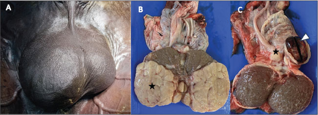
AbstractBackground: Testicular tumors are rare in horses, with seminoma being the most frequently described in the literature. In Morocco, many stallions used for Tbourida are intentionally kept intact, as uncastrated horses are traditionally viewed as symbols of prestige and cultural pride. This cultural practice increases the likelihood of detecting testicular pathology in mature stallions. Case Description: A 15-year-old Arabian–Barbe stallion used in Tbourida was presented for evaluation after the owner noticed a slow, progressive enlargement of the right testis over a period of more than 1 year. Clinical examination revealed a firm, enlarged right testicle, while the left testis appeared normal. Post-castration assessment revealed multiple firm, lobulated masses occupying roughly two-thirds of the right testis, and a small nodular lesion near the epididymal head on the left, associated with focal hemorrhage. Histopathology confirmed the coexistence of a seminoma and a leiomyoma within the same testis. Conclusion: This report describes the rare coexistence of seminoma and leiomyoma within the same equine testis, along with an epididymal hematoma. Together, these findings provide valuable insight into the spectrum of testicular lesions observed in stallions and highlight the importance of a comprehensive clinical assessment supported by histopathological examination for accurate diagnosis and appropriate management of testicular abnormalities in intact horses. Keywords: Seminoma, Leiomyoma, Testis tumor, Epididymal hematoma, Arabian–Barbe stallion. IntroductionTesticular tumors in horses are rare, and their true prevalence remains poorly defined in the literature (Leidinger et al., 2018). These neoplasms are classified as germ cell tumors (e.g., seminoma and teratoma), sex cord-stromal tumors (e.g., Leydig and Sertoli cell tumors), or tumors of other origins, such as lymphoma or smooth muscle neoplasms (Cooper and Valentine, 2002; MacLachlan and Kennedy, 2002; Valentine, 2009). Among germ cell tumors, seminoma is the most commonly reported in equids (Valentine, 2009; Govaere et al., 2010). Knowledge regarding equine seminomas remains limited and is largely derived from individual case reports, partly because most horses are castrated at a young age, restricting long-term observation of intact stallions (Schumacher, 1999; Valentine, 2009; Peeters et al., 2024). Interestingly, the number of published case reports relative to the small population of intact adult stallions suggests that testicular tumors may be more frequent than traditionally assumed (Brinsko, 1998; Valentine, 2009). Equine seminoma is typically observed in adult and older stallions, with the majority of published cases involving horses older than 10 years of age; reports in younger animals remain uncommon (Trigo et al., 1984; Brinsko, 1998; Weiermayer and Richter, 2009; Batista et al., 2024; Peeters et al., 2024). These tumors may develop in both descended and cryptorchid testes, with cryptorchidism considered a potential predisposing factor (Weiermayer and Richter, 2009; De Lange et al., 2015; Varner et al., 2015; Pasolini et al., 2016; Turner, 2019). Although equine seminomas are frequently classified as benign, malignant behavior and metastatic spread have been reported in horses, suggesting a more aggressive biological profile compared with other domestic species (Valentine, 2009; Agnew and MacLachlan, 2016; Meuten, 2020; Peeters et al., 2024). Although uncommon, testicular tumors can present significant clinical and management challenges, especially in regions where stallions are traditionally left intact. In Morocco, stallions are kept uncastrated to participate in traditional equestrian performance known as Tbourida, a practice deeply rooted in historical and cultural heritage (Talley and G, 2017). Tbourida was officially recognized as a national heritage sport during the reign of King Hassan II (1961–1999) and is performed during moussems (saints’ day celebrations), weddings, and national festivals. During these spectacular displays, teams of horsemen (sorba) ride in synchronized formations and fire gunpowder rifles in unison, showcasing discipline, coordination, and strength (Talley, 2017). In 2021, Tbourida was inscribed on UNESCO (2021) Representative List of the Intangible Cultural Heritage of Humanity, further highlighting its cultural significance. Owning an intact stallion carries social prestige and reflects tradition; therefore, castration is totally avoided. This management preference increases the likelihood of observing chronic testicular pathology in this special population. This report describes an unusual case of coexisting seminoma and leiomyoma in the testis of an Arabian–Barbe stallion used for Tbourida. Due to the limited information available on how these lesions present clinically, ultrasonographically, and histopathologically in horses, this case provides practical insight for veterinarians. Case DetailsA 15-year-old Arabian–Barbe stallion used for Tbourida was referred to the Equine Clinic of the Hassan II Agronomic and Veterinary Institute (Rabat, Morocco) after the owner noticed a gradual enlargement of the right testicle. The swelling had been apparent for more than 1 year but had become more obvious in recent months. This change began to affect the horse’s appearance during Tbourida performances, posing practical concerns, and the stallion had recently started to show mild signs of discomfort. During the physical examination, the stallion was calm, alert, and all vital parameters were within normal limits. The right testicle was markedly enlarged and firm on palpation, while the left testicle appeared normal in size and consistency (Fig. 1A). No scrotal edema was observed, and the horse showed no signs of pain during manipulation. Ultrasonographic evaluation of the right testis revealed heterogeneous tissue with multiple poorly defined hypoechoic regions, mainly in its caudal portion. On the left side, a small hypoechoic area was identified near an echogenic structure at the epididymal head, suggestive of a localized hematoma. Considering the chronicity of the lesion history and the imaging characteristics, testicular neoplasia was considered the most likely diagnosis.
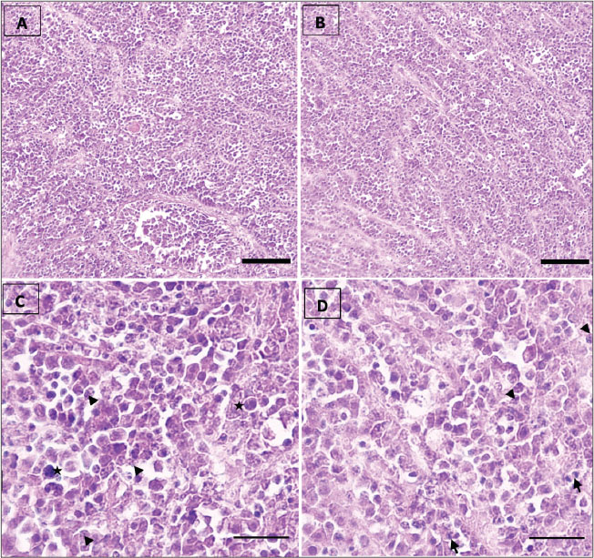
Fig. 1. Marked scrotal asymmetry in an adult stallion, characterized by a pronounced enlargement of the right testicle (A). Post-castration gross examination shows multiple lobulated masses of variable size and beige to dark beige color occupying most of the right testicular parenchyma (B) (asterisks), with whitish fibrous areas visible at the center of the section (white arrows). A well-demarcated hemorrhagic lesion is observed cranially to the left testis at the level of the epididymal head (C) (arrowhead), consistent with a localized hematoma, with an adjacent nodular lesion (asterisk). Anesthesia and surgical procedureThe stallion was premedicated with acepromazine (Calmivet®, Vetoquinol; 0.02 mg/kg body weight, IV). After approximately 20 minutes, sedation was induced using xylazine (Rompun®, Bayer; 0.8 mg/kg body weight, IV). General anesthesia was induced with ketamine hydrochloride (Ketamine®; 2.2 mg/kg body weight, IV) combined with diazepam (Diaphram®; 0.05mg/kg body weight, IV) and subsequently maintained with isoflurane (Aerrane®, Baxter) delivered via inhalation, as previously described by Muir et al. (2018). Preoperative analgesia was provided using flunixin meglumine (Norbrook®, Norbrook Laboratories; 1.1 mg/kg body weight, IV). Prophylactic antibiotic therapy consisted of benzylpenicillin sodium (Norbrook®, Norbrook Laboratories; 22.000 IU/kg body weight, IM). Castration was performed using a standard inguinal approach under routine aseptic conditions as previously described by Sedrish and Leonard (2001). The spermatic cord and cremaster muscle were isolated by blunt dissection, the testis was exteriorized, and the ligament of the tail of the epididymis was transected. An emasculator was applied to the spermatic cord for approximately 5 minutes prior to transection, followed by placement of a security ligature. The surgical site was closed routinely in two layers. Histopathological examinationRepresentative tissue samples from the testicular masses were fixed in 10% neutral buffered formalin for 48 hours to 5 days. Tissue specimens were processed routinely by dehydration through a graded series of ethanol solutions (70%–100%), cleared, and embedded in paraffin. Paraffin-embedded tissues were sectioned at a thickness of 3–5 µm, deparaffinized, and stained with hematoxylin and eosin (H&E) according to standard histological techniques as described by Bancroft and Gamble (2008). Histological sections were examined using a light microscope to evaluate the morphological characteristics of the testicular tissue and associated lesions. Post-castration examinationPost-castration examination of the right testis showed multiple firm, lobulated masses of varying sizes, occupying approximately the caudal two-thirds of the parenchyma (Fig. 1B). The combined diameter of the masses was approximately 14 cm. The remaining parenchyma contained scattered pale, irregular areas interspersed with zones that appeared relatively preserved (Fig. 1B). On the left side, a small, well-circumscribed 3-cm nodule was found above the left testis, along with a localized hematoma at the epididymal head. The cut surface of the neoplastic tissue was beige to dark beige in color and firm in texture, while the hemorrhagic area appeared dark red-brown, with partly clotted blood and mild compression of the adjacent parenchyma (Fig. 1C). These observations confirmed bilateral involvement, with lobulated neoplastic tissue in the right testis and a peri-epididymal nodule with focal hemorrhage on the left (Fig. 1B and C). Histopathological findingsExamination of the testicular parenchyma revealed a neoplastic proliferation with a diffuse architecture and trabecular architecture (Fig. 2A, B). The neoplastic cells exhibited enlarged, anisokaryotic, hyperchromatic, and nucleolated nuclei, with clear cytoplasm (Fig. 2C, asterisk). The stroma contained fibrous septa, infiltrated by lymphocytes occasionally forming lymphoid aggregates (Fig. 2B, white arrow). No vascular emboli were observed, and the epididymis, tunica albuginea, rete testis, and spermatic cord were free of neoplastic invasion. These morphological features were consistent with a seminoma.
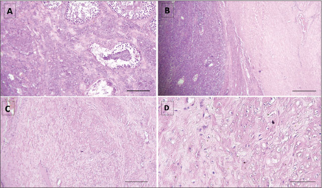
Fig. 2. Histological sections of seminoma in the testis of an Arabian-Barbe stallion. A: Overview of seminoma showing the general lobular architecture of the tumor (H&E, ×10; bar=160 µm). B: Presence of fibrous trabeculae (white arrows) separating tumor lobules, illustrating the structural organization within the seminoma (H&E, ×10). C: Nuclear atypia within tumor cells, including karyorrhectic nuclei (arrowhead) and anisokaryosis (asterisk), indicating cellular pleomorphism and active tumor proliferation (H&E, ×40; bar=90 µm). D: Mitotic figure (black arrow) highlighting active cell division, a feature of tumor growth and malignancy (H&E, ×40; bar=90 µm). The nodular lesion was well-circumscribed and, on cut section, displayed a whitish, fasciculated appearance without evidence of gross necrosis. Histological evaluation of H&E-stained sections revealed a benign mesenchymal neoplasm composed of interlacing fascicles of spindle-shaped cells. The neoplastic cells exhibited elongated, uniform nuclei with finely dispersed chromatin and moderately eosinophilic cytoplasm, consistent with smooth muscle differentiation (Fig. 3C, D). The fascicular architecture was prominent, and the tumor stroma contained scattered thick-walled blood vessels. Mitotic activity was low, with two to three mitotic figures per ten high-power fields. No areas of tumor necrosis, cellular atypia, or other features suggestive of malignancy were observed, supporting the diagnosis of leiomyoma.
Fig. 3. Histological features of seminoma and leiomyoma within the same testis in an Arabian-Barbe stallion. A–B: Interface between seminiferous tubules and the leiomyoma, showing distinct tissue separation (H&E, ×10; A: bar=200 µm, B: bar=340 µm). C: Well-circumscribed leiomyoma composed of spindle-shaped smooth muscle cells with elongated, regular nuclei and no signs of malignancy (H&E, ×10; bar=340 µm). D: Higher magnification of smooth muscle tumor cells, highlighting uniform nuclei and fascicular architecture (H&E, ×40; bar=160 µm). Both neoplastic components were located within the same testis. The seminoma occupied the central parenchyma, while the leiomyoma formed a well-circumscribed, septate proliferation. The two lesions were adjacent but remained distinctly separated, with no intermingling of cell populations (Fig. 3A, B). Ethical approvalEthical approval was not required, as this work reports a single clinical case managed within normal veterinary practice. DiscussionAlthough equine testicular tumors are considered uncommon and their true prevalence remains unclear (Leidinger et al., 2018), available data are largely derived from individual case reports, partly because most horses are castrated at a young age, limiting long-term monitoring of intact stallions (Schumacher, 1999; Valentine, 2009; Peeters et al., 2024). Among equine testicular neoplasms, seminoma is the most frequently reported tumor type (Brinsko, 1998; Valentine, 2009). Seminomas arise from spermatogenic cells and are typically unilateral, with a reported predilection for the right testis, although bilateral involvement has been described (MacLachlan and Kennedy, 2002; Scott et al., 2015). Advanced seminomas may become diffuse and replace most of the testicular parenchyma (Farjanikish et al., 2016; Leidinger et al., 2018). In the present case, the tumor involved the right testis, occupying approximately two-thirds of the parenchyma, with a small additional neoplastic lesion identified in the contralateral testis. Histopathological examination revealed the coexistence of a seminoma and a leiomyoma within the same testis, with seminoma representing the predominant component. Furthermore, equine seminomas are reported to grow relatively rapidly and may exhibit a higher metastatic potential than in other domestic species (Brinsko, 1998; Agnew and MacLachlan, 2016; Meuten, 2020). Palpation of the affected testis is often non-painful, which may contribute to delayed recognition and postponement of surgical intervention. As a result, clinical signs are frequently vague or intermittent, including mild discomfort, occasional colic-like episodes, or pain during ejaculation, and may therefore go unnoticed for prolonged periods (Christensen et al., 2007). In horses, although malignant presentations of seminoma are considered less frequent, this tumor appears to behave more aggressively than in other species, with metastatic spread reported primarily to abdominal and regional structures (Brinsko, 1998; Beck et al., 2001; Valentine, 2009; Knottenbelt et al., 2015; Agnew and MacLachlan, 2016; Meuten, 2020). Delayed diagnosis and treatment may therefore allow progressive tumor growth and increase the risk of metastatic disease, which has been associated with poor prognosis. When metastasis occurs, reported survival time ranges between 10 and 24 months (Christensen et al., 2007). This aggressive behavior has been illustrated in a case of intra-abdominal seminoma arising from a cryptorchid testis, in which extensive metastatic disease was identified at necropsy 18 months after surgery (Peeters et al., 2024). In the present case, castration was postponed until progressive tumor enlargement resulted in visible discomfort and aesthetic concerns during public appearances. This delay, influenced by the cultural context in which stallions are traditionally maintained intact, highlights a potential conflict between cultural practices and animal welfare considerations. Prolonged maintenance of an affected testis may expose the animal to chronic discomfort and increase the risk of advanced or metastatic disease, emphasizing the importance of timely clinical evaluation and intervention. These findings underscore the need to prioritize animal welfare and clinical decision-making over aesthetic or cultural considerations in the management of intact stallions. In cases of seminoma without evidence of metastasis, bilateral orchiectomy remains the recommended treatment, as affected stallions should not be maintained for breeding purposes (Govaere et al., 2010; De Lange et al., 2015; Farjanikish et al., 2016). Early surgical management is therefore essential not only for diagnostic confirmation but also to reduce welfare compromise and improve clinical outcome. A similar association of testicular neoplasms has rarely been described in horses. Weiermayer and Richter (2009) reported a Barb stallion with two distinct testicular tumors, a seminoma and a leiomyoma, each in a different testis. In contrast, the current case demonstrated both neoplastic lesions occurring within the same testis, with seminoma predominating. Previously, Cooper and Valentine (2002) described four cases of equine testicular leiomyomas, though the precise site of origin within the testicular architecture was unclear. According to Sisson (1975), testicular leiomyomas or leiomyosarcomas may originate from various smooth muscle elements, including the septula testis, testicular blood vessels, or the peritubular contractile cells of the convoluted and straight seminiferous tubules. In the case reported by Weiermayer and Richter (2009), the origin of the leiomyoma was considered unlikely to be peritubular, because the epididymis was not involved, as it typically would be if that were the case. In our case, determining the exact origin of the leiomyoma was challenging due to the close proximity of the two tumor types. In addition to the neoplastic lesions, a well-circumscribed hemorrhagic mass was identified adjacent to the epididymal region. This lesion was consistent with an epididymal hematoma, a finding not previously reported in horses. Such hematomas are likely to arise from rupture of small epididymal vessels, either after blunt trauma or secondary to venous congestion caused by the adjacent tumor. Stallions used for Tbourida are subjected to repeated physical exertion and scrotal impact during performances, which may contribute to local vascular injury and subsequent hematoma formation. An epididymal hematoma next to a testicular mass can make interpretation difficult, as it may mimic tumor extension or metastatic spread on ultrasound or during gross examination. Traumatic injuries of the external genitalia are considered reproductive emergencies because prompt management is essential to preserve fertility. Most cases occur during breeding, typically from a kick by the mare, but they may also result from attempts to jump fences or from impalement injuries sustained at pasture (DeVries, 1993; Perkins and Frazer, 1994; Sprayberry and Lu, 2021). To the authors’ knowledge, epididymal hematoma has not previously been reported in horses, and only a few isolated cases have been documented in humans, confirming the rarity of this lesion. Less than 1% of trauma-related injuries involve the scrotum, most of which result from athletic activity (≈50%), motor vehicle accidents (9%–17%), or assault (1%–2%). Among these injuries, those affecting only the epididymis are particularly uncommon and generally occur in association with testicular lesions (Guichard et al., 2008). Reported cases of epididymal or extratesticular hematomas in humans are scarce and mainly associated with blunt trauma or perioperative vascular compromise (Bonardi et al., 2011; Qi-Huang et al., 2021; Anastasiadis et al., 2022). ConclusionThe coexistence of two distinct neoplastic processes within a single gonad highlights the complexity and variability of testicular pathology in equines, and, in the present case, this was further associated with a well-circumscribed epididymal hematoma. This case underscores the importance of thorough histopathological evaluation to ensure accurate diagnosis, as mixed tumors may otherwise be overlooked or misclassified. Furthermore, this report contributes to the broader understanding of equine testicular disease by providing valuable insights into the spectrum of testicular lesions that may be encountered in stallions. Although the prognostic and reproductive implications of such combined lesions cannot be fully established on the basis of a single case, their recognition remains relevant for clinical decision-making and reproductive management in intact stallions. Awareness of these rare presentations may help guide appropriate therapeutic interventions and emphasize the need for further research into the pathogenesis, biological behavior, and long-term outcomes of mixed testicular tumors in horses. FundingNo external funding was received for this work. Authors’ contributionsZineb El Brini: Conceptualization; Investigation; Clinical examination; Data curation; Writing – Original Draft. Charifa Drissi Touzani: Investigation – Histopathology; Review & Editing. Abderrahmane Al Bouzidi: Writing – Review & Editing (histological sections). Mohammed Piro: Investigation; Clinical examination; Writing – Review & Editing; Final Approval of the Manuscript. All authors have read and approved the final version of the manuscript. Conflict of interestThe authors have no conflict of interest to declare. Data availabilityAll data supporting the findings of this study are available within the manuscript. ReferencesAgnew, D.W. and MacLachlan, N.J. 2016. Tumors of the genital systems.In Tumors in Domestic Animals. Meuten, D.J Anastasiadis, K., Godosis, D., Kepertis, C., Mouravas, V., Lampropoulos, V., Demiri, C., Tsopozidi, M. and Spyridakis, I. 2022. Partial epididymal rupture and spermatic cord haematoma with an associated secondary testicular torsion due to blunt scrotal injury in a 12-year-old boy. Afr. J. Paediatric Surg. 19(3), 183–185; doi:10.4103/ajps.AJPS_29_21 Bancroft, J.D. and Gamble, M. 2008. Theory and Practice of Histological Techniques. Batista, L.A.S., Santos Júnior, D.A., Rodrigues, A.S., Menezes, A.A., Nascimento, M.J.R., Galiza, G.J.N., Dantas, A.F.M. and Frade, M.T.S. 2024. Morphological and immunohistochemical characteristics of diffuse seminoma in horses: a case report. Reprod. Domestic Anim. 59(8), e14706; doi:10.1111/rda.14706 Beck, C., Charles, J.A. and Maclean, A.A. 2001. Ultrasound appearance of an equine testicular seminoma. Vet. Radiol. & Ultrasound. Off. J. Am. Coll. Vet. Radiol. Int. Vet. Radiol. Assoc. 42(4), 355–357; doi:10.1111/j.1740-8261.2001.tb00954.x Bonardi, M., Dellabianca, C. and Alessi, S. 2011. Post-traumatic hematoma of the epididymis: case report. J. Ultrasound 14, 196–198; doi:10.1016/j.jus.2011.09.001 Brinsko, S.P. 1998. Neoplasia of the Male Reproductive Tract. Vet. Clin. North Am. Equine Pract. 14, 517–533; doi:10.1016/S0749-0739(17)30184-0 Christensen, B.W., Ernst, N.S., Powe, J.R., Pozor, M.A., Morton, A.J. and Reinhard, M.K. 2007. Theriogenology Question of the Month. J. Am. Vet. Med. Assoc. 231(4), 531–534; doi:10.2460/javma.231.4.531 Cooper, B.J. and Valentine, B.A. 2002. Tumors of muscle. 4th ed. In Tumors in domestic animals. Ed., Meuten, D.J. Ames, IA: Iowa State Press, pp: 319–363. De Lange, V., Chiers, K., Lefère, L., Cools, M., Ververs, C. and Govaere, J. 2015. Malignant seminoma in two unilaterally cryptorchid stallions. Reprod. Domestic. Animals. 50(3), 510–513; doi:10.1111/rda.12488 Devries, P.J. 1993. Diseases of the testes, penis, and related structures.In Equine Reproduction. McKinnon JL Voss. and Eds. Philadelphia, PA: Lea & Febiger, pp: 873–97. Farjanikish, G., Sayari, M., Raisi, A. and Shirian, S. 2016. Diffuse type testicular seminoma in a stallion. Comp. Clin. Pathol. 25, 1133–1136; doi:10.1007/s00580-016-2316-z Govaere, J., Ducatelle, R., Hoogewijs, M., De Schauwer, C. and De Kruif, A. 2010. Case of bilateral seminoma in a trotter stallion. Reprod. Domestic Animals 45(3), 537–539; doi:10.1111/j.1439-0531.2008.01212.x Guichard, G., El Ammari, J., Del Coro, C., Cellarier, D., Loock, P.Y., Chabannes, E., Bernardini, S., Bittard, H. and Kleinclauss, F. 2008. Accuracy of Ultrasonography in Diagnosis of Testicular Rupture After Blunt Scrotal Trauma. Urology 71, 52–56; doi:10.1016/j.urology.2007.09.014 Knottenbelt, D.C., Patterson-Kane, J.C. and Snalune, K.L. 2015. Clinical equine oncology. London, UK: Elsevier. Leidinger, E., Springler, G., Furman, E. and Wallner, A. 2018. What Is Your Diagnosis? Testicular tumor in a horse. Vet. Clin. Pathol. 47, 166–167; doi:10.1111/vcp.12577 MacLachlan, N.J. and Kennedy, P.C. 2002. Tumors of the genital systems. 4th ed. In Tumors in domestic animals. Ed., Meuten, D.J. Ames, IA: Iowa State Press, pp: 547–573. Meuten, D.J. 2020. Tumors in domestic animals, 5th ed. Hoboken, NJ: John Wiley & Sons. Muir, W.W., Hubbell, J.A.E., Skarda, R.T. and Bednarski, R.M. 2018. Handbook of Veterinary Anesthesia. Pasolini, M.P., Della Valle, G., Pagano, T.B., Miele, F., Paciello, O., Fatone, G. and Greco, M. 2016. Mature teratoma arising from an undescended testis in a horse: comparison between ultrasonographic and morphological features. Folia Morphologica (Warsz) 75(2), 211–215; doi:10.5603/FM.a2015.0088 Peeters, C.M.P., Sterk, T., Grinwis, G., Giglia, G. and Rijkenhuizen, A.B.M. 2024. Colic signs caused by an unilateral abdominal seminoma in a Friesian stallion. Equine. Vet. Educ. 36, e176–e184; doi:10.1111/eve.13956 Perkins, N.R. and Frazer, G.S. 1994. Reproductive Emergencies in the Stallion. Vet. Clinics. North. Amer. Equine. Pract. 10(3), 671–683. Qi-Huang, S., Danilenko, A., Watson, C. and Krumenacker, J. 2021. Isolated extratesticular hematoma from intraoperative positioning during lumbar spinal surgery. Radiol. Case Rep. 16, 3746–3750; doi:10.1016/j.radcr.2021.09.011 Schumacher, J. 1999. Testicular neoplasia of horses: an underreported condition. Equine Vet. J. 31, 270–272; doi:10.1111/j.2042-3306.1999.tb03815.x Sedrish, S.A. and Leonard, J.M. 2001. How to perform a primary closure castration using an inguinal incision. In: Proceedings of the American Association of Equine Practitioners (AAEP), American Association of Equine Practitioners, Lexington, KY, 2001, Vol. 47. pp 423–424. Scott, C.J., Christensen, B.W., Dechant, J.E., Espinosa, P. and LaDouceur, E.E.B. 2015. Theriogenology question of the month. Neoplasms of the penis and testis. J. Am. Vet. Med. Assoc. 247(10), 1105–1108; doi:10.2460/javma.247.10.1105 Sisson, S. 1975. Urogenital system. 5th ed. In Sisson and Grossman’s the anatomy of domestic animals. Ed., Getty, R. Philadelphia, PA: W.B. Saunders, pp: 567–578. Sprayberry, K.A. and Lu, K.G. 2021. Managing Reproduction Emergencies in the Field: part 1: Injuries in Stallions; Injury of the External Portion of the Reproductive Tract and Gestational Conditions in the Mare. Vet. Clin. North. Am. Equine. Pract. 37, 339–366; doi:10.1016/j.cveq.2021.04.007 Talley, G. 2017. The gunpowder games: traditional equestrianism as Moroccan invented heritage tourism. In Equestrian cultures in global and local contexts. Eds., Adelman, M. and Thompson, K. Cham, Switzerland: Springer International Publishing, pp: 219 -240. Trigo, F.J., Miller, R.A. and Torbeck, R.L. 1984. Metastatic equine seminoma: report of two cases. Vet. Pathol. 21, 259–260; doi:10.1177/030098588402100223 Turner, R.M. 2019. Declining testicular function in the aging stallion: management options and future therapies. Anim. Reprod. Sci. 207, 171–179; doi:10.1016/j.anireprosci.2019.06.009 UNESCO. 2021. Tbourida (Morocco) – Representative List of the Intangible Cultural Heritage of Humanity. Available at: https://ich.unesco.org/en/RL/tbourida-01483 (accessed 12 November 2025). Valentine, B.A. 2009. Equine testicular tumors. Equine Vet. J. 41(5), 490–496; doi:10.2746/095777309X419342 Varner, D.D., Gibb, Z. and Aitken, R.J. 2015. Stallion fertility: a focus on the spermatozoon. Equine Vet. J. 47(1), 16–24; doi:10.1111/evj.12308 Weiermayer, P. and Richter, B. 2009. Simultaneous presence of a seminoma and a leiomyoma in the testes of a horse. Equine. Vet. Educ. 21, 172–176; doi:10.2746/095777309X400306
| ||


| How to Cite this Article |
| Pubmed Style El-brini Z, Touzani CD, Al-bouzidi A, Piro M. Coexistence of seminoma and leiomyoma in the testis of an Arabian–Barbe stallion: A case report. doi:10.5455/OVJ.2026.v16.i3.50 Web Style El-brini Z, Touzani CD, Al-bouzidi A, Piro M. Coexistence of seminoma and leiomyoma in the testis of an Arabian–Barbe stallion: A case report. https://www.openveterinaryjournal.com/?mno=296395 [Access: March 31, 2026]. doi:10.5455/OVJ.2026.v16.i3.50 AMA (American Medical Association) Style El-brini Z, Touzani CD, Al-bouzidi A, Piro M. Coexistence of seminoma and leiomyoma in the testis of an Arabian–Barbe stallion: A case report. doi:10.5455/OVJ.2026.v16.i3.50 Vancouver/ICMJE Style El-brini Z, Touzani CD, Al-bouzidi A, Piro M. Coexistence of seminoma and leiomyoma in the testis of an Arabian–Barbe stallion: A case report. doi:10.5455/OVJ.2026.v16.i3.50 Harvard Style El-brini, Z., Touzani, . C. D., Al-bouzidi, . A. & Piro, . M. (2026) Coexistence of seminoma and leiomyoma in the testis of an Arabian–Barbe stallion: A case report. doi:10.5455/OVJ.2026.v16.i3.50 Turabian Style El-brini, Zineb, Charifa Drissi Touzani, Abderrahmane Al-bouzidi, and Mohammed Piro. 2026. Coexistence of seminoma and leiomyoma in the testis of an Arabian–Barbe stallion: A case report. doi:10.5455/OVJ.2026.v16.i3.50 Chicago Style El-brini, Zineb, Charifa Drissi Touzani, Abderrahmane Al-bouzidi, and Mohammed Piro. "Coexistence of seminoma and leiomyoma in the testis of an Arabian–Barbe stallion: A case report." doi:10.5455/OVJ.2026.v16.i3.50 MLA (The Modern Language Association) Style El-brini, Zineb, Charifa Drissi Touzani, Abderrahmane Al-bouzidi, and Mohammed Piro. "Coexistence of seminoma and leiomyoma in the testis of an Arabian–Barbe stallion: A case report." doi:10.5455/OVJ.2026.v16.i3.50 APA (American Psychological Association) Style El-brini, Z., Touzani, . C. D., Al-bouzidi, . A. & Piro, . M. (2026) Coexistence of seminoma and leiomyoma in the testis of an Arabian–Barbe stallion: A case report. doi:10.5455/OVJ.2026.v16.i3.50 |

