


| Research Article | ||
Open Vet. J.. 2026; 16(3): 1885-1892 Open Veterinary Journal, (2026), Vol. 16(3): 1885-1892 Research Article Cholesterol and testosterone dynamics in a polycystic ovary syndrome animal modelYudit Oktanella1*, Amelia Dea Suryadi2, Risa Yuliana3, Jamilaturrosyidah Jamilaturrosyidah2, Andre Giovanni4, Nabilla Rizky Mahalita2 and Anna Listya Poetranto51Department of Veterinary Reproduction, Faculty of Veterinary Medicine, Universitas Brawijaya, Malang, Indonesia 2Faculty of Veterinary Medicine, Universitas Brawijaya, Malang, Indonesia 3Dream Veterinary Clinic, Malang, Indonesia 4International Degree Program of Ornamental Fish Technology and Aquatic Animal Health, International College, Neipu, Pingtung, Taiwan 5National Research and Innovation Agency, Bogor, Indonesia *Corresponding Author: Yudit Oktanella. Department of Veterinary Reproduction, Faculty of Veterinary Medicine, Universitas Brawijaya, Malang, Indonesia. Email: yudito [at] ub.ac.id Submitted: 21/12/2025 Revised: 19/02/2026 Accepted: 27/02/2026 Published: 31/03/2026 © 2026 Open Veterinary Journal
AbstractBackground: Polycystic ovary syndrome (PCOS) is a complex endocrine disorder characterized by hyperandrogenism, elevated luteinizing hormone, reduced follicle-stimulating hormone, increased estrogen/estradiol levels, anovulation/oligoovulation, and dyslipidemia, including elevated blood cholesterol levels. Understanding the interplay between hormonal imbalances and metabolic dysregulation in patients with PCOS remains a critical area of research. Aim: This study aimed to identify the impact of testosterone propionate and estradiol benzoate on cholesterol and testosterone levels in a rat model of PCOS. Methods: This study employed a testosterone propionate (TP) and estradiol benzoate (EB) induced female Wistar rat model of PCOS to explore a possible link between cholesterol and testosterone levels in the context of hormonal and metabolic interactions inherent to the syndrome. Six-month-old female Wistar rats (130–150 g) were randomly allocated into three experimental groups: Animals were divided into three groups: NC (corn oil, 100 µl, i.p.), TP (testosterone propionate 100 mg/kg BW, i.p., 12 days), and EB (estradiol benzoate 2 mg/kg BW, i.p., 2 days). Results: Serum cholesterol and testosterone levels were measured and compared across the groups. Induction with TP and EB did not significantly alter cholesterol levels in the PCOS model compared with the negative control group (p ≥ 0.05). However, while the testosterone levels in the TP group showed no significant difference from the control (p ≥ 0.05), the testosterone levels in the EB group were significantly higher than those in the control (p ≤ 0.05). Conclusion: The findings indicate that estradiol benzoate, but not testosterone propionate, significantly elevates testosterone levels in this PCOS model, without a corresponding impact on cholesterol levels. These results underscore the differential effects of hormonal induction on androgen production and lipid metabolism, providing insights into PCOS’s complex pathophysiology. Further research is required to explore the mechanisms underlying these hormonal and metabolic interactions. Keywords: PCOS, Testosterone, Estradiol benzoate, Endocrine disorder, Cholesterol. IntroductionPolycystic Ovary Syndrome (PCOS) is a multifaceted endocrine disorder that mainly affects women in their reproductive years. Its worldwide prevalence varies from 6% to 20%, depending on the diagnostic criteria and population characteristics (Teede et al., 2018). The syndrome manifests itself in a variety of symptoms, including hyperandrogenemia, ovulatory dysfunction, and polycystic ovarian morphology. These symptoms are often associated with metabolic issues such as insulin resistance, dyslipidemia, and obesity (Sparić et al., 2024). The combination of these symptoms, such as type 2 diabetes, cardiovascular diseases, and infertility, poses serious health risks in the long run, which is why PCOS constitutes a major health concern at the population level (Joham et al., 2022). Despite being a common disorder with wide-ranging effects, the pathogenesis of PCOS remains unclear. Therefore, there is an urgent need for the creation of appropriate animal models to explore its basic mechanisms. However, creating animal models that accurately replicate the multifaceted nature of human PCOS presents significant challenges. First, the heterogeneity of PCOS phenotypes in humans complicates the translation of findings from animal models, as no single model can fully capture the spectrum of hormonal, metabolic, and reproductive abnormalities seen in patients (Rosenfield et al., 2016). Second, while rodent models are commonly used due to their cost-effectiveness and genetic manipulability, they often fail to sufficiently mimic key aspects of human PCOS, such as insulin resistance and hyperandrogenism (Walters et al., 2018). Third, the induction methods used in animal models, such as testosterone propionate or estradiol benzoate administration, may not fully replicate the chronic hormonal imbalances observed in human PCOS, leading to discrepancies in study outcomes (Kurniawati et al., 2023). Additionally, the lack of standardized protocols for inducing and evaluating PCOS in animal models further limits the research findings’ comparability and reproducibility (Rocha et al., 2019). These challenges underscore the need for more refined and translational animal models to advance PCOS understanding and develop targeted therapies. Hormonal imbalances associated with PCOS include hyperandrogenism, increased luteinizing hormone (LH), decreased follicle-stimulating hormone (FSH), elevated estrogen/estradiol levels, decreased progesterone, anovulation, and increased insulin levels. Hyperandrogenism, indicated by excess free testosterone and hirsutism, contributes to menstrual cycle irregularities, insulin resistance, and obesity. Approximately 80% of women with hyperandrogenism are diagnosed with PCOS, with estrogen (estradiol) playing a critical role in regulating estrus and the menstrual cycle; excessive levels of estrogen can trigger the onset of PCOS (Oyebanji et al., 2018). It is still unclear which mechanisms cause the different effects of hormonal induction (for example, testosterone propionate vs. estradiol benzoate) on cholesterol and testosterone levels in animal models of PCOS. Although testosterone propionate is the most common to induce hyperandrogenism in PCOS models, studies have shown conflicting results concerning its effect on cholesterol metabolism. Conversely, estradiol benzoate, which mimics elevated estrogen levels, has been shown to disrupt steroidogenesis and androgen production, but its specific effects on cholesterol levels in patients with PCOS are underexplored (Goodarzi et al., 2011). Furthermore, the interplay between these hormones and their metabolic consequences, such as dyslipidemia, is not well-characterized in rodent models, limiting the translational relevance of the findings to human PCOS. Addressing this gap is critical for understanding the hormonal and metabolic interactions that drive the pathogenesis of PCOS. The effects of hormonal induction on cholesterol and testosterone levels in PCOS models are crucial for several reasons. Understanding the hormonal and metabolic interactions in PCOS can provide insights into the disorder’s pathophysiology, which is essential for developing targeted therapies. Second, the refinement of animal models for PCOS to more accurately mirror human phenotypes is crucial for enhancing the translational value of preclinical research, ultimately bridging the gap between laboratory discoveries and clinical practice. Third, addressing the inconsistencies in hormonal induction methods and their metabolic outcomes will improve the reproducibility and reliability of research on PCOS. Materials and MethodsMaterialsThis research was carried out at Universitas Brawijaya in Indonesia, using several of its facilities for different phases of the study—including the Animal Laboratory, Veterinary Anatomy Laboratory, Anatomical Pathology Laboratory, and the Animal Disease Diagnosis Laboratory. Eighteen female Wistar rats (Rattus norvegicus, aged 5–6 months, weighing 130–150 g) were obtained from the Animal Experiment Laboratory of the university. Rats were housed in standard polypropylene cages (40 cm × 30 cm × 15 cm) with wood shavings as bedding. They were maintained under controlled conditions: a 12-hour light/dark cycle, temperatures between 22°C and 24°C, and humidity levels of 50%–60%. The rats had unlimited access to pellet feed (Comfeed, Indonesia) and drinking water throughout the study. Testosterone propionate (100 mg/kg BW) and estradiol benzoate (2 mg/kg BW) were administered intraperitoneally to induce hormonal changes (Mukilan et al., 2019). Vaginal cytology was performed to assess the stages of the estrous cycle. Serum testosterone and cholesterol levels were measured using standardized commercial kits. The study followed a completely randomized design using female Wistar rats (Rattus norvegicus) that were housed under standard laboratory conditions and divided into three treatment groups (n=6). Animal modelThe study included three treatment groups: (1) the negative control (NC) group, which received 100 µl of corn oil via intraperitoneal (i.p.) injection; (2) the testosterone propionate (TP) group, which received testosterone propionate at a dose of 100 mg/kg body weight via i.p. injection. injection once daily for 12 consecutive days; and (3) the EB group, which received EB at a dose of 2 mg/kg body weight via i.p. Injection for 2 consecutive days. Testosterone propionate and estradiol benzoate were dissolved in corn oil and intraperitoneally administered to induce hormonal alterations consistent with the PCOS models, as previously described (Mukilan et al., 2019). Vaginal swab procedureThe estrous cycle was monitored via vaginal cytology to assess the reproductive status and confirm the induction of PCOS. Based on epithelial cell patterns, the cycle was classified into prooestrus, estrus, metestrus, and diestrus. Persistent diestrus or prolonged estrus—indicative of disrupted ovarian function—confirmed successful induction of PCOS in the hormonal treatment groups (Singh, 2005; Oktanella et al., 2023). The results of vaginal cytology confirmed the successful induction of PCOS in the hormonal treatment groups, validating the experimental model (Singh, 2005; Oktanella et al., 2023). Quantification of serum testosterone using ELISATo determine serum testosterone levels, a Rat Testosterone ELISA Kit (Bioenzy®, Indonesia) was used according to the manufacturer's protocol. The test is said to have an analytical sensitivity of 0.05 ng/ml, with the testosterone range ranging from 0.1 to 16 ng/ml. The intra- and inter-assay coefficients of variation were <8% and <10%, respectively, as indicated by the manufacturer. Briefly, reagents, standards, and serum samples were prepared at room temperature before analysis. Serum samples (40 µl) were added to each well, followed by 10 µl of anti-testosterone antibody, and incubated for 60 minutes at 37°C for 60 minutes. After incubation, the wells were washed five times with the provided wash buffer to remove unbound components. Subsequently, 50 µl each of Substrate A and Substrate B was added, and the plate was incubated in the dark for 10 minutes. The enzymatic reaction was terminated by adding 50 µl of stop solution, resulting in a color change from blue to yellow. Within 10 minutes, the optical density was read at 450 nm with an ELISA microplate reader (Bio-Rad®, USA) within 10 minutes. Testosterone concentrations were calculated from a standard curve generated using known testosterone standards. All samples and standards were analyzed in duplicate, and internal quality control samples provided by the manufacturer were included in each assay run to ensure assay reliability and reproducibility. Cholesterol measurementSerum cholesterol levels were measured using the NESCO® Multi-Check Cholesterol Meter according to the manufacturer’s protocol. The procedure involved inserting a NESCO® cholesterol test strip into the meter, followed by the application of 10 µl of serum onto the designated test area. The device was then allowed to process the sample for 60–90 second, after which the cholesterol concentration was displayed on the screen. Each sample was analyzed in duplicate to ensure accuracy and minimize variability, and the mean value was used for further statistical analysis. Data analysisAll statistical analyses were performed using the Statistical Package for the Social Sciences, version 29.0 (IBM Corp., USA). A normality check of the data was performed using the Shapiro–Wilk test, and the homogeneity of variances was tested using Levene’s test before performing inferential analysis. For datasets that met the assumptions of normality and homogeneity of variance, including serum testosterone levels, comparisons among groups were conducted using one-way analysis of variance. When a significant main effect was detected, pairwise comparisons were performed using the least significant difference post hoc test. The least significant difference (LSD) test was selected to detect biologically relevant differences among predefined experimental groups, and results were interpreted cautiously, given the controlled experimental design. For data that did not meet parametric assumptions, including serum cholesterol levels, group comparisons were conducted using the Kruskal–Wallis test as a non-parametric alternative to one-way analysis of variance (ANOVA). When significant differences were observed, post hoc pairwise comparisons were performed using Dunn’s test with Bonferroni adjustment. All statistical tests were conducted at a 95% confidence level, with statistical significance set at p < 0.05. Data are presented as mean ± standard deviation for parametric data and median (interquartile range) for nonparametric data. Ethical approvalThis study followed all national guidelines and institutional policies for animal care and use. The study was reviewed and approved by the Research Ethics Committee of Brawijaya University (Approval No. 079-KEP-UB-2024). ResultsDetermination of PCOS in animal modelsMonitoring the estrous cycle was essential to confirm successful PCOS induction, as irregular cycles are a hallmark of the condition. The cycle was tracked by taking daily vaginal swabs and analyzing them under a light microscope (Olympus CX23, Japan). Persistent estrus—identified by the presence of cornified epithelial cells for two or more consecutive days (Fig. 1)—confirmed PCOS induction. This finding aligns with the hormonal imbalances and lack of ovulation that are typical of PCOS, supporting the validity of our experimental model. Cholesterol levels in PCOS animal models
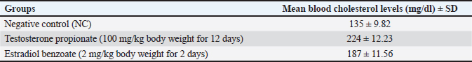
Fig. 1. Vaginal cytology of rats following hormonal induction at 12, 24, and 36 hours after injection. Representative photomicrographs (1,000× magnification) show the predominance of cornified epithelial cells, indicating persistent estrus (estrous arrest) in the testosterone propionate (TP) and estradiol benzoate (EB)–treated groups, which is consistent with the ovulatory dysfunction characteristic of PCOS models. *Notes: TPa=TP group, 12 hours post-TP injection; TPb=TP group, 24 hours post-TP injection; TPc=TP group, 36 hours post-TP injection; EBa=EB group, 12 hours post-EB injection; EBb=EB group, 24 hours post-EB injection; EBc=EB group, 36 hours post-EB injection. Cholesterol levels were analyzed as a primary precursor for androgen hormone synthesis to determine the impact of testosterone propionate and estradiol benzoate-induced PCOS on rats. Table 1 presents the average cholesterol levels for each treatment group. The results of the Kruskal–Wallis test for the groups induced with testosterone propionate (P1) at a dose of 100 mg/kg BW for 12 days and estradiol benzoate (P2) at a dose of 2 mg/kg BW for 2 days showed no significant difference (p ≥ 0.05) when compared to the NC group. Acute PCOS induction did not result in a statistically significant change in serum cholesterol levels in rats under the conditions of this study. Table 1. Cholesterol levels in rats with PCOS from all treatment groups.
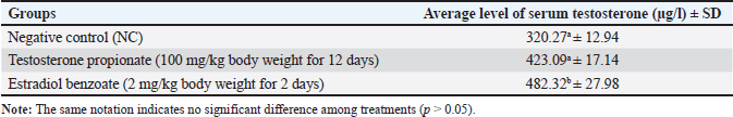
Testosterone levels in the PCOS animal modelTable 2 presents the results of the one-way ANOVA that indicate a significance level of less than 0.05, leading to further analysis using the LSD test. The average testosterone level in the NC group was 30.27%. Statistical analysis indicated that testosterone levels in the group induced with testosterone propionate at a dose of 100 mg/kg did not differ significantly (p ≥ 0.05) compared with the NC group. In contrast, the group induced with estradiol benzoate at a dose of 2 mg/kg for 2 days exhibited a significant difference (p ≤ 0.05) compared with the NC group. Table 2. Serum testosterone levels in rats with PCOS from all treatment groups.
DiscussionThis study successfully induced PCOS in rats, as shown by both persistent estrus and higher testosterone levels in both the TP and EB groups. Rats treated with TP (100 mg/kg body weight) showed extended estrus phases lasting 24 hours, whereas rats treated with EB (2 mg/kg) similarly displayed persistent estrus. This contrasts sharply with normal control rats, which typically have much shorter estrus phases (12 hours) followed by 48-hour diestrus periods (Cora et al., 2015). These results are consistent with those of other researchers, who found that TP-induced high androgen levels can throw off the normal estrous cycle, frequently causing prolonged diestrus (Osuka et al., 2019). The extended estrus and ovarian problems we observed are consistent with those observed in other rat models of PCOS with elevated androgen levels (Dumesic et al., 2015). Testosterone propionate remains biologically active for approximately 5 days due to its esterified structure, which gives it a 33-hour half-life (Turza et al., 2022). During the induction period, most rats transitioned to diestrus, likely because the slow absorption rate of testosterone propionate permitted continued FSH secretion and follicular development (Allan et al., 2006). The mechanism appears to involve testosterone propionate binding to hypothalamic androgen receptors, disrupting the normal pulsatile release of GnRH. This disruption triggers LH surges that ultimately lead to persistent estrus (Böttner et al., 2010). Other studies have documented similar hormonal disturbances, where excessive androgen levels impair GnRH secretion, resulting in anovulation and prolonged estrus phases (Walter et al., 2018). The analysis revealed distinct hormonal patterns between the treatment groups. Testosterone levels showed significant variation across groups (p ≤ 0.05), with the negative control animals maintaining average levels of 320,27µg/l. Interestingly, the TP group did not demonstrate a statistically significant increase in testosterone levels (p ≥ 0.05). This observation might reflect the metabolic processing of external testosterone by the body, as the esterified form of TP is gradually released and cleared (Walters et al., 2018; Turza et al., 2022). In stark contrast, rats treated with EB displayed substantially elevated testosterone levels (p ≤ 0.05). This finding supports existing research showing the capacity of EB to inhibit aromatase activity while promoting androgen buildup (Ishikawa et al., 2006). The estrogenic properties of EB appear to interfere with normal hypothalamic-pituitary-ovarian function, triggering excessive LH production, suppressing FSH, and ultimately creating a hyperandrogenic state, which is characteristic of PCOS (Palomba et al., 2017; Jin et al., 2019). This study uncovered important insights about cholesterol metabolism in patients with PCOS-like conditions. As the fundamental building block for steroid hormones, cholesterol regulation is disrupted in these hormonal imbalances. Treatment with both TP and EB altered normal GnRH patterns, causing LH spikes and FSH suppression (Imamichi et al., 2017). This hormonal shift stimulates LDL and HDL receptor activity in ovarian theca cells, essentially pulling more cholesterol into androgen production pathways (Strauss, 2019). However, the TP group's sustained diestrus phase-characterized by elevated progesterone, appears to interfere with cholesterol processing. This hormonal environment inhibits cholesterol esterification and reduces liver uptake, potentially explaining why the overall cholesterol measurements did not show dramatic changes (Jiang and Tian, 2017). Meanwhile, EB's induction of persistent estrus seems to downregulate the critical cholesterol-producing enzyme HMG-CoA reductase, effectively putting the brakes on cholesterol synthesis (Murphy and O'Brien, 2009). An unexpected finding emerged from the control group, where cholesterol readings averaged 135 mg/dl, which was notably higher than the established normal range of 20.4–87.6 mg/dl for female rats (Delwatta et al., 2018). This discrepancy might stem from not fasting before blood collection. Despite this anomaly, the data clearly showed that TP-treated rats maintained higher cholesterol levels than their EB-treated counterparts, which is consistent with previous research demonstrating the influence of testosterone on HDL breakdown and lipid oxidative stress (Gunness et al., 2016; Harris et al., 2020). Figure 2 captures the complex endocrine and metabolic disturbances characteristic of PCOS, building on the experimental findings discussed earlier. LH hypersecretion and FSH suppression—mirroring the hormonal patterns observed in both TP- and EB-induced models—drive ovarian theca cell hyperactivity, resulting in excessive androgen production (including testosterone). This aligns with the elevated testosterone levels measured in EB-treated rats, despite the more subtle effects of TP due to its metabolic clearance.
Fig. 2. Pathophysiological interplay of hormonal and metabolic dysregulation in patients with PCOS. The hormonal alterations observed in this study closely align with contemporary validated PCOS models that emphasize combined functional, endocrine, and ovarian endpoints. Recent work by Gökçek et al. (2025) demonstrated that the induction of PCOS in Wistar rats is characterized by elevated testosterone levels, disrupted estrous cyclicity, inflammatory imbalance, and impaired ovarian reserve, underscoring the importance of integrating hormonal parameters into the validation of the PCOS model. Our findings parallel this framework by confirming the presence of hyperandrogenism alongside estrous cycle disruption, thereby strengthening the model’s translational relevance. The illustration also highlights the lipid metabolism disruptions that explain the cholesterol variations observed in the study. Increased LDL/HDL receptor activity—consistent with the elevated cholesterol seen in TP-treated rats—reflects enhanced steroidogenesis and cholesterol uptake. Meanwhile, the insulin resistance depicted in the figure offers additional context for the metabolic dysregulation, as hyperinsulinemia can further stimulate ovarian androgen production while impairing glucose uptake. Notably, the figure’s depiction of reduced aromatase activity connects directly to the mechanism of action of EB, where estrogenic disruption leads to androgen accumulation. The clinical manifestations (hirsutism, insulin resistance, and so on) echo the phenotypic hallmarks of both human PCOS and the animal models studied. The experimental findings demonstrate a complex bidirectional relationship between cholesterol metabolism and hyperandrogenism in the pathophysiology of PCOS. The observed LH-mediated theca cell stimulation–evident in both TP- and EB-induced models not only enhances androgen synthesis but also increases cholesterol use for steroidogenesis. This metabolic interplay creates a self-reinforcing cycle in which dyslipidemia provides an abundant substrate for further testosterone production, whereas hyperandrogenism perpetuates metabolic disturbances. Notably, the elevated cholesterol levels in the TP group despite modest androgenic effects suggest that cholesterol dysregulation may precede and facilitate subsequent hormonal imbalances in PCOS progression. These mechanistic insights have important therapeutic implications. The inextricable link between endocrine and metabolic dysfunction in PCOS necessitates comprehensive treatment strategies that simultaneously address both disorder components. Isolated interventions targeting either hyperandrogenism or metabolic disturbances yield suboptimal outcomes, as residual pathological mechanisms continue to drive disease progression. Therefore, future therapeutic development should focus on multimodal approaches that simultaneously regulate gonadotropin secretion, improve insulin sensitivity, and normalize lipid metabolism. Such integrated strategies may prove more effective in breaking the PCOS pathogenesis vicious cycle and achieving sustained clinical improvement. ConclusionOur investigation of cholesterol-testosterone dynamics in a Rattus norvegicus PCOS model yielded several key insights. Contrary to expectations, neither testosterone propionate (100 mg/kg BW) nor estradiol benzoate (2 mg/kg BW) administration significantly altered cholesterol levels compared to controls—a finding that challenges conventional assumptions about hormonal regulation of lipid metabolism in PCOS. The hormonal profiles revealed a particularly intriguing pattern: while testosterone propionate-maintained androgen levels were comparable to controls (likely due to its gradual metabolic clearance), estradiol benzoate induction markedly increased testosterone levels. This paradoxical hyperandrogenic response to estrogenic stimulation reflects disruption of normal hypothalamic–pituitary–gonadal axis feedback, potentially involving aromatase inhibition or altered gonadotropin secretion. AcknowledgmentsThe authors express sincere thanks for the monetary support from the internal funding of Universitas Brawijaya for 2024. The assistance enabled the completion of the research variables in this article to be completed. Conflict of interestWe confirm that the submitted manuscript has no conflicts of interest of any kind, including (but not limited to) financial, personal, or other relationships with individuals or organizations that could be perceived as biasing the research. FundingResearch funding was provided through an internal grant from the Faculty of Veterinary Medicine, Universitas Brawijaya, in 2024. Authors' contributionsY. Oktanella designed and supervised the study and drafted the manuscript. Amelia D. Suryadi, Risa Yuliana, and Jamilaturrosyidah conducted the experiments and collected the data. Andre Giovannie performed the statistical analysis. N. R. Mahalita contributed to the molecular analysis. A. L. Poetranto reviewed and edited the manuscript. All authors have approved the final version. Data availabilityAll the data used to support the results of the study are included in the article, and there is no need for any other data sources. ReferencesAllan, C.M., Wang, Y., Jimenez, M., Marshan, B., Spaliviero, J., Illingwsorth, P. and Handelsman, D.J. 2006. Follicle-stimulating hormone increases the reserve of the primordial follicle in mature female hypogonadal mice. J. Endocrinol. 188, 549–557. Böttner, M., Leonhardt, S., Wuttke, W., Wedel, T. and Jarry, H. 2010. Expression of estrogen receptors in the hypothalamo-pituitary-ovarian axis in middle-aged rats after re-instatement of estrus cyclicity. Biogerontology 11, 75–85. Cora, M.C., Kooistra, L. and Travlos, G. 2015. Vaginal cytology of the laboratory rat and mouse: review and criteria for the staging of the estrous cycle using stained vaginal smears. Toxicol. Pathol. 43, 776–793. Delwatta, S.L., Gunatilake, M., Baumans, V., Seneviratne, M.D., Dissanayaka, M.L.B., Batagoda, S.S. Udagedara, A.H. and Walpola, P.B. 2018 Reference values for selected hematological, biochemical, and physiological parameters of Sprague-Dawley rats at the Animal House, Faculty of Medicine, University of Colombo, Sri Lanka. Animals 1, 250–254. Dumesic, D.A., Oberfield, S.E., Stener-Victorin, E., Marshall, J.C., Laven, J.S. and Legro, R.S. 2015. Scientific statement on the diagnostic criteria, epidemiology, pathophysiology, and molecular genetics of PCOS. Endocr. Rev. 36, 487–525. Gökçek, I. and Uyanık, G. 2025. Protective effects of thymol with hormonal, antiinflammatory and antioxidant pathways in lipopolysaccharide-induced ovarian damage in rats. Vet. Sci. Practices 20(1), 1–7; doi:10.1016/j.vsp.2025 Goodarzi, M.O., Dumesic, D.A., Chazenbalk, G. and Azziz, R. 2011. Polycystic ovary syndrome: etiology, pathogenesis and diagnosis. Nat. Rev. Endocrinol. 7, 219–231. Gunness, P., Williams, B.A., Gerrits, W.J.J., Bird, A.R., Kravchuk, O. and Gidley, M.J. 2016. Circulating triglycerides and bile acids are reduced by a soluble wheat arabinoxylan via modulation of bile concentration and lipid digestion rates in a pig model. Mol. Nutr. Food Res. 60, 642–651. Harris, K., Herati, A., Andriole, G., Castro-Santamaria, R., Freedland, S. and Moreira, D. 2020. Relationship between endogenous testosterone and lipid parameters: insight from REDUCE Study Group database. J. Sexual. Med. 17(Suppl. 1), S22–S23. Imamichi Y, Sekiguchi T, Kitano T, Kajitani T, Okada R, Inaoka Y, Terashima M, Kuwahara A, Yamada, K., and Miyamoto, K. 2017. Diethylstilbestrol administration inhibits theca cell androgen and granulosa cell estrogen production in immature rat ovary. Sci Rep 2017;7:8374. Ishikawa, T., Glidewell-Kenney, C. and Jameson, J. L. 2006. Aromatase-independent testosterone conversion into estrogenic steroids is inhibited by a 5α-reductase inhibitor. J. Steroid Biochem. Mol. Biol. 98(2), 133–138. Jiang, Y. and Tian, W. 2017. The effects of progesterones on blood lipids in hormone replacement therapy. Lipids Health Dis. 16, 219. Jin, J., Hu, Q., Xu, W., Zhu, W., Liu, B., Liu, J., Wang, W. and Zhou, H. 2019. Tanshinone IIA attenuates estradiol-induced polycystic ovarian syndrome in mice by ameliorating FSHR expression in the ovary. Exp. Ther. Med. 17, 3505–3507. Joham, A.E., Norman, R.J., Stener-Victorin, E., Legro, R.S., Franks, S., Moran, L.J., Boyle, J., Teede, H.J. and the International PCOS Network. 2022. Polycystic ovary syndrome. Lancet. Diabetes Endocrinol. 10, 668–680. Kurniawati, E.Y., Pramono, N., Hidayat, S.T. and Mahati, E. 2023. Assessment and experimental procedure of the polycystic ovary syndrome (PCOS) rat model: a review. J. Sain. P. Indones. 18, 242–256. Mukilan, R., Vijayaraman, M. and Subbiah, A.J. 2019. Dose determination of estradiol benzoate for PCOS induction. Int. J. Pharm. Biol. Sci. 9, 1131–1136. Murphy, M.S. and O'Brien, T. 2009. Dyslipidemias.In Pharmacology and Therapeutics. Eds., Waldman, S.A. and Terzic, A. Amsterdam, The Netherlands: Elsevier. Oktanella, Y., Untari, H., Wuragil, D., Agustina, G. and Pratama, D. 2023. Evaluation of renal disturbance in animal models of polycystic ovary syndrome. Open Vet. J. 13, 1003. Osuka, S., Nakanishi, N., Murase, T., Nakamura, T., Goto, M., Iwase, A. and Kikkawa, F. 2019. Animal models of polycystic ovary syndrome: a review of hormone-induced rodent models focused on hypothalamus–pituitary–ovary axis and neuropeptides. Reprod. Med. Biol. 18, 151–160. Oyebanji, O.G., Asaolu, M.F. and Amonimo, E.O. 2018. Hormonal imbalance in polycystic ovarian syndrome in teaching hospitals in Ekiti, Nigeria. Open. J. Obstet. Gynecol. 8, 1456–1464. Palomba, S., Daolio, J. and La Sala, G.B. 2017. Oocyte competence in polycystic ovary syndrome. Trends Endocrinol. Metab. 28, 186–198. Rocha, A.L., Oliveira, F.R., Azevedo, R.C., Silva, V.A., Peres, T.M., Candido, A.L., Gomes, K.B. and Reis, F.M. 2019. Recent advances in understanding and managing polycystic ovary syndrome F1000Res. 8, 565. Rosenfield, R.L. and Ehrmann, D.A. 2016. Pathogenesis of polycystic ovary syndrome (PCOS): revision of the hypothesis of PCOS as functional ovarian hyperandrogenism. Endocr. Rev. 37, 467–520. Sparić, R., Zlatar, J., Nikolić, L., Opalić-Palibrk, M., Radić, L., Bjekić-Macut, J., Ognjanović, S. and Macut, D. 2024. Clinical manifestations of polycystic ovary syndrome. Medicinska Istraživanja 57(4), 93–102. Singh. 2005. Persistent estrus rat models of polycystic ovary disease: an update. Fertil. Steril. 84, 1228–1234. Strauss JF. 2019. Organization of ovarian steroidogenic cells and cholesterol metabolism, 3rd ed. In The Ovary. Eds., Leung, P.C.K. and Adashi, E.Y. London,UK: Academic Press, pp. 83–94. Teede, H.J., Misso, M.L., Costello, M.F., Dokras, A., Laven, J.S.E., Moran, L.J., Piltonen, T., Norman, R.J. and International PCOS Network. 2018. Recommendations from the International Evidence-Based Guideline for the Assessment and Management of Polycystic Ovarian Syndrome. Hum. Reprod. 33, 1602–1618. Turza, A., Popescu, V., Mare, L. and Borodi, G. 2022. Structural aspects and intermolecular energy for some short testosterone esters. Mater. (Basel). 15, 7245. Walters, K.A., Gilchrist, R.B., Ledger, W.L., Teede, H.J., Handelsman, D.J. and Campbell, R.E. 2018. New Perspectives on the Pathogenesis of PCOS: neuroendocrine Origins. Trends Endocrinol. Metab. 29, 841–852. | ||


| How to Cite this Article |
| Pubmed Style Oktanella Y, Suryadi AD, Yuliana R, Jamilaturrosyidah J, Giovanni A, Mahalita NR, Poetranto AL. Cholesterol and testosterone dynamics in a polycystic ovary syndrome animal model. Open Vet. J.. 2026; 16(3): 1885-1892. doi:10.5455/OVJ.2026.v16.i3.44 Web Style Oktanella Y, Suryadi AD, Yuliana R, Jamilaturrosyidah J, Giovanni A, Mahalita NR, Poetranto AL. Cholesterol and testosterone dynamics in a polycystic ovary syndrome animal model. https://www.openveterinaryjournal.com/?mno=303929 [Access: March 31, 2026]. doi:10.5455/OVJ.2026.v16.i3.44 AMA (American Medical Association) Style Oktanella Y, Suryadi AD, Yuliana R, Jamilaturrosyidah J, Giovanni A, Mahalita NR, Poetranto AL. Cholesterol and testosterone dynamics in a polycystic ovary syndrome animal model. Open Vet. J.. 2026; 16(3): 1885-1892. doi:10.5455/OVJ.2026.v16.i3.44 Vancouver/ICMJE Style Oktanella Y, Suryadi AD, Yuliana R, Jamilaturrosyidah J, Giovanni A, Mahalita NR, Poetranto AL. Cholesterol and testosterone dynamics in a polycystic ovary syndrome animal model. Open Vet. J.. (2026), [cited March 31, 2026]; 16(3): 1885-1892. doi:10.5455/OVJ.2026.v16.i3.44 Harvard Style Oktanella, Y., Suryadi, . A. D., Yuliana, . R., Jamilaturrosyidah, . J., Giovanni, . A., Mahalita, . N. R. & Poetranto, . A. L. (2026) Cholesterol and testosterone dynamics in a polycystic ovary syndrome animal model. Open Vet. J., 16 (3), 1885-1892. doi:10.5455/OVJ.2026.v16.i3.44 Turabian Style Oktanella, Yudit, Amelia Dea Suryadi, Risa Yuliana, Jamilaturrosyidah Jamilaturrosyidah, Andre Giovanni, Nabilla Rizky Mahalita, and Anna Listya Poetranto. 2026. Cholesterol and testosterone dynamics in a polycystic ovary syndrome animal model. Open Veterinary Journal, 16 (3), 1885-1892. doi:10.5455/OVJ.2026.v16.i3.44 Chicago Style Oktanella, Yudit, Amelia Dea Suryadi, Risa Yuliana, Jamilaturrosyidah Jamilaturrosyidah, Andre Giovanni, Nabilla Rizky Mahalita, and Anna Listya Poetranto. "Cholesterol and testosterone dynamics in a polycystic ovary syndrome animal model." Open Veterinary Journal 16 (2026), 1885-1892. doi:10.5455/OVJ.2026.v16.i3.44 MLA (The Modern Language Association) Style Oktanella, Yudit, Amelia Dea Suryadi, Risa Yuliana, Jamilaturrosyidah Jamilaturrosyidah, Andre Giovanni, Nabilla Rizky Mahalita, and Anna Listya Poetranto. "Cholesterol and testosterone dynamics in a polycystic ovary syndrome animal model." Open Veterinary Journal 16.3 (2026), 1885-1892. Print. doi:10.5455/OVJ.2026.v16.i3.44 APA (American Psychological Association) Style Oktanella, Y., Suryadi, . A. D., Yuliana, . R., Jamilaturrosyidah, . J., Giovanni, . A., Mahalita, . N. R. & Poetranto, . A. L. (2026) Cholesterol and testosterone dynamics in a polycystic ovary syndrome animal model. Open Veterinary Journal, 16 (3), 1885-1892. doi:10.5455/OVJ.2026.v16.i3.44 |

