| Original Article | ||
Open Vet. J.. 2023; 13(4): 443-450 Open Veterinary Journal, (2023), Vol. 13(3): 443–450 Original Research A retrospective histopathological survey on canine lymphomas subtypes of Porto DistrictKatia Pinello1*, Marta Santos2, Patrícia Dias-Pereira3, João Niza-Ribeiro1, and Augusto de-Matos41Departamento de Estudo de Populações, Vet-OncoNet, ICBAS, Instituto de Ciências Biomédicas Abel Salazar, Universidade do Porto, Porto, Portugal 2Departamento de Microscopia, Laboratório de Histologia e Embriologia, Serviços de Diagnóstico Citológico e Hematológico, ICBAS, Instituto de Ciências Biomédicas Abel Salazar, Universidade do Porto, Porto, Portugal 3Departamento de Patologia e Imunologia Molecular, ICBAS, Instituto de Ciências Biomédicas Abel Salazar, Universidade do Porto, Porto, Portugal 4Departamento de Clínicas Veterinárias, ICBAS, Instituto de Ciências Biomédicas Abel Salazar, Universidade do Porto, Porto, Portugal *Corresponding Author: Katia Pinello. Departamento de Estudo de Populações, Vet-OncoNet, ICBAS, Instituto de Ciências Biomédicas Abel Salazar, Universidade do Porto, Porto, Portugal. Email: kcpinello [at] icbas.up.pt Submitted: 12/07/2022 Accepted: 15/03/2023 Published: 13/04/2023 © 2023 Open Veterinary Journal
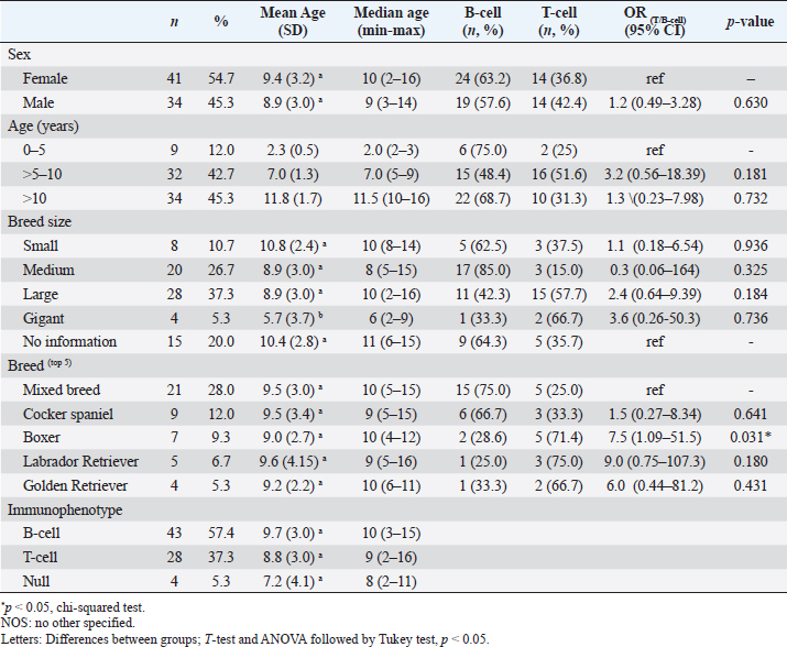
AbstractBackground: Lymphomas are dogs' most common hematopoietic neoplasms and represent a heterogeneous group, as occurs in humans. Considering the role of dogs as models of human lymphomas and the geographical correlation of the cases of canine and human lymphoma, it is important to continuously assess the epidemiological distribution of lymphoma subtypes in dogs. Aim: This study aimed to provide a survey of canine lymphoma subtypes diagnosed from 2005 to 2016 in the academic veterinary pathology laboratory of the University of Porto. Methods: A total of 75 canine lymphomas diagnosed by histopathology in the Porto district were included. All cases were immunophenotyped by CD3 and PAX5, classified according to the current classification WHO and coded with Vet-ICD-O-canine-1. Results: Mixed breed dogs were most common (28%), followed by Cocker Spaniels (12%), Boxers (9%), and Labrador Retrievers (6%). The mean age was 9.2 years (SD=3.3) (10.7 years for small, 8.9 years for medium and large, and 5.7 years for giant breed dogs, p < 0.05). Regarding sex, there was no difference in frequencies or mean age. B-cell lymphomas were more common (57.4%) than T-cell lymphomas (37.3%), and 5.3% were classified as non-B/non-T-cell lymphomas. Of the cases, 49% had a multicentric distribution, followed by splenic (22%), cutaneous (12%), alimentary (12%), and extranodal (3%) forms. The most common B-cell subtypes were diffuse large B-cell lymphoma (DLBCL) (16.3%) and large immunoblastic lymphoma (14%), while T-zone lymphoma (21.4%) and intestinal lymphoma (18%) were the most common T-cell lymphoma subtypes. Conclusion: Our study shows that the Porto district follows the international trend of higher prevalence of B-cell lymphomas in dogs, especially of the DLBCL subtype. Keywords: Canine, Lymphoma, Histopathology, Portugal, Vet-ICD-O-canine-1. IntroductionLymphomas are one of the most prevalent neoplasias in dogs worldwide and represent a significant practical challenge because they are a heterogeneous group of tumors with distinct biological behavior, clinical course, and treatment response, thus paralleling with human lymphomas (Vail et al., 2019). Such heterogeneity resulted in more than 30 entities listed in the World Health Organization (WHO) classification, each with several morphologic features and clinical outcomes (Valli et al., 2011, 2015). Hence, the simple diagnosis of “lymphoma” is clearly insufficient when correct treatment is envisaged. The accurate diagnosis and classification of lymphoma require (1) an exact location of the lesion, (2) appropriate selection and handling of sampled tissues, (3) evaluation of tissue microscopic architecture, (4) immunophenotyping, (5) clonality assessment, (6) assessment of cell size and nucleolus features, (7) mitotic index, (8) evaluation of invasion, and (9) clinical course (Valli et al., 2011, 2013, 2015). Several studies documented geographical differences in the prevalence of non-Hodgkin lymphoma (NHL) in humans (Muller et al., 2005; Chiu and Hou, 2015). While follicular lymphomas are more prevalent in Western countries, T-cell lymphomas prevail in Asia, while in the Middle East, intestinal lymphomas are particularly common (Muller et al., 2005). Epidemiologically, studies concerning geographical differences in canine lymphoma subtypes are increasingly relevant, both for the benefit of animals and for their characterization as models of human lymphoma (Marconato et al., 2013; Ito et al., 2014; Pinello et al., 2019). This study describes canine lymphomas diagnosed in the Porto district according to their main types and epidemiological characteristics to compare with the international distribution of this cancer. Material and MethodsIn a cross-sectional study, 75 formalin-fixed paraffin wax-embedded tissue samples of canine lymphomas were retrieved from the archives of the Laboratory of Veterinary Pathology of the School of Medicine and Biomedical Sciences, ICBAS—University of Porto. The cases were diagnosed between 2005 and 2016 and were chosen only from the samples from the District of Porto. Immunohistochemical evaluation of the lesions was performed in all samples with antibody anti-CD3 (rabbit polyclonal, Dako) for T-cells and antibody anti-PAX5 (monoclonal mouse, Leica Biosystems, Nussloch, Germany) for B-cells (Willmann et al., 2009) using an indirect immunoperoxidase assay Novolink Polymer® (Leica Mycrosystems, Nussloch, Germany). All cells with the expected staining pattern (membranous/cytoplasmic for CD3 and nuclear for PAX-5) were considered positive, regardless of the staining intensity. Lymphomas were classified as B- or T-cell types if at least 70% of neoplastic cells were labeled for the respective immunomarkers or null (O) when there was no immunolabelling (Ponce et al., 2010a). Histological subtypes were assigned based on tissue architecture, cell morphological characteristics (size, nucleus, and nucleoli characteristics), and mitotic index, according to the criteria defined by the WHO classification (Valli et al., 2015). The cases in which it was impossible to determine a classification were denominated No Other Specified (NOS). Each record was classified according to the anatomical localization (topography) and histological type (morphology) using the Vet-ICD-O-canine-1 classification system (Pinello et al., 2022). Data were collected on the breed, sex, and age of the animals. Pedigreed dogs were categorized by size (small, medium, and large). Mixed breed dogs were categorized by weight when available: small <11 kg, medium: 11.1–23 kg, large: 23.1–40 kg, and giant: >40 kg. Descriptive analysis was performed for the breed, sex, breed size, immunophenotype, morphology, and topography. Continuous data were tested for normality by the Shapiro–Wilk test. Numerical summaries included frequencies, percentages, mean with respective standard deviation (SD), and median with minimum and maximum values. T-test (for two categories) and analysis of variance (ANOVA) (more than two categories) followed by multiple comparison Tukey tests to compare differences between age groups were performed to describe differences in the mean age. B- and T-cell proportions were calculated, and the crude odds ratios (OR) were used to find the over-representation of immunophenotype (T-cell/B-cell). A Chi-squared test was used to assess p-values. Statistical significance was considered for a p-value less than 0.05. The analyses were performed using IBM SPSS Statistics software (Version 27). Ethical approvalThe study was approved by the Organism responsible for the Animal Welfare of the Institute of Biomedical Sciences of Abel Salazar of the University of Porto (ORBEA ICBAS-UP) on October 12, 2015 (approval number 066/2014). ResultsThe overall mean age of lymphomas was 9.2 years (SD=3.3), with no difference between the sexes. In addition, the distribution of lymphomas in dogs (Table 1) showed that there was no sex predisposition, and there were no differences in the age of diagnosis or the proportion of B- and T-cell proportions. Lymphomas were evenly distributed in three age categories: 0–5 years, 5–10 years, and older than 10 years. More than 88% occur after 5 years old. The highest T-cell proportion occurs between 5 and 10 years old, however not statically significant (Table 1). Regarding breed size, the highest proportion of the cases belongs to large dogs (Table 1). Compared to the group without size information (mixed breed dogs), there were no differences in the T-/B-cell proportions. However, when grouped, small and medium-sized dogs had a higher proportion of B-cell lymphomas, whereas the opposite was true in large and giant dogs (p=0.004). In addition, gigantic dogs had a statistically lower mean age at the time of the diagnosis than all others (p < 0.05) (Table 1). Regarding breeds, mixed-breed dogs were the most common, followed by Cocker spaniels, Boxers, and Labrador retrievers (Table 1). The mean and median ages were similar in all breeds but differed in proportions of B- and T-cell lymphomas, with Boxers presenting 7.5-fold higher odds of having a T-cell lymphoma. B-cell lymphomas were diagnosed most frequently (57.4%), followed by T-cell lymphomas (37.3%). The mean ages of dogs affected by B-cell lymphomas were slightly higher than for T-cell and null lymphomas; however, the difference did not reach statistical significance (Table 1). The predominant anatomic types (Table 2) were multicentric (49.3%), followed by splenic (22.7%), cutaneous and gastrointestinal lymphomas (12.0% each), and extranodal with fewer cases (3.0%). The mean age of dogs with splenic lymphomas (11 years old) was significantly higher than that of the other anatomic forms, except for the alimentary form (p < 0.05). Table 1. Descriptive analysis of canine lymphomas subtypes (B and T cells) cases by sex, age, breed size, and breeds.
Table 2. Descriptive analysis of canine lymphoma by anatomic forms: frequency, percentage, mean age with respect to SD, median age with the minimum and maximum values.
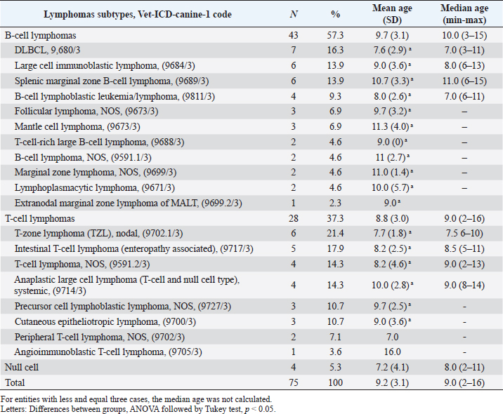
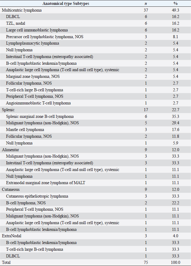
Regarding the subtypes of lymphomas (Table 3), the most common subtype of B-cell lymphoma was large diffuse cell (DLBCL) (16.3%), followed by large cell immunoblastic lymphoma and splenic marginal zone lymphoma (8.0%) and NOS (6.7%). In the T-cell lymphoma group, the most common were T-zone lymphoma (TZL) (21.4%), intestinal lymphoma (17.9%), NOS (14.3%), and large anaplastic lymphoma (14.3%). Considering the subtypes per anatomic types (Table 4), DLBCL, large cell immunoblastic lymphoma, and TZL were the most predominant in MC lymphomas (n =6, 16.2% each). Splenic marginal zone B-cell lymphoma was the most frequent in the splenic (n=6, 35.3%). Intestinal T-cell lymphoma (enteropathy associated) and malignant lymphoma (non-Hodgkin), NOS subtypes were the most frequent diagnosis in the alimentary lymphoma type (n=3, 33.3% each). And cutaneous epitheliotropic lymphoma was the most frequent subtype in the skin. Table 3. Descriptive analysis of canine lymphoma subtypes: frequencies, proportions, mean age with respect to SD, and median age with minimum and maximum values.
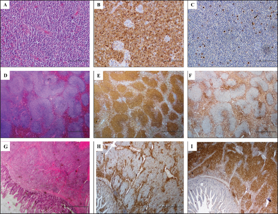
Figure 1–C shows the unusual diagnosis of angioimmunoblastic T-cell lymphoma with prominent capillaries through the node and the corresponding immunostaining. Figure 1D–F shows a case diagnosed as follicular B-cell lymphoma, grade 2. Figure 1G–I represents an intestinal marginal zone lymphoma of the mucosa-associated lymphoid tissue type (MALT). DiscussionThis study shows that in dogs from the district of Porto, Portugal, diagnosed with lymphoma between 2005 and 2016, (1) there was no sex predisposition and no difference in mean age between the sexes, (2) the size/breed of the dogs was inversely related to age at diagnosis, (3) the highest mean age at diagnosis was found in splenic lymphomas, (4) DLBCL lymphomas predominated, followed by TZLs. The WHO classification for canine lymphomas (Valli et al., 2015) used in this study requires information on the cell phenotype, location, and histologic architectural pattern of the lesion (Valli et al., 2015). These parameters correspond to those of the WHO classification for human lymphomas and thus allow comparative oncological studies. Unlike previous studies that verified a higher male proportion (Gavazza et al., 2001; Ponce et al., 2010b; Kimura et al., 2011; Vail et al., 2012), our series consisted mainly of females, although there was no statistical difference. In addition, the mean age at diagnosis was slightly higher than that reported by other authors (9.2 years as opposed to a range of 5.9–9 years) (Ponce et al., 2010b; Vezzali et al., 2010; Vail et al., 2012; Aupperle-Lellbach et al., 2022). Table 4. Distribution of canine lymphoma subtypes diagnosed per anatomical location.
Another difference from previous results was the fact that Cocker Spaniels were the second most common dog breed after mixed breeds, similar to a recent German study (Aupperle-Lellbach et al., 2022). However, this position is usually occupied by Boxers (Ponce et al., 2010b; Kimura et al., 2011). It is important to note that such differences in breed disposition between studies could be due to differences in the popularity and number of certain breeds in different countries, an issue that only will be dismissed with the animal census.
Fig. 1. Photomicrographs of canine lymphoma. A, B, and C: T-cell large angioimmunoblastic lymphoma. Lymph node. A: Prominent capillaries spread through the node. B: CD3 positive immunolabelling. C: PAX5 immunolabeling. D, E and F: B-cell center follicular lymphoma (grade 2). Lymph node. D: Large follicles, lack of mantle cell cuff. E: Neoplastic B-cells uniformly immunolabelled for anti-PAX5. F: CD3 immunolabeling. G, I, and H: B-cell MALT lymphoma. Intestine. G: Mucosa-associated lymphoid tissue composed predominantly of sheets and coalescent nodules of small lymphoid cells. I: PAX 5 positive immunolabeling of more than 90% of the neoplastic. cells. H: CD3 immunolabelling of a few T cells presented in the margins of the neoplastic lymphoid nodules. Consistent with previous studies (Priester, 1967), most boxers had T-cell lymphomas, with the multicentric form being the most common (Ponce et al., 2010b; Vezzali et al., 2010; Kimura et al., 2011). However, the second most common anatomic location was the splenic form; in contrast to most publications, the cutaneous and alimentary forms were more common than the splenic form (Vezzali et al., 2010). However, it is important to note that some authors did not consider the classification of splenic lymphomas (Vail et al., 2012; Valli et al., 2013, 2015). The immunophenotypic differentiation of B or T lymphomas has become a fundamental classification and prognostic tool (Ponce et al., 2004, 2010b; Valli et al., 2013). Traditionally, T-cell lymphomas are inevitably linked to a poor prognosis, whereas B-cell lymphomas were associated with a better prognosis (Ponce et al., 2010b; Valli et al., 2013). This generalization was, however, losing strength as other characteristics, namely subtypes, locations, and histological grades, were shown to have a higher prognostic value (Valli et al., 2006; Aresu et al., 2015). However, immunophenotypes remain of great importance in epidemiological studies, and human lymphomas have shown different risk factors among subtypes (Fisher and Fisher, 2004; Ekstrom-Smedby, 2006). The most frequent B-cell lymphoma was diffuse large cell (DLBCL), in agreement with the literature (Valli et al., 2015). The DLBCL category consists of a mixture of immunoblast and centroblast populations and includes the centroblastic, immunoblastic, NOS, and “T-rich” variants, differentiable by the number of immunoblasts (Valli et al., 2011, 2015). In Vet-ICD-O-canine-1, the centroblastic form is coded as “related” under the DLBCL, while the immunoblastic form has its own code. The “T-rich” variant, most common in horses and cats although poorly reported in dogs (Valli et al., 2015), can be mistaken for T-cell lymphoma due to its high number of T-cells and the fact that centroblasts and immunoblasts (B-cells) may represent less than 10% of the cell population (Valli et al., 2015). Among T lymphomas, the predominant subtype was the T-zone, followed by the intestinal lymphoma, similar to previous studies (Vezzali et al., 2010; Valli et al., 2011, 2013). The cutaneous T-cell lymphomas of this study were classified as epitheliotropic, characterized by being indolent, sometimes multifocal, and locally widespread, sometimes referred to as mycosis fungoides (Valli et al., 2015). Finally, the results found for null lymphomas (noB/noT) are in agreement with the literature, representing less than 5% of the cases (Caniatti et al., 1996; Fournel-Fleury et al., 1997; Zandvliet, 2016). In conclusion, the results of this study follow the international trend of a higher proportion of B-cell lymphomas in dogs, especially of the DLBCL subtype. AcknowledgmentsThe authors thank Professor Maria Fatima Gärtner for her invaluable support and collaboration. The authors would also thank for the techniques of the Laboratory of Veterinary Pathology of the University of Porto Alexandra Rêma and Fátima Carvalho. This research was supported by an international Ph.D. fellowship from CAPES (Coordenação de Aperfeiçoamento de Pessoal de Nível Superior), Brazil (CSF 0342-13-0). Conflict of interestThe authors declare that there is no conflict of interest. Author ContributionsKP, JNR, and AJM participated in the study’s inception and design. MS, PDP, and KP performed the lymphomas diagnostic and classification. In addition, KP procured digital photographs, data, and statistical analyses. KP, JNR, AJM, MS and PDP were involved in manuscript preparation. ReferencesAresu, L., Martini, V., Rossi, F., Vignoli, M., Sampaolo, M., Arico, A., Laganga, P., Pierini, A., Frayssinet, P., Mantovani, R. and Marconato, L. 2015. Canine indolent and aggressive lymphoma: clinical spectrum with histologic correlation. Vet. Comp. Oncol. 13, 348–362. Aupperle-Lellbach, H., Grassinger, J. M., Floren, A., Torner, K., Beitzinger, C., Loesenbeck, G. and Muller, T. 2022. Tumour incidence in dogs in Germany: a retrospective analysis of 109,616 histopathological diagnoses (2014-2019). J. Comp. Pathol. 198, 33–55. Caniatti, M., Roccabianca, P., Scanziani, E., Paltrinieri, S. and Moore, P. F. 1996. Canine lymphoma: immunocytochemical analysis of fine-needle aspiration biopsy. Vet. Pathol. 33, 204–212. Chiu, B.C. and Hou, N. 2015. Epidemiology and etiology of non-Hodgkin lymphoma. Cancer Treat. Res. 165, 1–25. Ekstrom-Smedby, K. 2006. Epidemiology and etiology of non-Hodgkin lymphoma—a review. Acta Oncol. 45, 258–271. Fisher, S.G. and Fisher, R.I. 2004. The epidemiology of non-Hodgkin's lymphoma. Oncogene 23, 6524–6534. Fournel-Fleury, C., Magnol, J.P., Bricaire, P., Marchal, T., Chabanne, L., Delverdier, A., Bryon, P.A. and Felman, P. 1997. Cytohistological and immunological classification of canine malignant lymphomas: comparison with human non-Hodgkin's lymphomas. J. Comp. Pathol. 117, 35–59. Gavazza, A., Presciuttini, S., Barale, R., Lubas, G. and Gugliucci, B. 2001. Association between canine malignant lymphoma, living in industrial areas, and use of chemicals by dog owners. J. Vet. Int. Med. 15, 190–195. Ito, D., Frantz, A.M. and Modiano, J.F. 2014. Canine lymphoma as a comparative model for human non-Hodgkin lymphoma: recent progress and applications. Vet. Immunol. Immunopathol. 159, 192–201. Kimura, K., Zanini, D., Nishiya, A., Dias, R. and Dagli, M. 2011. Morphology and immunophenotypes of canine lymphomas: a survey from the service of animal pathology, school of veterinary medicine and animal science, university of São Paulo, Brazil. Brazilian J. Vet. Pathol. 4, 199–206. Marconato, L., Gelain, M. E. and Comazzi, S. 2013. The dog as a possible animal model for human non-Hodgkin lymphoma: a review. Hematol. Oncol. 31, 1–9. Muller, A. M., Ihorst, G., Mertelsmann, R. and Engelhardt, M. 2005. Epidemiology of non-Hodgkin's lymphoma (NHL): trends, geographic distribution, and etiology. Annal. Hematol. 84, 1–12. Pinello, K., Baldassarre, V., Steiger, K., Paciello, O., Pires, I., Laufer-Amorim, R., Oevermann, A., Niza-Ribeiro, J., Aresu, L., Rous, B., Znaor, A., Cree, I.A., Guscetti, F., Palmieri, C. and Dagli, M.L.Z. 2022. Vet-ICD-O-Canine-1, a system for coding canine neoplasms based on the human ICD-O-3.2. Cancers 14, 1529. Pinello, K.C., Niza-Ribeiro, J., Fonseca, L. and de Matos, A.J. 2019. Incidence, characteristics and geographical distributions of canine and human non-Hodgkin’s lymphoma in the Porto region (North West Portugal). Vet. J. 245, 70–76. Ponce, F., Magnol, J.P., Ledieu, D., Marchal, T., Turinelli, V., Chalvet-Monfray, K. and Fournel-Fleury, C. 2004. Prognostic significance of morphological subtypes in canine malignant lymphomas during chemotherapy. Vet. J. 167, 158–166. Ponce, F., Marchal, T., Magnol, J.P., Turinelli, V., Ledieu, D., Bonnefont, C., Pastor, M., Delignette, M.L. and Fournel-Fleury, C. 2010a. A morphological study of 608 cases of canine malignant lymphoma in France with a focus on comparative similarities between canine and human lymphoma morphology. Vet. Pathol. 47, 414–433. Ponce, F., Marchal, T., Magnol, J.P., Turinelli, V., Ledieu, D., Bonnefont, C., Pastor, M., Delignette, M.L. and Fournel-Fleury, C. 2010b. A morphological study of 608 cases of canine malignant lymphoma in France with a focus on comparative similarities between canine and human lymphoma morphology. Vet. Pathol. 47, 414–433. Priester, W.A. 1967. Canine lymphoma: relative risk in the boxer breed. J. Natl. Cancer Inst. 39, 833–845. Vail, D.M., Pinkerton, M.E. and Young, K.M. 2012. Canine lymphoma and lymphoid leukemia. In Withrow and MacEwen´s small animal clinical oncology. St. Louis, MO: Elseviers, pp: 608–638. Vail, D.M., Thamm, D.H. and Liptak, J.M. 2019. 33—Hematopoietic tumors. In Withrow and MacEwen's small animal clinical oncology, 6th ed. Eds., Vail, D. M., Thamm, D. H. and Liptak, J. M. St. Louis, MO: W.B. Saunders, pp: 688–772. Valli, T., Kiupel, M. and Bienzle, D. 2015. Hematopoietic system. In Jubb, Kennedy & Palmer's pathology of domestic animals. Ed., Grant Maxie, M. St. louis, MO: Elsevier, Vol. 3, pp: 102–268. Valli, V.E., Kass, P.H., San Myint, M. and Scott, F. 2013. Canine lymphomas: association of classification type, disease stage, tumor subtype, mitotic rate, and treatment with survival. Vet. Pathol. 50, 738–748. Valli, V.E., San Myint, M., Barthel, A., Bienzle, D., Caswell, J., Colbatzky, F., Durham, A., Ehrhart, E.J., Johnson, Y., Jones, C., Kiupel, M., Labelle, P., Lester, S., Miller, M., Moore, P., Moroff, S., Roccabianca, P., Ramos-Vara, J., Ross, A., Scase, T., Tvedten, H. and Vernau, W. 2011. Classification of canine malignant lymphomas according to the World Health Organization criteria. Vet. Pathol. 48, 198–211. Valli, V.E., Vernau, W., de Lorimier, L.P., Graham, P.S. and Moore, P.F. 2006. Canine indolent nodular lymphoma. Vet. Pathol. 43, 241–256. Vezzali, E., Parodi, A. L., Marcato, P.S. and Bettini, G. 2010. Histopathologic classification of 171 cases of canine and feline non-Hodgkin lymphoma according to the WHO. Vet. Comp. Oncol. 8, 38–49. Willmann, M., Mullauer, L., Guija de Arespacochaga, A., Reifinger, M., Mosberger, I. and Thalhammer, J.G. 2009. PAX5 immunostaining in paraffin-embedded sections of canine non-Hodgkin lymphoma: a novel canine pan pre-B- and B-cell marker. Vet. Immunol. Immunopathol. 128, 359–365. Zandvliet, M. 2016. Canine lymphoma: a review. Vet. Q. 36, 76–104. | ||
| How to Cite this Article |
| Pubmed Style Pinello K, Santos M, Dias-pereira P, Niza-ribeiro J, De-matos A. A retrospective histopathological survey on canine lymphomas subtypes of Porto District. Open Vet. J.. 2023; 13(4): 443-450. doi:10.5455/OVJ.2023.v13.i4.6 Web Style Pinello K, Santos M, Dias-pereira P, Niza-ribeiro J, De-matos A. A retrospective histopathological survey on canine lymphomas subtypes of Porto District. https://www.openveterinaryjournal.com/?mno=67730 [Access: January 25, 2026]. doi:10.5455/OVJ.2023.v13.i4.6 AMA (American Medical Association) Style Pinello K, Santos M, Dias-pereira P, Niza-ribeiro J, De-matos A. A retrospective histopathological survey on canine lymphomas subtypes of Porto District. Open Vet. J.. 2023; 13(4): 443-450. doi:10.5455/OVJ.2023.v13.i4.6 Vancouver/ICMJE Style Pinello K, Santos M, Dias-pereira P, Niza-ribeiro J, De-matos A. A retrospective histopathological survey on canine lymphomas subtypes of Porto District. Open Vet. J.. (2023), [cited January 25, 2026]; 13(4): 443-450. doi:10.5455/OVJ.2023.v13.i4.6 Harvard Style Pinello, K., Santos, . M., Dias-pereira, . P., Niza-ribeiro, . J. & De-matos, . A. (2023) A retrospective histopathological survey on canine lymphomas subtypes of Porto District. Open Vet. J., 13 (4), 443-450. doi:10.5455/OVJ.2023.v13.i4.6 Turabian Style Pinello, Katia, Marta Santos, Patrícia Dias-pereira, João Niza-ribeiro, and Augusto De-matos. 2023. A retrospective histopathological survey on canine lymphomas subtypes of Porto District. Open Veterinary Journal, 13 (4), 443-450. doi:10.5455/OVJ.2023.v13.i4.6 Chicago Style Pinello, Katia, Marta Santos, Patrícia Dias-pereira, João Niza-ribeiro, and Augusto De-matos. "A retrospective histopathological survey on canine lymphomas subtypes of Porto District." Open Veterinary Journal 13 (2023), 443-450. doi:10.5455/OVJ.2023.v13.i4.6 MLA (The Modern Language Association) Style Pinello, Katia, Marta Santos, Patrícia Dias-pereira, João Niza-ribeiro, and Augusto De-matos. "A retrospective histopathological survey on canine lymphomas subtypes of Porto District." Open Veterinary Journal 13.4 (2023), 443-450. Print. doi:10.5455/OVJ.2023.v13.i4.6 APA (American Psychological Association) Style Pinello, K., Santos, . M., Dias-pereira, . P., Niza-ribeiro, . J. & De-matos, . A. (2023) A retrospective histopathological survey on canine lymphomas subtypes of Porto District. Open Veterinary Journal, 13 (4), 443-450. doi:10.5455/OVJ.2023.v13.i4.6 |


