


| Research Article | ||
Open Vet J. 2023; 13(5): 569-575 Open Veterinary Journal, (2023), Vol. 13(5): 569–575 Original Research Variation in palmaromedial articulations of carpometacarpal joints in Thoroughbred and Standardbred racehorsesAiman H. Oheida1*, Aiman A. Shalgum1, Abdulrhman M. Alrtib1, Ali O. Booker1, Khaled M. Ben-Naser2 and Helen M.S. Davies31Department of Anatomy, Histology and Embryology, Faculty of Veterinary Medicine, University of Tripoli, Tripoli, Libya 2Department of Animal Production, Faculty of Agriculture, University of Tripoli, Tripoli, Libya 3Department of Veterinary BioSciences, The University of Melbourne, Parkville, Australia *Corresponding Author: Aiman H. Oheida. Department of Anatomy, Histology and Embryology, Faculty of Veterinary Medicine, University of Tripoli, Tripoli, Libya. Email: a.oheida [at] uot.edu.ly Submitted: 27/02/2023 Accepted: 10/04/2023 Published: 10/05/2023 © 2023 Open Veterinary Journal
AbstractBackground: Carpal conformation is an important factor in relation to joint soundness. The equine carpometacarpal joint (CMCJ) was reported to have variations in its three palmaromedial articulations. Lacking one or more of these articulations has not been radiographically evaluated in Thoroughbred (TB) and Standardbred (SB) racehorses. Aim: The study aimed to identify the prevalence of the variation in the palmaromedial articulation of the carpometacarpal joint (PM-CMCJ) in TB and SB horses. Additionally, to detect the probability of having each of the three articulations within and between the breeds. Finally, to establish an anatomical description for the different patterns of the articulations in these horses. Methods: 313 dorsopalmar radiographs from 174 horses (117 TB and 57 SB) were used. Three articulations at PM-CMCJ were evaluated based on their presence or absence: the articulations between the second and third carpal bones (C2-C3), the second carpal-second metacarpal (C2-Mc2), and the second and third metacarpal (Mc2-Mc3) bones. The probability of each articulation was determined in the breeds. Depending on the presence/absence of one or more of these articulations in each horse, each group of horses that had the same patterns of articulation was gathered into one category. Results: Prevalence of variation in articulations of PM-CMCJ was identified in about 28% of the horses. SB showed a higher variation than TB throughout the comparisons. C2-C3 articulation was significantly the most common articulation, especially in TB (98%). The most common pattern of articulations (73%) was found in category I, which had three articulations whereas three horses grouped in category VI had no palmaromedial articulations. Conclusion: The variations in the articulations of PM-CMCJ in TB and SB racehorse might show a breed association. C2-C3 articulation was considerably the most frequent feature and category I was the common pattern of articulations in PM-CMCJ. The potential clinical effects of the varied patterns of the articulations require investigation. Keywords: Carpometacarpal joint, Radiology, Standardbreds, Thoroughbreds, Variation. IntroductionCarpal morphology was considered one of the factors that related to the incidence of carpal damage (Palmer, 1986; Oheida et al., 2022). Several undesirable conformational traits and defects have been identified and suggested to be associated with carpal pathology (Rooney and Prickett, (1966; Schneider, 1979; Stashak and Hill, 2002; Malone et al., 2003). Simultaneously, there were some preferable morphological characteristics that were considered normal or even a protective conformational trait from carpal injuries (McIlwraith et al., 2003; Weller et al., 2006; Oheida et al., 2022). However, most of the carpal conformational studies focused on the commonly damaged bones and joints, such as the third carpal bone, radial carpal bone, and distal extremity of the radius (McIlwraith et al., 1987; Garvican and Clegg, 2007; Oheida et al., 2022). Unlike these bones, which form the radiocarpal and the middle carpal joints (MCJ), the carpometacarpal bony components are rarely injured and thus had the least conformational studies, especially of their palmar aspects. The carpus is composed of three main articulations: the radiocarpal joint, the MCJ or intercarpal joint and the carpometacarpal joint (CMCJ) (Nickel et al., 1986; Pasquini et al., 1997; Frandson et al., 2003). CMCJ was formed by the articulation between the distal articular surfaces of the distal carpal row and the proximal articular surfaces of the metacarpal bones. The distal carpal row included the second (C2), third (C3), and the fourth (C4) carpal bones while the metacarpal bones were the second (Mc2), third (Mc3), and fourth (Mc4) metacarpal bones (Sisson, 1975; Nickel et al., 1986). At the palmaromedial region of the carpometacarpal joint (PM-CMCJ) there were small articulations between C2, C3, Mc2, and Mc3 via their articular facets (Sisson, 1975; Abdunnabi, 2006). These articulations were found to have some variations in their presence by several authors. Abdunnabi (2006), for instance, stated in his gross study that the small palmar facets which were for the articulation between C2 and C3 were absent in 21.4% (6/28) of horses. In addition, the small palmar facet of the distal articular surface of C2, which articulated with Mc3 (Riegel and Hakola, 1996; Budras et al., 2009) was absent from about 14.3% of the horses in that study (Abdunnabi, 2006). Regarding the palmar articulation between Mc2 and Mc3 proximally, Malone et al. (2003) reported that 42 horses out of a total of 177 horses had no palmar articulation. Despite the limited gliding movement of CMCJ (Frandson et al., 2003) a lack of the small facets and their articulations between the bones would have some effect on the stability of the joint and the soundness of its soft tissues (Malone et al., 2003). If so, then how would this be in carpi of racing horses such as Thoroughbreds (TB) and Standardbreds (SB) that are subject to enormous loading during high-speed racing? There is no doubt that understanding the biomechanical outcome of the variations in that part of PM-CMCJ needs to start with specific and adequate anatomical details. However, the information in the literature about the articulations of PM-CMCJ is scant especially in TB and SB racehorses that had differences in their carpal morphology (Oheida et al., 2019), racing gait/speed (Bramlage, 1983) and accordingly carpal pathology (Palmer, 1986; Pasquini et al., 1997). The current study hypothesized that the prevalence and the pattern of the articulations in PM-CMCJ varied considerably between horses. Therefore, the aims of the study were first to identify the prevalence of the variation in the articulations of PM-CMCJ in TB and SB racehorses using Dorsopalmar (DP) radiographs. The second purpose was to evaluate the probability of having each articulation despite the presence or absence of the others. Finally, to establish an anatomical description for the different patterns of the articulations and their probabilities of occurrence in these horses. Materials and MethodsArticulations of PM-CMCJFour boiled carpi from different horses were used for a gross study. It included locating articular facets and their articulations between the bones of interest as well as the related bones and tissues (Fig. 1). Two of the boiled carpi were mounted and radiographed for the same purpose. The three articulations which were evaluated between C2, C3, Mc2 and Mc3 in the PM-CMCJ were: 1. C2-C3 articulation: It was formed between the small facet of the lateral surface of the second carpal bone and the small palmar facet of the third carpal bone. 2. C2-Mc3 articulation: It was formed between the small facet of the distal articular surface of the second carpal bone and the small facet at the palmaromedial angle of the proximal articular surface of the third metacarpal bone. 3. Mc2-Mc3 articulation: It was formed between the small palmar facet of the lateral aspect of the proximal extremity of the second metacarpal bone and the small facet of the medial side of the proximal extremity of the third metacarpal bone.
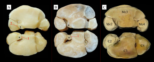
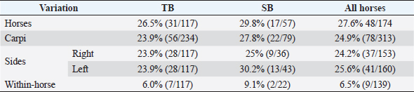
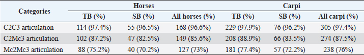
Fig. 1. Bony components of the CMCJ in horses. They include the proximal articular surfaces of the metacarpal bones (upper) and the distal articular surfaces of the distal row of the carpal bones (lower). A: CMCJ of a horse that had the three palmaromedial articulations. B: CMCJ of a horse without the palmar articulation between the second and the fourth metacarpal bones. C: CMCJ without any articulation at its palmaromedial region. C2: Second carpal bone; C3: Third carpal bone; C4: Fourth carpal bone; Mc2: Second metacarpal bone; Mc3: Third metacarpal bone; Mc4: Fourth metacarpal bone; 1: C2-C3 articulation; 2: Mc2-Mc3 articulation; *: Articular facets for C2-Mc3 articulation. Horses and radiographsThe DP carpal view was selected to be used in this retrospective study as it was precise and helpful in identifying carpal conformation (Bramlage and Auer, 2006; Oheida et al., 2016) including bone shape, size, and locating joint spaces (Morgan, 1993). 313 DP carpal radiographs from 174 racehorses (117 TB and 57 SB) were used. 153 of these radiographs were from the right carpi (117 TB and 36 SB) and 160 were from the left side (117 TB and 43 SB). All TB had DP radiographs of their right and left carpi. In 22 SB, radiographs of their two sides were available, whereas 35 horses had radiographs of only one side (14 right and 21 left). The gender of all the horses was reported (111 males and 6 females) except in 3 TB. All the horses were adults and their mean ages were 4.7 ± 2.48 years old (4.7 ± 2.69 in TB and 4.8 ± 2.0 in SB). The radiographs were collected from the Veterinary Clinic and Hospital in The Faculty of Veterinary and Agricultural Sciences, The University of Melbourne, The University Veterinary Teaching Hospital at The University of Sydney, and private racehorses. The radiographs were of good quality and include all carpal bones, distal radius, and proximal metacarpal bones. All horses were free from any radiographically discernible pathology orclinical signs related to the CMCJ based on their medical reports. Prevalence and comparisons of variationSince there was a possibility of variation in one side of a horse and also because of missing one of the two sides in some SB horses, the evaluations and comparisons were designed to be at the levels of horses (TB, SB, and all horses) and carpi (TB, SB and all horses) to obtain a more representative result. The prevalence of the anatomical variations in the palmaromedial articulation of CMCJ was evaluated in each breed (117 TB and 57 SB) and in all the horses (174). Prevalence was also evaluated in the carpi of each breed (234 TB and 79 SB) and all the carpi (313). Evaluating the variations in PM-CMCJ also included the differences between the right and the left carpi in each breed and all the horses. In the 139 horses (117 TB and 22 SB) where both their right and left carpal DP radiographs were available, the difference in the palmaromedial articulation between the two sides in each individual horse (within-horse) was tested on all those horses. Individual articulationsThe incidence of each of the three articulations was separately determined on the horses and the carpi. Comparisons of the probability between the three articulations were conducted within and between the breeds. CategoriesIn order to organize the widely different patterns of the three articulations anatomically each group of horses that had the same form of the articulation/s was gathered in one category. The probability of each category was determined in the horses and the carpi. Articulation between the dorsal articular facet of C2 and the facet of the distal surface of C3 (Rooney and Prickett type-B carpometacarpal configuration) was excluded from the study, as the DP view was not useful in the accurate evaluation of this articulation. Statistical analysisStatistical analysis was conducted using the statistical analysis system (SAS, 2002). Chi-squared test for categorical data was used to compare the variations in the articulations of PM-CMCJ between and within breeds and between individual articulations. Statistical results were considered significant when the p-value < 0.05. Ethical approvalNot needed as this was a retrospective study. ResultsThe three articulations in palmaromedial part of the CMCJ showed anatomical variation in about 28% (48/174) of all the horses and 25% (78/313) of all the examined carpi (Table 1). In the breeds, the variation was found in 26.5% of TB and in 29.8% of SB racehorses. Although there was no difference between the right and the left sides in TB, the statistical analysis revealed that the left PM-CMCJ articulation in SB was more susceptible to variation (30%) than the right side (25%). The difference between the right and the left sides (within-horse) was only found in 6.5% of the total 139 racehorses. This means that about 93.5% of the horses showed a bilateral morphological similarity in this part of the joint regardless of what variations were present. Individual articulationsIdentifying the prevalence of each of the three articulations showed that the incidence of the articulation between C2 and C3 was significantly the highest in both the horses and the individual carpi in the two breeds (Table 2). In the TB carpi comparison, for instance, C2-C3 articulation was found in almost 98% ( p < 0.0001) of the carpi and in SB horses comparison it was found in about 97% (p < 0.001) of the horses. Whereas the palmar articulation between Mc2 and Mc3 had the lowest probability, especially in SB horses in which it was detected in 70% (40/57). Table 1. Prevalence of the variation in the articulation of the PM-CMCJ using the carpal DP radiographs of 174 racehorses (117 TB and 57 SB).
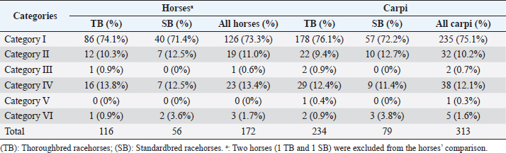
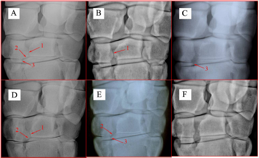
CategoriesThe three articulations, C2-C3, C2-Mc2, and Mc2-Mc3, in the palmaromedial part of CMCJ in TB and SB racehorses exhibited different patterns of articulations (Table 3). These patterns were grouped into six categories (Fig. 2): Category I: PM-CMCJ had all three articulations which were C2-C3, C2-Mc3, and Mc2-Mc3 articulations. Category II: PM-CMCJ had only C2-C3 articulation. Category III: PM-CMCJ had only Mc2-Mc3 articulation. Category IV: PM-CMCJ had only C2-C3 and C2-Mc3 articulations. Category V: PM-CMCJ had only C2-Mc3 and Mc2-Mc3 articulations. Category VI: PM-CMCJ did not have any of the three articulations. The probabilities of the six categories are shown in Table 3. The most common pattern of the articulation in PM-CMCJ was category I with percentages of 76% in all the examined horses and 75% in all the carpi. It was followed by category IV with percentages of 13% and 12% in the horses and the carpi, respectively. The rarest type of articulation was category V which was only identified in the right carpus of one TB. In five carpal radiographs from one TB and two SB, the three individual articulations of the palmaromedial part of CMCJ were absent (Fig. 2), category VI. DiscussionAlthough variations in the palmaromedial articulations of the CMCJ have been reported in two studies (Malone et al., 2003; Abdunnabi, 2006), neither of them included all three palmar articulations in PM-CMCJ in one investigation. The current study has not only detected the prevalence of the variation in PM-CMCJ in TB and SB but also the differences in the incidence of each articulation as well as describing their variant pattern of articulations. The prevalence of the variation in the articulation of PM-CMCJ was found in about 28% (48/174) of the horses. This means that the three articulations were consistently found in most of the horses (72%), especially in TB racehorses. Perhaps the presence of the three articulations was not unexpected as a common feature, but the study obviously highlighted a relatively high and wide level of variation. Thus, the potential morphometrical and biomechanical circumstances of the varied PM-CMCJ in more than a quarter of the horses are questionable, especially with the fact that all the evaluated CMCJ were free from any pathology. Nevertheless, the presence of the three articulations in PM-CMCJ seems to be necessary to guarantee a more functional interlocking wedge arrangement, which was assumed to dissipate the axial loading (Bramlage et al., 1988; Deane and Davies, 1995), promote joint stability, and protect soft tissues. If so, then it might be reasonable to consider it as the ideal bony conformation in terms of functional anatomy for these racing horses. However, the comparison between the two breeds identified a slightly higher probability of variation in SB (29.8%) than in TB (26.5%). This might reflect the specific anatomical response in each breed to the different racing gaits. Forelimbs of TB were loaded differently because of the asymmetrical gait during galloping (Barrey, 2001) whereas forelimbs of SB were loaded similarly during trotting (Bramlage, 1983). Accordingly, as long as the joint was subjected to a greater and varied loading, it may need to have more consistent anatomical features. This would explain why the comparison between the right and left sides in each breed presented a higher similarity in TB than in SB, where the two sides were sometimes different. Variation in C2-C3, C2-Mc3, and Mc2-Mc3 articulations has been previously evaluated. The first two articulations were studied by Abdunnabi (2006) while the third one was studied by Malone et al. (2003). Unlike these investigations, the present study was designed to evaluate the three articulations in order to obtain a more comprehensive vision of their articular relationships. Among these articulations, C2-C3 articulation was considerably the highest existing articulation in both horses and carpi comparisons. In the carpi of TB, for instance, it was found in almost 98% of the carpi ( p < 0.0001). This finding differed from the other study, which found C2-C3 articulation in about 86% (24/28) of the horses (Abdunnabi, 2006). Perhaps the difference between the two studies was because of the relatively small sample size and/or using horses from different breeds (10 TB, 3 SB, 2 Quarter horses, and 13 Ponies) in Abdunnabi’s study (2006). Theoretically, it could be assumed that C2-C3 articulation was the most important feature among the three articulations. Besides its potential role in dissipating the axial load, it may also play a more important role among the three articulations in preventing any abnormal movement during flexion of the joint. When the carpal joint was flexed, the proximal carpal row was displaced palmarly and laterally in relation to the distal carpal row (Olusa et al., 2020) with the involvement of collateral and intercarpal ligaments (Sledge, 1993; Whitton and Rose, 1997). As the distal surface of the radial carpal bone glided on the proximal and then on the palmar surface of C2, a greater force might be applied to the bone during the displacement. Hence, the presence of the connection between C2 and C3 would prevent any abnormal movements to support stability in the joint. However, in the actual status of the joint, the anatomical and biomechanical functions of any articulation would be more complicated depending on all of its related structures. Unexpectedly, the least incidence of articulation in PM-CMCJ was in the Mc2-Mc3 articulation, which was found in only 73% of the horses. Malone et al. (2003) identified an even lower incidence of this articulation (41%) in Arab horses and, thus, it was interpreted as a breed association. Despite the differences between the incidences of the three articulations, assuming the breed association would be also possible in the current study, as their probabilities were always higher in TB than in SB. Table 2. Probability of the individual-articulation in all horses and carpi.
Table 3. Incidence of the six different categories of the PM-CMCJ in the horses (116 TB and 56 SB) and in all carpi (234 from 117 TB and 79 from 57 SB).
Fig. 2. DP radiographs of horses. They show the six patterns or categories of the articulations in the PM-CMCJ. 1. C2-C3 articulation; 2. C2-Mc3 articulation; 3. Mc2-Mc3 articulation; A. Category I; B. Category II; C. Category III; D. Category IV; E. Category V; F. Category VI. The wide range in the patterns of variation between horses in PM-CMCJ has motivated this study to establish an objective anatomical database depending on the presence or absence of one or more of the three articulations in each single joint. Six different forms or categories of articulations were defined. Category I, which had all three articulations, was the most common pattern of articulations with percentages of 73% (126/172) in horses and 75% (235/313) in carpi comparisons. Contrary to this pattern, one TB and two SB that grouped in category VI were interestingly with no palmaromedial articulation. It was not clear whether this major lack of articulation was in relation to other changes in the surrounding structures or not. Presumably, there were some changes in the morphometry of the related bones and soft tissues because the size and shape of a bone in a healthy joint is unlikely to have the same morphometry with variations in the presence or absence of some features that determine its articulation with the surrounding bones. In terms of biomechanical function, it was even more difficult to understand how these joints compensated for the absence of all three articulations, especially in horses subjected to enormous forces during high-speed racing. In the necropsy study of Malone et al. (2003), lacking the palmar articulation between Mc2 and Mc3 was assumed to be highly associated with the incidence of severe carpometacarpal osteoarthritis in Arab horses. If this assumption was made based on the absence of only one of the three palmar articulations, then losing them all would be reasonably identified as a conformational defect. The stability of such joints in relation to uncommon anatomical and perhaps biomechanical properties would be uncertain, but if they remain undamaged under load as in the horses in the current study, further investigation of such carpal joints might assist in identifying more of the essential underlying anatomical aspects of this complex joint. ConclusionThere is no doubt that the study has offered an additional example of the anatomical complexity of the equine carpal joint that should be considered when evaluating radiographs. This retrospective study identified several variations in the articulations of PM-CMCJ in TB and SB racehorses. Some breed association was present as all the articulations showed less variation in TB than in SB. The C2-C3 articulation was unexpectedly the most frequent articulation and category I, where all three articulations were present was the common pattern of articulations in PM-CMCJ. Clinically relevant potential effects of the varied pattern of articulation remain to be investigated via morphometrical and biomechanical studies on sound and injured horses. This may help to increase the understanding of the joint biomechanics, soundness, and/or the horse's performance. AcknowledgmentsThe authors would like to thank the Libyan government for financial support; Brendan Kehoe, staff of the Radiology departments at The Faculty of Veterinary and Agricultural Sciences—The University of Melbourne and The University Veterinary Teaching Hospital—The University of Sydney for their assistance. Conflict of interestThe authors declare that there is no conflict of interest. ReferencesAbdunnabi, A. 2006. Morphometrical study of carpal bones in thoroughbreds, ponies and other breeds of horses. M. S. thesis, The University of Melbourne, Melbourne, Australia. Barrey, E. 2001. Inter-limb coordination. In Equine locomotion. Eds., Back, W. and Clayton, H. London, UK: W.B. Saunders, pp: 77–94. Bramlage, L.R. 1983. Surgical diseases of the carpus. Vet. Clin. North. Am. Large. Anim. Pract. 5, 261–274. Bramlage, L.R. and Auer, J.A. 2006. Diagnosis, assessment, and treatment strategies for angular limb deformities in the foal. Clin. Tech. Equine. Pract. 5, 259–269. Bramlage, L.R., Schneider, R.K. and Gabel, A.A. 1988. A clinical perspective on lameness originating in the carpus. Equine. Vet. J. Suppl. 6, 12–18. Budras, K.D., Sack, W.O. and Rock, S. 2009. Anatomy of the horse, 5th ed. Hannover, Germany: Schlutersche GmbH and Co. KG. Deane, N.J. and Davies, A.S. 1995. The function of the equine carpal joint: a review. N. Z. Vet. J. 43, 45–47. Frandson, R.D., Wilke, W.L. and Fails, A.D. 2003. Anatomy and physiology of farm animals, 6th ed. Philadelphia, PA: Lippincott Williams and Wilkins. Garvican, E. and Clegg, P. 2007. Clinical aspects of the equine carpal joints. U. K. Vet. 12, 1–5. Malone, E.D., Les, C.M. and Turner, T.A. 2003. Severe carpometacarpal osteoarthritis in older Arabian horses. Vet. Surg. 32, 191–195. McIlwraith, C.W., Anderson, T.M. and Sanschi, E.M. 2003. Conformation and musculoskeletal problems in the race horse. Clin. Tech. Equine. Pract. 2, 339–347. McIlwraith, C.W., Yovich, J.V. and Martin, G.S. 1987. Arthroscopic surgery for the treatment of osteochondral chip fractures in the equine carpus. J. Am. Vet. Med. Assoc. 191, 531–540. Morgan, J.P. 1993. Techniques of veterinary radiology, 5th ed. Ames, IA: Lowa State University Press. Nickel, R., Schummer, A., Seiferle, E., Frewein, J., Wilkens, H. and Wille, K.H. 1986. The locomotor system of the domestic mammals. Berlin, Germany: Verlag Paul Parey. Oheida, A.H., Alrtib, A.M., Abushhiwa, M.H., Philip, C.J. and Davies, H.M.S. 2022. Carpal morphometry in normal horses and horses with carpal bone pathology. AJVS 72(1), 1–8. Oheida, A.H., Alrtib, A.M., Shalgum, A.A., Shemla, M.E., Marzok, M.A. and Davies, H.M.S. 2019. Radiographic comparison of carpal morphometry in thoroughbred and standardbred race horses. AJVS 61(1), 74–82. Oheida, A.H., Anderson, G.A., Alrtib, A.M., Abushhiwa, M.H., Philip, C.J. and Davies, H.M.S. 2016. Carpal parameters on dorsopalmar radiographs of the equine carpus. J. Vet. Adv. 6(6), 1258–1268. Olusa, T.A.O., Akbar, Z., Murray, C.M. and Davies, H.M.S. 2020. Morphometric analysis of the intercarpal ligaments of the equine proximal carpal bones during simulated flexion and extension of cadaver limbs. Anat. Histol. Embryol. 50(1), 1–10. Palmer, S.E. 1986. Prevalence of carpal fractures in thoroughbred and standardbred racehorses. J. Am. Vet. Med. Assoc. 188, 1171–1173. Pasquini, C., Spurgeon, T. and Pasquini, S. 1997. Anatomy of domestic animals, 9th ed. Pilot Point, TX: Sudz Publishing. Riegel, R.J. and Hakola, S.E. 1996. Illustrated atlas of clinical equine anatomy and common disorders of the horse. Marysville, OH: Equistar publications, Ltd. Rooney, J.R. and Prickett, M.E. 1966. Foreleg splints in horses. Cornell. Vet. 56, 259–269. SAS. 2002. Statistical analysis system (SAS) 9.00. Cary, NC: SAS Institute Inc. Schneider, R.K. 1979. Incidence and location of fractures within the carpus. In Proceedings of the Annual Convention of the American Association of Equine Practitioners, pp: 145–146. Sisson, S. 1975. Equine syndesmology. In The anatomy of the domestic animals, 5th ed. Ed., Getty, R. Philadelphia, PA: W. B. Saunders Company, pp: 355–358. Sledge, C.B. 1993. Biology of the joint. In Textbook of rhenumatology. Eds., Kelley, W.N., Harris, E.D., Ruddy, S. and Sledge, C.B. Philadelphia, PA: W.B. Saunders Company, pp: 1–21. Stashak, T.S. and Hill, C. 2002. Conformation and movement. In Adam’s lameness in horses, 5th ed. Ed., Stashak, T.S. Philadelphia, PA: Lea and Febiger, pp: 80–92. Weller, R., Pfau, T., May, S.A. and Wilson, A.M. 2006. Variation in conformation in a cohort of national hunt racehorses. Equine. Vet. J. 38, 616–621. Whitton, R.C. and Rose, R.J. 1997. The intercarpal ligaments of the equine midcarpal joint, part 2: the role of the palmar intercarpal ligaments in the restraint of dorsal displacement of the proximal row of the carpal bones. Vet. Surg. 26, 367–373. | ||
| How to Cite this Article |
| Pubmed Style Oheida AH, Shalgum AA, Alrtib AM, Booker AO, Ben-naser KM, Davies HM. Variation in palmaromedial articulations of carpometacarpal joints in Thoroughbred and Standardbred racehorses. Open Vet J. 2023; 13(5): 569-575. doi:10.5455/OVJ.2023.v13.i5.9 Web Style Oheida AH, Shalgum AA, Alrtib AM, Booker AO, Ben-naser KM, Davies HM. Variation in palmaromedial articulations of carpometacarpal joints in Thoroughbred and Standardbred racehorses. https://www.openveterinaryjournal.com/?mno=144714 [Access: July 01, 2025]. doi:10.5455/OVJ.2023.v13.i5.9 AMA (American Medical Association) Style Oheida AH, Shalgum AA, Alrtib AM, Booker AO, Ben-naser KM, Davies HM. Variation in palmaromedial articulations of carpometacarpal joints in Thoroughbred and Standardbred racehorses. Open Vet J. 2023; 13(5): 569-575. doi:10.5455/OVJ.2023.v13.i5.9 Vancouver/ICMJE Style Oheida AH, Shalgum AA, Alrtib AM, Booker AO, Ben-naser KM, Davies HM. Variation in palmaromedial articulations of carpometacarpal joints in Thoroughbred and Standardbred racehorses. Open Vet J. (2023), [cited July 01, 2025]; 13(5): 569-575. doi:10.5455/OVJ.2023.v13.i5.9 Harvard Style Oheida, A. H., Shalgum, . A. A., Alrtib, . A. M., Booker, . A. O., Ben-naser, . K. M. & Davies, . H. M. (2023) Variation in palmaromedial articulations of carpometacarpal joints in Thoroughbred and Standardbred racehorses. Open Vet J, 13 (5), 569-575. doi:10.5455/OVJ.2023.v13.i5.9 Turabian Style Oheida, Aiman H., Aiman A. Shalgum, Abdulrhman M. Alrtib, Ali O. Booker, Khaled M. Ben-naser, and Helen M.s. Davies. 2023. Variation in palmaromedial articulations of carpometacarpal joints in Thoroughbred and Standardbred racehorses. Open Veterinary Journal, 13 (5), 569-575. doi:10.5455/OVJ.2023.v13.i5.9 Chicago Style Oheida, Aiman H., Aiman A. Shalgum, Abdulrhman M. Alrtib, Ali O. Booker, Khaled M. Ben-naser, and Helen M.s. Davies. "Variation in palmaromedial articulations of carpometacarpal joints in Thoroughbred and Standardbred racehorses." Open Veterinary Journal 13 (2023), 569-575. doi:10.5455/OVJ.2023.v13.i5.9 MLA (The Modern Language Association) Style Oheida, Aiman H., Aiman A. Shalgum, Abdulrhman M. Alrtib, Ali O. Booker, Khaled M. Ben-naser, and Helen M.s. Davies. "Variation in palmaromedial articulations of carpometacarpal joints in Thoroughbred and Standardbred racehorses." Open Veterinary Journal 13.5 (2023), 569-575. Print. doi:10.5455/OVJ.2023.v13.i5.9 APA (American Psychological Association) Style Oheida, A. H., Shalgum, . A. A., Alrtib, . A. M., Booker, . A. O., Ben-naser, . K. M. & Davies, . H. M. (2023) Variation in palmaromedial articulations of carpometacarpal joints in Thoroughbred and Standardbred racehorses. Open Veterinary Journal, 13 (5), 569-575. doi:10.5455/OVJ.2023.v13.i5.9 |

