


| Research Article | ||
Open Vet J. 2023; 13(10): 1239-1250 Open Veterinary Journal, (2023), Vol. 13(10): 1239–1250 Original Research Echocardiographic characterization of cardiac chambers and vasculatures in buffaloes (Bubalus bubalis) during diastolic and systolic phasesArafat Khalphallah1, Enas Elmeligy2, Khaled A. Khesruf3, Haitham H. Mohammed4, Abdulrahman Abdulkarim5*, Ashraf M. Abu-Seida6 and Al-lethie A. Al-lethie71Division of Internal Medicine, Department of Animal Medicine, Faculty of Veterinary Medicine, Assiut University, Assiut 71526, Egypt 2Veterinary Teaching Hospital, Faculty of Veterinary Medicine, Assiut University, Assiut 71526, Egypt 3Department of Animal diseases, Faculty of Veterinary Medicine, Aleppo University, Aleppo, Syria 4Department of Rangeland, Wildlife and Fisheries Management, Texas A&M University, College Station, Texas 77843, USA 5Faculty of Veterinary Medicine, Omar Almukhtar University, Bayda, Libya 6Department of Surgery, Anesthesiology and Radiology, Faculty of Veterinary Medicine, Cairo University, P.O. Box 12211, Giza, Egypt 7Department of Surgery, Anaesthesiology and Radiology, Faculty of Veterinary Medicine, Aswan University, Aswan 81528, Egypt *Corresponding Author: Abdulrahman Abdulkarim. Faculty of Veterinary Medicine, Omar Almukhtar University, Bayda, Libya. Email: abdulrahman.tahir [at] omu.edu.ly Submitted: 27/05/2023 Accepted: 06/09/2023 Published: 31/10/2023 © 2023 Open Veterinary Journal
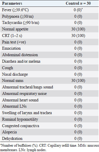
AbstractBackground: Ultrasonography had diagnostic importance in the evaluation of different diseases in buffaloes, including cardiovascular diseases. Aim: The current work describes the normal echocardiographic findings in healthy buffaloes, along with establishing reference values for echocardiographic dimensions for both sides of the heart, i.e., left and right ones. Methods: About 30 healthy adult buffaloes that belonged to private farms in Assiut, Egypt, were included in this study. Each animal underwent a complete clinical evaluation as well as hematological analyses, lipid profile indices, liver functions, cardio-thoracic radiography, and echocardiography to confirm no diseased conditions were detected. The study was conducted on healthy buffaloes (n=30) in Assiut Governorate, Egypt. Results: The obtained results reported healthy buffaloes with normal clinical findings as well as indices of blood pictures and serum biochemicals that were within the reference intervals. Radiography revealed a free reticulum and a well-defined diaphragm. The heart was seen as a typical radio-opaque organ. Ultrasonographically, using grayscale B-mode and M-mode, the heart was commonly imaged from the left fourth intercostal space. Different echocardiographic views were described, including the four chamber view, i.e., right atrium (RA), right ventricle (RV), tricuspid valve (TCV), left atrium, left ventricle, mitral valve, and interventricular septum (IVS), and the right ventricular outflow tract, i.e., RA, TCV, RV, pulmonary artery (PA), and pulmonary valve. Cross sections in each of the apex and base of the heart were described. Echocardiographic dimensions during cardiac diastole and systole, including diameters and wall thickness of each of the atria and ventricles, were demonstrated. Interventricular septal thickness wall thickness as well as diameters of the aorta and PA, were stated. Conclusion: The work tried to put reference values on the normal echocardiographic dimensions using 2-D B-mode gray scale ultrasonography in healthy adult buffaloes. These echocardiographic reference dimensions with normal echocardiographic imaging will be very helpful in enhancing the diagnostic efficacy of ultrasounds for recognizing abnormal findings related to cardiac disorders. Keywords: Buffaloes, Cardiac diastole and systole, Clinical findings, Echocardiographic dimensions, Right ventricular out flow tract. IntroductionThe water buffalo (Babalus bubalis) has been reported to constitute an important source for the production of milk and meat in Egypt. Buffaloes constituted approximately 185 million worldwide, 5.3 million of them being present in Egypt, according to FAO estimates from 2011 (Fooda et al., 2011). Early diagnosis of cardiac disorders in large ruminants was an essential issue for animal productivity and had economic importance. To reach a decision in most of these cases, a clinical examination alone was insufficient (Braun et al., 2007a). In recent decades, several diagnostic imaging modalities have been available in veterinary practice, including ultrasonography. Ultrasonography was a reliable, non-invasive, and extensively used method for diagnosing a variety of disorders in cattle (Braun et al., 2007b; Khalphallah et al., 2018) and horses (Hughes, 2006) in particular cardiac diseases. Moreover, ultrasonography had a wide range of diagnostic applications in buffaloes, including the respiratory system (Hussein et al., 2018), digestive and cardiovascular diseases (Acorda and Pilapil, 2008; Khalphallah et al., 2016a, 2016b, 2016c, 2016d, 2016e; Constante et al., 2017; Khalphallah et al., 2017, 2018; Chavez et al., 2021). Echocardiography was used to diagnose a variety of bovine cardiac diseases, including pericarditis (Braun, 2009; Khalphallah et al., 2018), endocarditis (Hollenberg et al., 2002), and cardiomyopathy (Schweizer et al., 2003). Echocardiography is an uncomplicated, simple technique to assess the size and function of the heart (Braun et al., 2001; Buczinski, 2009). Understanding normal echocardiographic anatomy provided the basis for recognizing and evaluating pathological changes (Schiller et al., 1989; Kriz and Rose, 1996; Braun, 2009). The spatial relationships between cardiac structures could be examined using the two-dimensional (2-D) display mode, and the motion of the chambers, valves, and septum could be examined using the M-mode (Carlsten, 1987). However, in order to discriminate between healthy animals and animals with cardiac abnormalities on the echocardiogram in any species, for each species, it was necessary to create accurate reference values (Boon, 2011). Echocardiography in cattle was primarily used to diagnose traumatic pericarditis (TP) (Braun et al., 2001; Braun, 2009; Khalphallah et al., 2015). There were earlier reports on the methods and standards for echocardiography in healthy cows (Hallowell et al., 2007), donkeys (Hassan and Torad, 2015), and camels (Tharwat et al., 2012). Radiographic technique was an effective method for detecting foreign metal objects, whereas metallic items like magnets were rarely seen by ultrasonography. Radiography was the best method for discovering metallic foreign objects outside or inside the reticulum. When diagnosing traumatic reticuloperitonitis (TRP), the foreign object’s location was the most obvious evidence. On the other hand, ultrasonography was the most suitable technique for imaging abscesses, fibrinous deposits, and inflammatory changes that could not be visualized radiographically. Though none of radiography or ultrasonography alone could provide a definitive diagnosis of TRP, both methods work well together (Braun et al., 1994, Kaske et al., 1994; Khalphallah et al., 2016c, 2016e, 2017). Several studies described the normal echocardiographic patterns in buffaloes (El-Khodery et al., 2010; Torad et al., 2017), but there were limitations, including the small number of examined buffaloes and the fact that some echocardiographic dimensions were not measured in healthy buffaloes. Furthermore, in comparison to other species of animals, cattle, and buffalo formed the largest proportion of the animal population. Approximately 90% of our overall patient cases at our veterinary teaching hospital were cattle and buffaloes, and because the data and literature about the ultrasonographic evaluation of the heart in normal Egyptian buffaloes were not commonly defined. From this point of view, the attention was mainly directed toward conducting this study that presented the normal sonographic findings of the heart. Consequently, the objective of the current study was to describe the normal echocardiographic findings in healthy buffaloes while establishing reference values for echocardiographic dimensions for both the right side and left side of the heart in healthy adult buffaloes. These findings will be very important in identifying any pathological alterations in the structure of this organ caused by other diseases. Materials and MethodsAnimalsThis study included thirty female non-pregnant buffaloes ranging in age from 5 to 7 years. These animals were selected from private farms in Assiut governorate, Egypt. Their total body weight varied from 330 to 400 kg. A total mixed ration was given to them. The farm used a free-stall method. SamplesA sample of whole blood was taken and kept on ethylenediamine tetraacetic acid at 4°C until analysis. Venous serum specimens were obtained on plain vacutainer tubes and stored at −20°C until analysis, according to Coles (1986). Clinical examinationAll of the animals were subjected to a complete clinical examination, as described by Cockcroft (2015). Clinical examination, includes the estimation of general signs of health such as temperature, respiratory rate, pulse rate, rumen movements, mucous membranes, lymph nodes, and skin. The digestive system and cardiothoracic organs were also clinically assessed. Complete blood countSeveral hematological indicators were estimated by a completely automated blood cell counter machine named Medonic CA620 Vet Hematology Analyzer (Stockholm, Sweden). They included red blood corpuscles (RBCs), hemoglobin (Hb), packed cell volume (PCV), and total leucocytic count (TLC). The differential leukocytic count (DLC) was calculated manually based on methods described by Coles (1986) and Latimer et al. (2011). Biochemical assaysThe Spectro Ultraviolet-Vis RS spectrophotometer (Labomed, Los Angeles, USA) was employed for determining serum levels of total protein, albumin, γ-glutamyl transferase (GGT), aspartate aminotransferase (AST), alkaline phosphatase (ALP), and triglycerides. Serum globulins were measured by subtracting albumins from total proteins; then, their values were used to measure the albumin/globulin ratio. In addition, all kits and reagents were bought in bulk from Gamma Trade Company in Egypt. Radiographic examinationRadiographic examination was performed for the control buffaloes based on techniques mentioned by Nägeli (1991) and Braun et al. (1993) using a fixed X-ray machine (Philips Super 80 CP, Germany). Ultrasonographical examinationAccording to Braun et al. (2001), El-Khodery et al. (2010), and Torad et al. (2017), ultrasonographic examination of the heart was performed on each animal using a 3.5 MHz sector transducer of apparatus [FF Sonic (Model UF-4000), Tokyo City, Japan]. After applying transmission gel, it was done on buffaloes that were standing up and not sedated. On the left side of the thorax, a region above the 2–6-intercostal spaces (ICSs) was clipped and shaved before being thoroughly cleaned. The skin was covered in coupling gel soon before the inspection. The left side caudal and cranial long axes of the heart were then sonographically inspected (Braun et al., 2001). The dimensions of the heart were estimated using B-mode 2-D grayscale ultrasonography via the caudal long-axis view (left side), while the dimensions of the aorta, as well as pulmonary artery (PA), were measured via the cranial long-axis view (left side). The transducer was put in the fourth left ICS mid-way between the elbow point and shoulder point. The echocardiographic techniques used in this study were an adaptation of those used in cattle (Weyman, 1994; Hallowell et al., 2007; Buczinski, 2009; Boon, 2011). The three 2-D images provided on the left side included the caudal long axis view (four-chamber view), the caudal long axis view of the left ventricular outflow tract (LVOT), and the caudal short axis view of the ventricles. The following measurements were reported as (2-D left parasternal images: left ventricular diameter during both diastole (LVDd) and systole (LVDs), interventricular septal thickness during both diastole (IVSd) and systole (IVSs), left ventricular wall thickness during both diastole (LVWd) and systole (LVWs), left atrial diameter during both diastole (LADd) and systole, left atrial wall thickness during both diastole (LAWd) and systole (LAWs), right ventricular diameter during both diastole (RVDd) and systole (RVDs), right ventricular wall thickness during both diastole (RVWd) and systole (RVWs), right atrial diameter during both diastole (RADd) and systole (RADs), right atrial wall thickness during both diastole (RAWd) and systole (RAWs), and PA diameter during both diastole (PAd) and systole (PAs). Statistical analysisThe Statistical Package for the Social Sciences Statistical Programme for Windows 10.0.1 was used to analyze the data (SPSS Inc., USA). The reported data were presented as mean ± standard deviation (SD). The paired t-test was used to detect the significance of the difference between means of echocardiographic measurements of diastolic status and those of systolic status either for the right or left side of the heart. The independent-sample t-test was used to determine the significance of differences between the means of echocardiographic measurements of the right side and those of the left side either in diastole status or in systole status. The significance of the differences was measured by Dunnett’s test at p < 0.05. The Kolmogorov–Smirnov normality test was used to determine whether the analyzed indices had a normal distribution. Every variable had a normal distribution. To determine the means, standard deviations, and range of clinical, laboratory, and echocardiographic values, descriptive statistics were used. Ethical approvalAll animal procedures carried out in this study complied with and were approved by the Faculty of Veterinary Medicine’s ethical committee, licensed number 06/2023/0107, as well as with Egyptian laws and the WOAH’s standards for the care and use of animals in research and education. ResultsThe examined buffaloes had no abnormal findings, whereas the authors did not report any abnormalities related to digestive disorders, respiratory problems, cardiovascular diseases, skin, lymph nodes, or mucous membranes (Table 1). The general signs of health, such as pulse, body temperature, respiratory rates, and ruminal motility, were within the reference intervals, respectively (Table 2). RBCs, Hb, PCV, TLC, and DLC, as complete blood picture components, were all within the reference ranges. A/G ratio, total proteins, albumins, globulins, GGT, ALP, AST, and triglycerides were among the biochemical measures whose serum values were also within normal ranges (Table 2). Radiography showed that the reticulum in healthy buffaloes was radio-opaque and free from any metal bodies. The reticulum and heart were radio-opaque structures, and the diaphragm was visualized as a prominent black line separating them. Adhesions between the heart and diaphragm or between the reticulum and diaphragm were not seen. The heart, as was a radio-opaque structure, had clear borders and a regular size and shape (Fig. 1a and b). Table 1. The common findings in the healthy buffaloes.
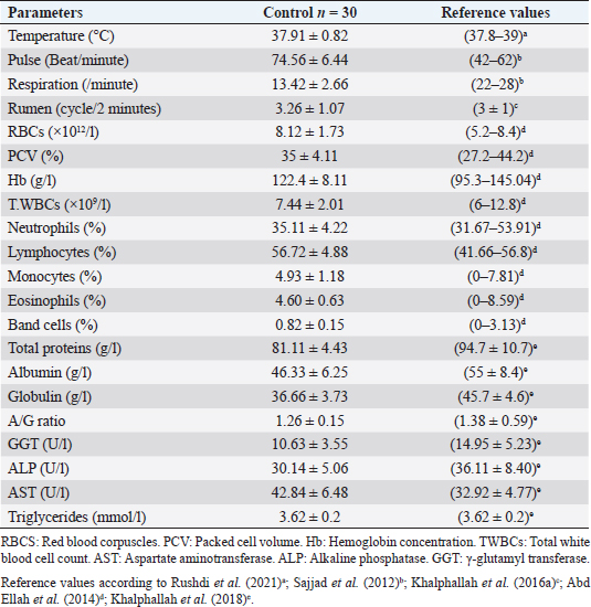
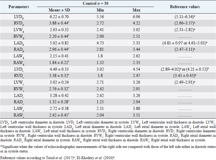
Ultrasonography of the normal heart in the healthy buffaloes was imaged from the left fourth ICS in midway between the elbow point and shoulder point as the transducer directed dorsally and slightly cranially toward the left fifth ICS showed the four chamber view either in diastole (Fig. 2a and b) or systole status (Fig 2c and d); right atrium (RA), right ventricle (RV), tricuspid valve (TCV), left atrium (LA), left ventricle (LV), mitral valve (MV), and interventricular septum (IVS). The heart was imaged from the left fourth ICS in midway between the elbow and shoulder points as the angle of the transducer directed dorsally and slightly cranially toward the third ICS, showing visualization of the right ventricular outflow tract (RVOT); RA, RV, TCV, PA, and pulmonary valve (PV) (Fig. 3). Cross sections at the apex of the heart, either in diastole (Fig. 4a) or systole status (Fig. 4b) showed RV and LV. Cross sections in the base of the heart showed RA, LA, TCV, and MV either in diastole (Fig. 5a) or systole status (Fig. 5b). Regular and strong myocardial contractility was clearly observed in both B-mode and m-mode 2-D-gray scale ultrasonography in all buffaloes. The echocardiographic measurements in healthy buffaloes reported significant changes between diastolic status and diastolic status, either for the right heart side dimensions or for the left heart side dimensions. They were within the reference intervals. Regarding the left heart side in buffaloes, the study mentioned that LVDd and LADd were significantly (p < 0.05) wider than LVDs and LADs, respectively. On the other side, LVWd and LAWd were significantly (p < 0.05) lower than LVWd and LAWd, respectively. The dimensions of the right side of the healthy heart in buffaloes reported that RVDd and RADd were significantly (p < 0.05) wider than RVDs and RADs, respectively. Moreover, RVWd and RAWd were significantly (p < 0.05) lower than RVWd and RAWd, respectively. IVSs were significantly (p < 0.05) thicker than IVSd. No significant changes were stated between Aortad and Aortad; however, PAd was significantly (p < 0.05) higher than PAs (Table 3). The healthy buffalo’s heart showed significant changes in echocardiographic measurements between the right side and left side of the heart, whereas values of LVDd, LVWd, LADd, LAWd, LVDs, LVWs, LADs, and LAWs were remarkably (p < 0.05) increased when they were compared with values of RVDd, RVWd, RADd, RAWd, RVDs, RVWs, RADs, and RAWs, respectively (Table 4). DiscussionEchocardiography was an effective technique for identifying inherited and acquired cardiac diseases in dairy cows and buffaloes (Acorda and Pilapil, 2008; Zarifi et al., 2012; Buczinski et al., 2013; Constante et al., 2017; Chavez et al., 2021). The current study did not report any abnormalities where the general signs of health, such as pulse, body temperature, respiration rates, and ruminal motility, were within the physiological intervals mentioned by Rushdi et al. (2021); Sajjad et al. (2012); Khalphallah et al. (2016a). The whole blood picture parameters and the estimated serum biochemical parameters were within the physiological intervals mentioned by Abd Ellah et al. (2014); Khalphallah et al. (2018). El-Khodery et al. (2010) advised that the dimensions and image or shape of the heart were crucial for determining reference values and comparing them to diseased cases. Furthermore, Strattner et al. (2002) concluded that clinical diagnosis alone was insufficient for diagnosing cardiac problems in cattle. Radiography in healthy buffaloes revealed an empty reticulum, a well-defined diaphragm, and a normal heart with a radio-opaque appearance. Braun et al. (1993) discussed the benefit of radiography in the diagnosis of TRP in cattle. At the same time, Braun et al. (1994) and Khalphallah et al. (2015, 2016c, 2016e, 2017) reported the diagnostic importance of ultrasonography and radiography in buffaloes and cows with TRP and/or TP. Table 2. Mean values of temperature, pulse rate, respiratory rate, rumen movements, whole blood picture indices, and serum biochemical parameters in healthy buffaloes.
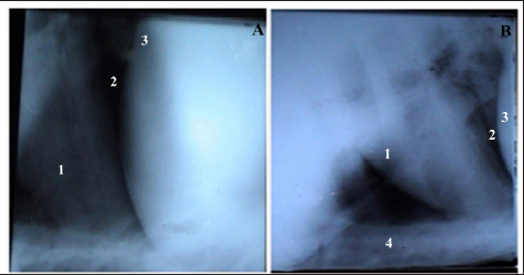
Fig. 1. Lateral radiographic view of the cranial abdomen (a) and the thorax (b) of a 3-year non-pregnant female buffalo showed the normal radiographic appearance of the reticulum, heart, and diaphragm. 1; Heart. 2; Diaphragm. 3; Reticulum. 4; Sternum.
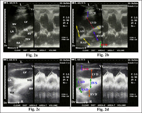
Fig. 2. Echocardiogram of caudal long axis view in adult healthy female non-pregnant (4-year-old) buffalo (left side) during diastole (a and b) and systole (c and d) using a 3.5 MHz sector 2-D-gray scale transducer (B-mode and M-mode). It was imaged from the left fourth ICS in midway between the elbow point and shoulder point as the transducer directed dorsally and slightly cranial toward the left fifth ICS. It showed a normal heart with regular, strong contractions and clear margins. The four chambers view was imaged where it included the RA, RV, TCV, IVS, LA, LV, LV, MV. Echocardiographic dimensions, either in diastole or in systole, were also visualized, including right ventricular diameter (RVD), right ventricular wall thickness (RVW), right atrial diameter (RAD), right atrial wall thickness (RAW), IVS, left ventricular diameter (LVD), left ventricular wall thickness (LVW), left atrial diameter (LAD) and left atrial wall thickness (LAW). Dr; Dorsal. Vt; Ventral. R; Right. L; Left. Buffalo echocardiography was a practical method that needed physical strength because buffaloes resisted the positioning and rotation of the transducer to produce uniform images (Torad et al., 2017; Chavez et al., 2021). The current study showed that the heart in healthy buffaloes was normally imaged ultrasonographically using 2-D B-mode and M-mode gray-scale from the left fourth ICS in the midway between shoulder point and elbow point as the transducer directed dorsally and slightly cranially toward the left fifth ICS where it showed the four-chamber view (RA, RV, LA, LV, TCV, MV, and IVS). These results coincided with the results reported by Braun et al. (2001) and Chavez et al. (2021) about echocardiography of the normal bovine heart. They also mentioned that RVOT of the heart was also imaged from the same site, but the angle of the transducer was pointed a little cranially and dorsally inward the third ICS. Braun et al. (2001) and El-Khodery et al. (2010) reported that in almost all cases, both atria, both ventricles, IVS, MV, and TCV could be seen in the caudal long-axis view of the heart from the left side. There was a better chance to visualize the heart from the right side than the left one. LVOT was also imaged in this view at the fourth ICS in all animals. The present results stated visualization of RVOT in which the heart was imaged through the left fourth ICS in midway between both shoulder point and elbow point as the angle of the transducer directed dorsally and slightly cranially toward the third ICS. Regular and strong myocardial contractility was observed in different modes of ultrasonography, i.e., B- and m-modes, in all examined healthy buffaloes. El-Khodery et al. (2010) reported the ultrasonographic appearance as well as dimensions of the heart in adult healthy buffaloes, in which the heart was examined through 3–6 ICSs on both sides of the thorax using a 3.5 MHz-convex transducer. They stated that in the caudal long-axis view from both sides, all of the heart’s chambers could be seen clearly. The right side’s caudal short-axis view allowed us to see the RVs and LVs. RVs and LVs as well as ventricular outflow, could be observed on the left side; however, the PA was difficult to see. On the other side, Torad et al. (2017) mentioned several limitations in their study about normal echocardiography in small and large buffaloes, including a small buffalo population was used, and the recommendation for further research on a larger population. Another limitation of the study was that it was impossible to verify that examined animals had normal heart features at post-mortem investigation because all animals survived and appeared to be in good health after a 6 months’ follow-up. In contrast, the current work avoided these limitations through the use of large numbers of animals as well as complete clinical and laboratory investigations to confirm a healthy heart. Torad et al. (2017) also added that the four chambers view was gotten from the left and right parasternal long axes views when the probe was placed at the fourth ICS from the right or the fifth ICS from the left. RVOT and LVOT were determined by rotating the probe caudally and cranially, respectively. Buffalo’s echocardiographic measurements appeared to be smaller than those previously recorded for cattle of comparable body weight (Hallowell et al., 2007).
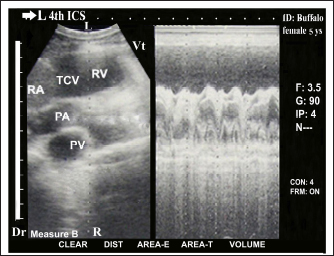
Fig. 3. Echocardiogram of cranial long axis view in adult healthy female non-pregnant (5-year-old) buffalo (left side) using a 3.5 MHz sector 2-D-gray scale transducer (B-mode and M-mode). It was imaged from the left fourth ICS in midway between the elbow point and shoulder point as the transducer directed dorsally and slightly cranial toward the left third ICS. It showed a normal heart with regular, strong contractions and clear margins. The right ventricular RVOT view was imaged whereas it included the RA, RV, TCV, PA, and PV. Dr; Dorsal. Vt; Ventral. R; Right. L; Left.
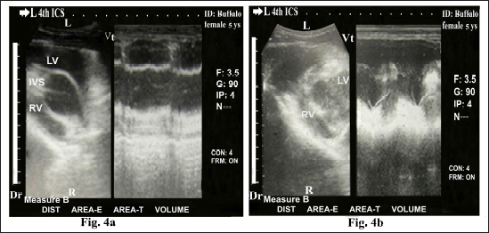
Fig. 4. Echocardiogram of caudal long axis view in adult healthy female non-pregnant (5-year-old) buffalo (left side) during diastole (a) and systole (b) using a 3.5 MHz sector 2-D-gray scale transducer (B-mode and M-mode). It was imaged from the left fourth ICS in the midway between the elbow point and shoulder point. It showed CS in the apex of the healthy heart with regular, strong contractions and clear margins. The RV, LV, and IVS were clearly visualized. Dr; Dorsal. Vt; Ventral. R; Right. L; Left.
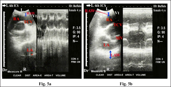
Fig. 5. Echocardiogram in adult healthy female non-pregnant (4-year-old) buffalo (left side) during diastole (a) and systole (b) using a 3.5 MHz sector 2-D-gray scale transducer (B-mode and M-mode). It was imaged from the left fourth ICS in the midway between the elbow point and shoulder point. It showed CS in the base of the heart with regular and strong contractions and clear margins. The RA, LA, TCV, and MV were clearly imaged. Echocardiographic dimensions, either in diastole or in systole, were also visualized, including right atrial wall thickness (RAW) and left atrial wall thickness (LAW). Dr; Dorsal. Vt; Ventral. R; Right. L; Left. Table 3. Echocardiographic measurements (cm) in healthy adult buffaloes between the diastolic phase and systolic phase, either on the left side or on the right side of the heart.
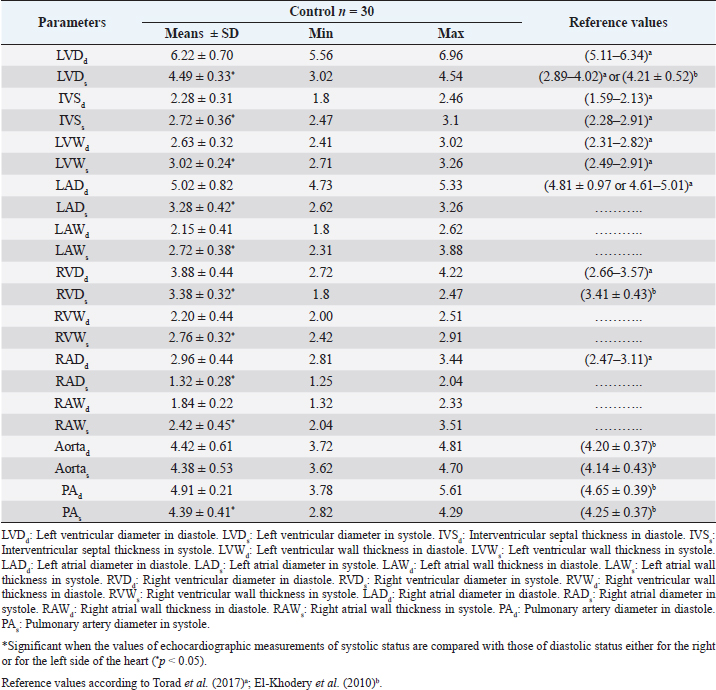
The echocardiographic measurements in healthy buffaloes reported in the present work reported significant changes between diastolic status and diastolic status either for the right heart side dimensions or for the left heart side dimensions. They were within the reference intervals mentioned by El-Khodery et al. (2010) and Torad et al. (2017). Regarding the left heart side in buffaloes, the current study mentioned that LVDd and LADd were significantly wider than LVDs and LADs, respectively. On the other side, LVWd and LAWd were significantly lower than LVWs and LAWs, respectively. The dimensions of the right side of the healthy heart in buffaloes reported that RVDd and RADd were significantly wider than RVDs and RADs, respectively. Moreover, RVWd and RAWd were significantly lower than RVWs and RAWs, respectively. IVSs were significantly thicker than IVSd. No significant changes were stated between Aortad and Aortad; however, PAd was significantly higher than PAs. These results are supported by Braun et al. (2001), El-Khodery et al. (2010), and Torad et al. (2017). El-Khodery et al. (2010) added that the size of the ventricles in healthy adult buffaloes was remarkably larger during diastole than their values in systole. Left ventricular wall and IVS were significantly greater in systole than diastole. The PA’s diameter showed a significant rise in diastole compared to systole, whereas the aorta’s diameter did not change significantly from systole to diastole. Table 4. Echocardiographic measurements (cm) in healthy adult buffaloes between the left and right sides of the heart, either in diastolic or systolic status.
In the present study, the healthy buffalo’s heart showed significant changes in echocardiographic measurements between the right and left sides of the heart, whereas values of LVDd, LVWd, LADd, LAWd, LVDs, LVWs, LADs, and LAWs were remarkably increased when they were compared with values of RVDd, RVWd, RADd, RAWd, RVDs, RVWs, RADs, and RAWs, respectively. El-Khodery et al. (2010) added that the dimensions or measurements of the LV during systole and diastole in normal female adult buffaloes were significantly higher than those of the RV during systole and diastole, respectively. Moreover, other studies revealed that little was known about buffalo heart disease. This could be explained by buffalo having the lowest prevalence of heart illness or by the lack of accurate diagnostic tools. Consequently, the detection and diagnosis of heart disorders were made easier by having a thorough understanding of normal heart dimensions and echocardiographic appearance. The initial stage of dealing with these diseases is an early diagnosis (Torad et al., 2017). ConclusionThe work set reference values for the normal echocardiographic dimensions were determined using 2-D of B-mode gray scale ultrasonography in healthy adult buffaloes. These echocardiographic reference dimensions with normal echocardiographic imaging will be very helpful in enhancing the diagnostic efficacy of ultrasounds for identifying the abnormal findings associated with cardiac diseases. The echocardiographic measurements in healthy buffaloes reported significant changes between diastolic status and diastolic status, either for the right heart side dimensions or for the left heart side dimensions. Moreover, the healthy buffaloes showed significant differences in echocardiographic measurements between the right and left sides of the heart. AcknowledgmentsNot applicable. Authors’ contributionsAll authors set the conception as well as the design of the study. AK, KAK, AAA, and AMA conducted the field work, ultrasonography, and animals’ investigations. HHM, AA, and EE collected laboratory samples and performed laboratory analyses. AK, KAK, HHM, and AAA adjusted the data and performed a statistical analysis. AMA, AA, EE, and AAA performed data curation, analysis, and interpretation. AK, EE, KAK, HHM, and AA drafted the manuscript. AK, AMA, AAA, and EE performed final writing, revision, and critical review. All authors have approved the final manuscript. FundingThe study did not receive any external fund. Data availabilityThe datasets used and/or analyzed during the current study are available from the corresponding author upon reasonable request. Data was also available after publishing in this journal. Conflict of interestThe authors declare that they have no competing interests. ReferencesAbd Ellah, M.R., Hamed, M.I. and Derar, R.I. 2014. Serum biochemical and hematological reference values for lactating buffaloes. Comp. Clin. Pathol. 23, 1179–1188. Acorda, J.A. and Pilapil, F.M.I.R. 2008. B-mode and M-mode ultrasonograpy of the heart in female buffaloes (Bubalus bubalis) in the Philippines. Philippine J. Vet. Med. 45(1), 7–13. Boon, J.A. 2011. Veterinary echocardiography, 2nd ed. Ames, IA: Wiley-Blackwell, pp: 610. Braun, U., Flückiger, M. and Götz, M. 1994. Comparison of ultrasonographic and radiographic findings in cows with traumatic reticuloperitonitis. Vet. Rec. 135(20), 470–478. Braun, U., Flückiger, M. and Nägeli, F. 1993. Radiography as an aid in the diagnosis of traumatic reticuloperitonitis in cattle. Vet. Rec. 132(5), 103–109. Braun, U., Lejeune, B., Schweizer, G., Puorger, M. and Ehrensperger, F. 2007a. Clinical findings in 28 cattle with traumatic pericarditis. Vet. Rec. 161(16), 558–563. Braun, U., Lejeune, B., Schweizer, G., Feller, B. and Flückiger, M. 2007b. Radiological findings in 28 heifers with pericarditis traumatica. Schweiz. Arch. Tierheilkd. 149(12), 563–565. Braun, U., Schweizer, T. and Pusterla, N. 2001. Echocardiography of the normal bovine heart: technique and ultrasonographic appearance. Vet. Rec. 148(2), 47–51. Braun, U. 2009. Traumatic pericarditis in cattle: clinical, radiographic and ultrasonographic findings. Vet. J. 182(2), 176–186. Buczinski, S., Tolouei, M., Rezakhani, A. and Tharwat M. 2013. Echocardiographic measurement of cardiac valvular thickness in healthy cows, cows with bacterial endocarditis, and cows with cardiorespiratory diseases. J. Vet. Cardiol. 15(4), 253–261. Buczinski, S. 2009. Cardiovascular ultrasonography in cattle. Vet. Clin. North Am. Food Anim. Pract. 25(3), 611–632. Carlsten, J.C. 1987. Two-dimensional real-time echocardiography in the horse. Vet. Radiol. 28, 76–87. Chavez M.J.B., Acorda, J.A. and Pilapil-Amante, F.M.I.R. 2021. B-mode, M-mode and Doppler echocardiographic features in dairy water buffalo calves. Philippine J. Vet. Med. 58(2),133–143. Cockcroft, P.D. 2015. Diagnosis and clinical reasoning in cattle practice. In Bovine medicine, 3rd ed. Eds., Cockcroft, P.D. West Sussex, Chichester, UK: Wiley-Blackwell, John Wiley & Sons Ltd., pp: 124–132. Coles, E.H. 1986. Veterinary clinical pathology, 4th ed. Philadelphia, PA: W.B. Saunders, pp: 46–47, 132–139. Constante J.L., Acorda, J.A., Tandang, A.G. and Pajas, A.M.G.A. 2017. B-Mode and M-mode ultrasonography of the heart of lactating dairy water buffaloes (Bubalus bubalis L.). Philippine J. Vet. Med. 54(1), 1–8. El-Khodery, S.A., Nassif, M.N. and Hassan, H.Y. 2010 Two dimension echocardiography in normal Buffaloes (Bubalus bubalis). J. Appl. Anim. Res. 37(1), 57–61. Fooda, T.A., Elbeltagy, A.R. and Laila, R.H. 2011. Assessment of Egyptian buffalo crossing with Pakistani and Italian buffaloes for some production traits. J. Am. Sci. 7(1), 269–276. Hallowell, G.D., Potter, T.J. and Bowen, I.M. 2007. Methods and normal values for echocardiography in adult dairy cattle. J. Vet. Cardiol. 9(2), 91–98. Hassan, E.A. and Torad, F.A. 2015. Two-dimensional and M-mode echocardiographic measurements in the healthy donkeys (Equus asinus). J. Equine Vet. Sci. 35(4), 283–289. Hollenberg, C., Strattner, A., Starke, A., Wohtsein, P. and Rehag, J. Efficiency of echocardiography in diagnosis of bovine vegetative endocarditis. In Proceeding of the XXII World Buatrics Congress, 2002, Germany, Hannover, pp: 179. Hughes, K. 2006. Diagnostic challenge: lethargy and weakness in an Arabian foal with cardiac murmurs. Ventricular septal defect (VSD). Aust. Vet. J. 84(6), 209–212. Hussein, H.A., Binici, C. and Staufenbiel, R. 2018. Comparative evaluation of ultrasonography with clinical respiratory score in diagnosis and prognosis of respiratory diseases in weaned dairy buffalo and cattle calves. J. Anim. Sci. Technol. 60, 29. Kaske, M., Midasch, A. and Rehage, J. 1994. Sonographic investigation of reticular contractions in healthy sheep, cows and goats and in cows with traumatic reticulo-peritonitis. Zentralbl. Veterinarmed. A 41(10), 748–756. Khalphallah, A., El-Sebaie, A.H. and Raghib, M.F. 2015. Approach for diagnosis of complicated traumatic reticuloperitonitis in cattle using ultrasonography. J. Adv. Vet. Res. 5(4), 157–164. Khalphallah, A., Elmeligy, E., Elsayed, H.K. and El-Hawari, S.F. 2016a. The ultrasonographic findings of the gastrointestinal tract and spleen in healthy Egyptian buffaloes (Bubalus bubalis). Assiut Vet. Med. J. 62(148), 39–47. Khalphallah, A., Abdelhakiem, M. and Elmeligy, E. 2016b. The ultrasonographic findings of the liver, gall bladder and their related vasculatures in healthy Egyptian buffaloes (Bubalus bubalis). Assiut Vet. Med. J. 62(149), 156–162. Khalphallah, A., Elmeligy, E., Elsayed, H.K., El-hawari, S.F. and Elrashidy, M.H. 2016c. Diagnostic significance of ultrasonography in complicated traumatic reticuloperitonitis in Egyptian buffaloes (Bubalus bubalis). Asian J. Anim. Vet. Adv. 11(6), 319–330. Khalphallah, A., Abu-Seida, A.M., Abdelhakiem, M., Elmeligy, E., Mahmoud, U.T. 2016e. Laboratory, radiographic and ultrasonographic findings of acute traumatic reticuloperitonitis in buffaloes (Bubalus Bubalis). Asian J. Anim. Vet. Adv. 11(11), 675–683. Khalphallah, A., Aref, N.M., Elmeligy, E. and El-Hawari, S.F. 2016d. Clinical and ultrasonographic observations of functional and mechanical intestinal obstruction in buffaloes (Bubalus bubalis). Vet. World 9(5), 475–480. Khalphallah, A., Aref, N.M., Abu-Seida, A.M., Elmeligy, E., Bayoumi, S.A., Al-Lethie, A.A. and Salman, D. 2018. Hepatobiliary diseases in buffalo (Bubalus bubalis): clinical, laboratory, and ultrasonographic findings. J. Vet. Sci. 19(4), 543–549. Khalphallah, A., Elmeligy, E., Elsayed, H.K., Abdellaah, A.B., Salman, D., Al-Lethie, A.A. and Bayoumi, S.A. 2017. Ultrasonography as a diagnostic tool in Egyptian buffaloes (Bubalus bubalis) with traumatic pericarditis. Int. J. Vet. Sci. 5(2), 159–167. Kriz, N.G. and Rose, R.J. 1996. Effect of detraining on cardiac dimensions and indices of cardiac function in horses. Proc. Annu. Conv. Am. Assoc. Equine Pract. 42, 96–97. Latimer, K.S., Mahaffey, E.A., Prasse, K.W. 2011. Duncan and Prasse’s veterinary laboratory medicine: clinical pathology, 5th ed. Ames, IA: Wiley-Blackwell, Blackwell Publishing, pp: 3–82. Nägeli, F. 1991. X-ray diagnostics in the traumatic reticuloperitonitis of cattle: technique, findings, interpretation and diagnostic significance. Dr Med Vet thesis, Faculty of Veterinary Medicine, University of Zurich, Switzerland. Rushdi, M., Hamed, M.I., Ibrahim, D.R. and Rateb, H.Z. 2021. Reference intervals for rectal temperature in water buffalo (Bubalus bubalis) Heifers. J. Adv. Vet. Res. 11(3), 180–182. Sajjad, A., Khan, M.S., Waqar-ul-Haq, F., Altaf, M. and Hussain, M.A. 2012. Incidence and therapeutic trials on primary and secondary bloats in buffaloes. Lasbela Univ. J. Sci. Technol. 1, 23–30. Schiller, N.B., Shah, P.M., Crawford, M., Schiller, N.B., Maurer, G., Ritter, S.B., Armstrong, W.F., Spotnitz, H., Cahalan, M., Quinones, M., Meltzer, R., Feinstein, S., Konstadt, S. and Seward, J. 1989. Recommendations for quantitation of the echocardiography committee on standards, subcommittee on quantitation of two-dimensional echocardiograms. J. Am. Soc. Echocard. 2, 358–367. Schweizer, Th., Sydler, T. and Braun, U. 2003. Kardiomyopathie, Endokarditis vulvularis thromboticans und perikarditis traumatic beim Rind- Klinische und echokardiographische Befunde an drei Fallberichten. Schweiz. Arch. Tierheilk. 145, 425–430. Strattner, A., Hollenberg, C., Starke, A., Wohtsein, P. and Rehage J. Two dimensional and m-mode echocardiography in German Holstein Friesian cattle. In Proceeding of XXII World Buiatrics Congress, 2002, Germany, Hannover, pp: 179. Tharwat, M., Al-Sobayil, F., Ali, A. and Buczinski, S. 2012. Echocardiography of the normal camel (Camelus dromedaries) heart: technique and cardiac dimensions. BMC Vet. Res. 8, 130. Torad, F.A., Amer, M.S., Shamaa, A.A. and Elsherpieny, E.A. 2017. Echocardiographic measurements and indices in normal adult buffalo (Bubalus bubalis). J. Appl. Anim. Res. 45(1), 336–341. Weyman, A.E. 1994. Cross-sectional scanning: technical principles and instrumentation. In Principles and practice of echocardiography, 2nd ed. USA: Philadelphia, PA, Lea & Febiger, pp: 29–56. Zarifi, M., Buczinski, S., Rezakhani, A., Mokhber Dezfouli, M.R. and Khonsha, A. 2012. Effect of lactation on functional and morphological echocardiographic variables in adult dairy cows. J. Vet. Cardiol. 14(3), 415–421. | ||
| How to Cite this Article |
| Pubmed Style Khalphallah A, Elmeligy E, Khesruf KA, Mohammed HHMH, Abdulkarim A, Abu-seida AM, Al-lethie AA. Echocardiographic characterization of cardiac chambers and vasculatures in buffaloes (Bubalus bubalis) during diastolic and systolic phases. Open Vet J. 2023; 13(10): 1239-1250. doi:10.5455/OVJ.2023.v13.i10.2 Web Style Khalphallah A, Elmeligy E, Khesruf KA, Mohammed HHMH, Abdulkarim A, Abu-seida AM, Al-lethie AA. Echocardiographic characterization of cardiac chambers and vasculatures in buffaloes (Bubalus bubalis) during diastolic and systolic phases. https://www.openveterinaryjournal.com/?mno=154347 [Access: July 06, 2025]. doi:10.5455/OVJ.2023.v13.i10.2 AMA (American Medical Association) Style Khalphallah A, Elmeligy E, Khesruf KA, Mohammed HHMH, Abdulkarim A, Abu-seida AM, Al-lethie AA. Echocardiographic characterization of cardiac chambers and vasculatures in buffaloes (Bubalus bubalis) during diastolic and systolic phases. Open Vet J. 2023; 13(10): 1239-1250. doi:10.5455/OVJ.2023.v13.i10.2 Vancouver/ICMJE Style Khalphallah A, Elmeligy E, Khesruf KA, Mohammed HHMH, Abdulkarim A, Abu-seida AM, Al-lethie AA. Echocardiographic characterization of cardiac chambers and vasculatures in buffaloes (Bubalus bubalis) during diastolic and systolic phases. Open Vet J. (2023), [cited July 06, 2025]; 13(10): 1239-1250. doi:10.5455/OVJ.2023.v13.i10.2 Harvard Style Khalphallah, A., Elmeligy, . E., Khesruf, . K. A., Mohammed, . H. H. M. H., Abdulkarim, . A., Abu-seida, . A. M. & Al-lethie, . A. A. (2023) Echocardiographic characterization of cardiac chambers and vasculatures in buffaloes (Bubalus bubalis) during diastolic and systolic phases. Open Vet J, 13 (10), 1239-1250. doi:10.5455/OVJ.2023.v13.i10.2 Turabian Style Khalphallah, Arafat, Enas Elmeligy, Khaled A. Khesruf, Haitham H. Mohammed H. Mohammed, Abdulrahman Abdulkarim, Ashraf M. Abu-seida, and Al-lethie A. Al-lethie. 2023. Echocardiographic characterization of cardiac chambers and vasculatures in buffaloes (Bubalus bubalis) during diastolic and systolic phases. Open Veterinary Journal, 13 (10), 1239-1250. doi:10.5455/OVJ.2023.v13.i10.2 Chicago Style Khalphallah, Arafat, Enas Elmeligy, Khaled A. Khesruf, Haitham H. Mohammed H. Mohammed, Abdulrahman Abdulkarim, Ashraf M. Abu-seida, and Al-lethie A. Al-lethie. "Echocardiographic characterization of cardiac chambers and vasculatures in buffaloes (Bubalus bubalis) during diastolic and systolic phases." Open Veterinary Journal 13 (2023), 1239-1250. doi:10.5455/OVJ.2023.v13.i10.2 MLA (The Modern Language Association) Style Khalphallah, Arafat, Enas Elmeligy, Khaled A. Khesruf, Haitham H. Mohammed H. Mohammed, Abdulrahman Abdulkarim, Ashraf M. Abu-seida, and Al-lethie A. Al-lethie. "Echocardiographic characterization of cardiac chambers and vasculatures in buffaloes (Bubalus bubalis) during diastolic and systolic phases." Open Veterinary Journal 13.10 (2023), 1239-1250. Print. doi:10.5455/OVJ.2023.v13.i10.2 APA (American Psychological Association) Style Khalphallah, A., Elmeligy, . E., Khesruf, . K. A., Mohammed, . H. H. M. H., Abdulkarim, . A., Abu-seida, . A. M. & Al-lethie, . A. A. (2023) Echocardiographic characterization of cardiac chambers and vasculatures in buffaloes (Bubalus bubalis) during diastolic and systolic phases. Open Veterinary Journal, 13 (10), 1239-1250. doi:10.5455/OVJ.2023.v13.i10.2 |

