


| Original Article | ||
Open Vet J. 2022; 12(6): 951-955 Open Veterinary Journal, (2022), Vol. 12(6): 951–955 Original Research Base-apex electrocardiographic examination in healthy cows of Chianina breedMaria Cicogna1, Piero Boni2, Tommaso Frigo3 and Domenico Caivano1*1Department of Veterinary Medicine, University of Perugia, Perugia, Italy 2Private Practitioner, Cannara, Italy 3Private Practitioner, Chiusi, Italy Submitted: 19/07/2022 Accepted: 10/11/2022 Published: 07/12/2022 *Corresponding Author: Domenico Caivano. Department of Veterinary Medicine, University of Perugia, Perugia, Italy. Email: domenico.caivano [at] unipg.it © 2022 Open Veterinary Journal
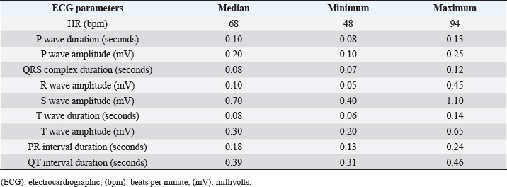
AbstractBackground: The electrocardiography is a useful diagnostic tool for the characterization of the cardiac rhythm in veterinary medicine. In cattle, standard electrocardiographic (ECG) exam is commonly performed using the base-apex lead system and reference values for ECG parameters have been reported for different breeds of cows. However, no ECG studies have been conducted in Chianina breed. Aim: To determine the feasibility and normal values for standard base-apex electrocardiography in clinically healthy Chianina cows. Methods: A standard base-apex ECG examination was acquired in 45 Chianina cows. All the cows were considered healthy based on history and physical examination. The following ECG parameters were evaluated: heart rate (HR); morphology of P wave, QRS complex, and T wave; amplitude and duration of P wave, QRS complex, and T wave; duration of PR and QT intervals. Correlations between ECG parameters and clinical variables were analyzed. Results: Morphology of the ECG waves/complexes and values for all ECG parameters recorded in the 45 clinically healthy Chianina cows were reported. Correlations between HR and body weight (BW), QT interval and BW, QT interval and HR, P wave amplitude and HR, PR interval and HR, R wave amplitude and age were observed. Statistical difference between HR in pregnant and non-pregnant cows was also found. Conclusion: Base-apex electrocardiogram is suitable for monitoring heart rhythm in clinically healthy Chianina cows and reference values for main ECG parameters have been reported for this breed. This study can be a useful contribution to the literature, updating current knowledge on the electrocardiography in cattle. Keywords: Arrhythmias, Cattle, Electrocardiogram, Heart. IntroductionThe electrocardiography is a non-invasive and inexpensive diagnostic tool that can be useful for the characterization of the cardiac rhythm in human and veterinary medicine. In adult cattle, a standard electrocardiographic (ECG) examination is commonly performed using the base-apex lead system (Rezakhani et al., 2004; Buczinski and Boerboom, 2010). This system allows to obtain clear waves/complexes and minimizes the effect of the animal movement during the recording (Deroth, 1980; Rezakhani et al., 2004). Therefore, base-apex ECG exam can be readily available in farms and useful to confirm or rule out the presence of cardiac arrhythmias in cattle (Buczinski and Boerboom, 2010). Previous studies reported reference values for ECG parameters for different breeds of cows such as Holstein (DeRoth, 1980; Rezakhani et al., 2004; Cedeno et al., 2016; Bonelli et al., 2019), Mithun (Sanyal et al., 2010), and crossbreed (Reddy and Sivajothi, 2016; Mohapatra et al., 2017; Areshkumar et al., 2018). To the authors’ knowledge, the feasibility and the reference values for ECG examination have not been previously reported in Chianina breed. The Chianina is an Italian cattle breed selected for its high-quality meat and it is present in different territories of the world (Italy, North and South America, and Australia). This breed is the largest and one of the oldest cattle breeds in the world. However, the scientific literature about cardiovascular assessment and cardiac diseases in Chianina cows is scarce (Caivano et al., 2021; Jacinto et al., 2022). Therefore, this study aims to establish the feasibility and normal values for base-apex ECG parameters in clinically healthy Chianina cows that could be different from normal range of other cattle breeds and useful to assess cardiac rhythm in this breed. Materials and MethodsAnimalsA total of 45 Chianina cows were recruited from four different farms of central Italy (Umbria). All the cows included in the study were considered healthy based on history and physical examination. Table 1. ECG parameters in 45 clinically healthy Chianina cows.
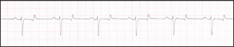
ECG examinationAll ECG examinations were recorded at the farms, the cows were kept in standing positions with helping of the owners and without sedation. The standard base-apex ECG examinations (ECG-1101G VET 5-lead, Carewell health care, Shenzhen, China) were acquired by positioning the positive electrode caudally to the olecranon, at the level of the fifth left intercostal space, and the negative electrode on the left jugular furrow in the lower 1/3 of the neck, as previously described (Rezakhani et al., 2004). The other electrodes were attached near the positive electrode. To obtain a good quality of the ECG tracings, a small amount of alcohol was used to optimize the contact. When the animals were quiet, ECG recording was acquired for 2 minutes and printed at 25 mm/second with a gain of 10 mm/mV. ECG parametersAll ECG tracings were analyzed, and the following parameters were assessed: heart rate (HR) (beats for minute, bpm), the morphology of P wave, QRS complex, and T wave (DeRoth, 1980; Rezakhani et al., 2004); amplitude (millivolts, mV) and duration (seconds) of P wave, amplitude (mV) and duration (seconds) of QRS complex, amplitude (mV) and duration (seconds) of T wave, and duration (seconds) of PR and QT intervals. Each parameter was calculated using five randomly selected heartbeats, and the mean of these five measurements was used for statistical analyses. Statistical analysisDistribution of the data was evaluated using the Shapiro–Wilk test. Because most of ECG parameters were not normally distributed, descriptive data regarding the ECG measurements were reported as median and range (minimum and maximum). Next, correlations of all ECG parameters with body weight (BW), HR, and age were examined by Spearman’s correlation test (Mukaka, 2012). Finally, the Mann–Whitney test was used to verify the difference between ECG parameters in pregnant and non-pregnant cows. Statistical analysis was performed using a commercial software (GraphPad Prism 5, La Jolla, CA) and statistical significance was set at p < 0.05. Fig. 1. ECG tracing recorded in a clinically healthy Chianina cow. Positive and “bifid” P waves, QRS complexes with “rS” configuration and “biphasic” T waves are evident in the electrocardiogram. HR is 60 bpm. Paper speed=25 mm/second; gain=10 mm/mV.
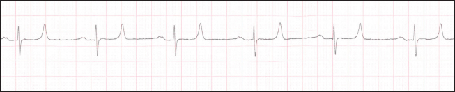
Ethical approvalThe study was approved by the Ethical Committee of the University of Perugia (19/2022), according to the D.L.116/92 and D. Lgs. 26/14. An owner’s written consent was obtained for ECG recording for all cows included in the study. ResultsCows were aged between 3 and 11 years (median age 6 years) and BW ranged between 710 and 980 kg (median 830 kg). All the cows have had at least one parturition and 24 of 45 cows were pregnant. ECG examination was carried out in all cows for a total of 45 ECG tracings and all animals tolerated the procedure well. All the ECG tracings were considered adequate for interpretation and no baseline artifacts were identified. All cows enrolled in the study showed sinus rhythm without presence of arrhythmias. ECG tracing analysis showed HR values between 48 and 94 bpm with a median of 68 bpm. Median, minimum, and maximum values for all ECG parameters are reported in Table 1. All cows showed a positive P wave with a “bifid” morphology in 10 of these. A QRS complex with “rS” morphology was recorded in 43 cows (Fig. 1), whereas the other 2 cows showed an “RS” morphology (Fig. 2). T wave polarity was positive in 38 cows and “biphasic” in 7 animals. Fig. 2. ECG tracing recorded in a clinically healthy Chianina cow. Positive and “bifid” P waves, QRS complexes with “RS” configuration, and positive T waves are evident in the electrocardiogram. HR is 60 bpm. Paper speed=25 mm/second; gain=10 mm/mV.
HR showed a moderate positive correlation (r=0.60, p < 0.0000) with BW, whereas the QT interval showed a moderate negative correlation (r=0.58, p < 0.0000) with BW and a high negative correlation with HR (r=0.86, p < 0.0000). Moreover, HR showed a low positive correlation with P wave amplitude (r=0.41, p < 0.0052) and a low negative correlation with PR interval duration (r=0.32, p < 0.0312). Finally, R wave amplitude showed a low positive correlation with age (r=0.30, p < 0.0431). All other ECG parameters showed no correlations with BW, HR, and age. Significant difference in HR was found between pregnant and non-pregnant cows (p=0.03): pregnant cows showed higher HR (median 72.5 bpm, range 52–94 bpm) than non-pregnant cows (median 63 bpm, range 48–90 bpm). All other ECG parameters did not differ between pregnant and non-pregnant cows (p > 0.07). DiscussionOur study demonstrates the feasibility of recording the cardiac rhythm by base-apex ECG examination in Chianina breed cows at the farms. We have also established the normal values for ECG parameters in clinically healthy cows of this breed. Chianina breed is an Italian cattle breed selected for producing top-quality beef, specifically the famous “Florentine steak”, and it is the largest cattle breed in the world (cows weigh 800–900 kg with a withers height >165 cm) (http://www.anabic.it/index_uk1.htm). However, few data about the cardiovascular assessment in this cattle breed have been reported in veterinary literature (Caivano et al., 2021; Jacinto et al., 2022). ECG examination was well tolerated in all Chianina cows included in the study and the quality of the ECG tracings was considered adequate for interpretation. As demonstrated in this study, this diagnostic tool can be easily performed with helping of the owners using a portable ECG machine in farms and can allow a rapid evaluation of the cardiac rhythm. Therefore, ECG examination could be useful to confirm the presence of cardiac arrhythmias suspected on the base of the clinical findings in Chianina cows. HR in Chianina cows showed values between 48 and 94 bpm with a median of 68 bpm. In previous studies, HR assessed by electrocardiography in cows showed values slightly higher: HR of 79 ± 12.9 bpm (Cedeno et al., 2016), 75.73 ± 9.13 bpm (Rezakhani et al., 2004) and 75 ± 7 bpm (DeRoth, 1980) has been reported in Holstein cows; similarly, Bonelli et al. (2019) recorded HR with a median of 80 bpm. We speculate that HR values recorded in Chianina cows could be due to inverse correlation between HR and BW in mammalian species (Noujaim et al., 2004): being Chianina cows larger than Holstein cows included in the previous studies (DeRoth, 1980; Rezakhani et al., 2004; Cedeno et al., 2016; Bonelli et al., 2019), their HR values are lower. Concerning the morphology of ECG waves and complexes, P wave was positive in all cows (in some animals were also “bifid”), in accordance with previous studies (DeRoth, 1980; Rezakhani et al., 2004; Cedeno et al., 2016; Bonelli et al., 2019). P wave duration was similar between Chianina and Holstein cows (DeRoth, 1980; Rezakhani et al., 2004; Cedeno et al., 2016; Bonelli et al., 2019), whereas a slight difference was recorded in P wave amplitude that was higher in Chianina cows (DeRoth, 1980; Rezakhani et al., 2004; Cedeno et al., 2016). P waves represent the atrial depolarization, and tall or wide P waves could indicate atrial enlargement. Therefore, we can only speculate that Chianina cows have a larger size of the heart, specifically the size of the atria, than Holstein cows. The QRS complex showed mostly a negative polarity with “rS” configuration (43 cows), whereas an “RS” configuration was found in two animals. Our results are in accordance with previous studies, even if we have not found “QS” configuration of QRS complex that has been previously recorded in Holstein cows (DeRoth, 1980; Rezakhani et al., 2004). Duration and amplitude of the QRS complex showed similar values reported in previous studies on Holstein cows (DeRoth, 1980; Rezakhani et al., 2004; Cedeno et al., 2016; Bonelli et al., 2019). The T wave was positive in most of Chianina Cows and “biphasic” in seven animals. Our results are somewhat consistent with some previous studies (DeRoth, 1980; Rezakhani et al., 2004), although also negative T waves have been recorded (Cedeno et al., 2016; Bonelli et al., 2019). Duration and amplitude of T wave did not differ from values reported in previous studies (DeRoth, 1980; Rezakhani et al., 2004; Cedeno et al., 2016). The ECG interval duration (PR and QT intervals) showed similar values reported in previous studies (DeRoth, 1980; Rezakhani et al., 2004; Cedeno et al., 2016; Bonelli et al., 2019). When we evaluated the correlations between ECG parameters and BW, HR, or age, we found a moderate correlation between HR and QT interval with BW; a high correlation between QT interval and HR was also observed. HR showed a moderate positive correlation with BW, this means that heavier cows showed higher HR. If HR is inversely correlated with BW (Noujaim et al., 2004), opposite results should be expected: lower HR should be recorded in heavier cows. However, some authors have already described this correlation in dairy cows: mean HR was higher in animals with higher BW (Hagen et al., 2005). Authors speculated about the reasons for their results, suggesting that BW could not be proportional to heart volume in the cows (Hagen et al., 2005). Further studies are needed to evaluate the relationships between BW and ECG parameters in cows in order to assess species-specific characteristics. Concerning the QT interval, this showed a high negative correlation with HR and a moderate negative correlation with BW. The QT interval is routinely used to measure the ventricular repolarization in human and veterinary medicine (Al-Khatib et al., 2003; Pedersen et al., 2013; Oliveira et al., 2014). In humans and animals, QT interval is negatively correlated with HR, leading to a decrease in the interval when the HR increases (Davey, 2002; Al-Khatib et al., 2003; Moss, 2003; Rajappan et al., 2003, Rezakhani et al., 2004). We have demonstrated similar correlation in Chianina cows. However, our results differ from those of previous studies with regard to the association between QT interval and BW (Bartlett et al., 2009; Schwarzwald et al., 2012; Oliveira et al., 2014). Indeed, a positive correlation has been reported between QT interval and BW in other animal species (Oliveira et al., 2014; Bartlett et al., 2009; Schwarzwald et al., 2012). Conversely, in this study, we have found a moderate negative correlation between QT interval and BW: to the authors’ knowledge, no relationships between QT interval and BW were previously reported in cattle. Therefore, further studies could be useful in order to have more information about normal QT interval in relation to body size in cows. With regard to low positive correlation between HR and P wave amplitude, our results are consistent with a previous study on Holstein cows (Rezakhani et al., 2004). In our study, we have also described a low negative correlation between HR and PR interval duration: although this correlation has never been described in cattle, a significantly negative relationship has been reported in humans and horses (Schwarzwald et al., 2012; Soliman and Rautaharju, 2012). Finally, a low positive correlation between age and R wave amplitude was found in Chianina cows and it has never been described in cattle. If this correlation is related to increased ventricular dimensions in older cows, remains undetermined. A similar number of pregnant and non-pregnant cows (24 vs. 21 animals) were included in our study. We have analyzed these subpopulations and we have found a significant difference only for HR. This is not surprising because higher HR has been previously reported in pregnant dairy and beef cows (Brosh et al., 2002; Trenk et al., 2015). In conclusion, we have demonstrated that base-apex ECG examination is suitable for monitoring heart rhythm in clinically healthy Chianina cows. We have also reported the reference values for ECG parameters and some specific ECG characteristics for this breed. Our study can be a useful contribution to the literature, updating current knowledge on the electrocardiography in cattle. AcknowledgmentsThe authors would like to thank the farm owners for their helping to perform this study. Conflict of interestThe authors declare that there were no conflicts of interest. Authors’ contributionAll authors conceived and performed the study, analyzed the data, and wrote the paper. All authors read and approved the final manuscript. ReferencesAl-Khatib, S.M., LaPointe, N.M., Kramer, J.M. and Califf, R.M. 2003. What clinicians should know about the QT interval. JAMA 289, 2120–2127. Areshkumar, M., Abiramy, A., Vijayalakshmi, P. and Selvi, D. 2018. Analysis of base apex lead electrocardiographic technique in normal Jersey cross-bred dairy cows. Int. J. Curr. Microbiol. App. Sci. 7, 1772–1776. Bartlett, S.L., Abou-Madi, N., Kraus, M.S., Wiedner, E.B., Starkey, S.R. and Kollias, G.V. 2009. Electrocardiography of the Asian elephant (Elephas maximus). J. Zoo Wildl. Med. 40, 466–473. Bonelli, F., Vezzosi, T., Meylan, M., Nocera, I., Ferrulli, V., Buralli, C., Meucci, V. and Tognetti, R. 2019. Comparison of smartphone-based and standard base-apex electrocardiography in healthy dairy cows. J. Vet. Intern. Med. 33, 981–986. Brosh, A., Aharoni, Y. and Holzer, Z. 2002. Energy expenditure estimation from heart rate: validation by long-term energy balance measurement in cows. Livestock Prod. Sci. 77, 287–299. Buczinski, S. and Boerboom, R.A. 2010. Heart disease in cattle: diagnostic, therapeutic approaches and prognosis. Vet. J. 184, 258–263. Caivano, D., Marchesi, M.C., Boni, P., Venanzi, N., Angeli, G., Porciello, F. and Lepri, E. 2021. Double-outlet right ventricle in a Chianina calf. Animals 11, 318. Cedeno, D.A, Lourenco, M.L.G., Daza, C.A.B., Filho, P.P. and Chiacchio, S.B. 2016. Electrocardiogram assessment using Einthoven and base-apex lead systems in healthy Holstein cows and neonates. Pesq. Vet. Bras. 36, 1–7. Davey, P. 2002. How to correct the QT interval for the effects of heart rate in clinical studies. J. Pharmacol. Toxicol. Methods 48, 3–9. Deroth, L. 1980. Electrocardiographic parameters in the normal lactating Holstein cow. Can. Vet. J. 21, 271–277. Hagen, K., Langbein, J., Schmied, C., Lexer, D. and Waiblinger, S. 2005. Heart rate variability in dairy cows-influences of breed and milking system. Physiol. Behav. 85, 195–204. Jacinto, J.G.P., Häfliger, I.M., Caivano, D. and Drögemüller, C. 2022. A germline de novo variant in NUMB associated with a double-outlet right ventricle in Chianina cattle. Anim. Genet. 53, 713–714. Mukaka, M.M. 2012. Statistics corner: a guide to appropriate use of correlation coefficient in medical research. Malawi Med. J. 24, 69–71. Mohapatra, S., Mohapatra, S.K., Sarangi, S., Jyotiranjan, T., Sahoo, P.R. and Kundu, A.K. 2017. A comparative evaluation of the Lead II Electrocardiogram in young and adult crossbred cows of Odisha. Explor. Anim. Med. Res. 7, 74–76. Moss, A.J. 2003. Long QT syndrome. JAMA 289, 2041–2044. Noujaim, S.F., Lucca, E., Muñoz, V., Persaud, D., Berenfeld, O., Meijler, F.L. and Jalife, J. 2004. From mouse to whale: a universal scaling relation for the PR interval of the electrocardiogram of mammals. Circulation 110, 2802–2808. Oliveira, M.S., Muzzi, R.A.L., Muzzi, L.A.L., Cherem, M. and Mantovani, M.M. 2014. QT interval in healthy dogs: which method of correcting the QT interval in dogs is appropriate for use in small animal clinics? Pesq. Vet. Bras. 34, 469–472. Pedersen, P.J., Kanters, J.K., Buhl, R. and Klaerke, D.A. 2013. Normal electrocardiographic QT interval in race-fit standardbred horses at rest and its rate dependence during exercise. J. Vet. Cardiol. 15, 23–31. Rajappan, K., O’Connell, C. and Sheridan, D.J. 2003. Changes in QT interval with exercise in elite male rowers and controls. Int. J. Cardiol. 87, 217–222. Reddy, B.S. and Sivajothi, S. 2016. Electrocardiographic parameters of normal dairy cows during different ages. J. Veter. Sci. Med. 4, 5. Rezakhani, A., Paphan, A.A. and Shekarfroush, S. 2004. Analysis of base apex lead electrocardiograms of normal dairy cows. Vet. Arhiv. 74, 351–358. Sanyal, S., Das, P.K., Ghosh, P.R., Das, K., Vupru, K.V., Rajkhowa, C. and Mondal, M. 2010. Electrocardiogram of clinically healthy mithun (Bos frontalis): variation among strains. Vet. Med. Int. 2010, 790310. Schwarzwald, C.C., Kedo, M., Birkmann, K. and Hamlin, R.L. 2012. Relationship of heart rate and electrocardiographic time intervals to body mass in horses and ponies. J. Vet. Cardiol. 14, 343–350. Soliman, E.Z. and Rautaharju, P.M. 2012. Heart rate adjustment of PR interval in middle-aged and older adults. J. Electrocardiol. 45, 66–69. Trenk, L., Kuhl, J., Aurich, J., Aurich, C. and Nagel, C. 2015. Heart rate and heart rate variability in pregnant dairy cows and their fetuses determined by fetomaternal electrocardiography. Theriogenology 84, 1405–1410. | ||
| How to Cite this Article |
| Pubmed Style Cicogna M, PB, TF, Caivano D, . Base-apex electrocardiographic examination in healthy cows of Chianina breed. Open Vet J. 2022; 12(6): 951-955. doi:10.5455/OVJ.2022.v12.i6.22 Web Style Cicogna M, PB, TF, Caivano D, . Base-apex electrocardiographic examination in healthy cows of Chianina breed. https://www.openveterinaryjournal.com/?mno=83623 [Access: November 08, 2024]. doi:10.5455/OVJ.2022.v12.i6.22 AMA (American Medical Association) Style Cicogna M, PB, TF, Caivano D, . Base-apex electrocardiographic examination in healthy cows of Chianina breed. Open Vet J. 2022; 12(6): 951-955. doi:10.5455/OVJ.2022.v12.i6.22 Vancouver/ICMJE Style Cicogna M, PB, TF, Caivano D, . Base-apex electrocardiographic examination in healthy cows of Chianina breed. Open Vet J. (2022), [cited November 08, 2024]; 12(6): 951-955. doi:10.5455/OVJ.2022.v12.i6.22 Harvard Style Cicogna, M., , P. B., , T. F., Caivano, D. & (2022) Base-apex electrocardiographic examination in healthy cows of Chianina breed. Open Vet J, 12 (6), 951-955. doi:10.5455/OVJ.2022.v12.i6.22 Turabian Style Cicogna, Maria, Piero Boni, Tommaso Frigo, Domenico Caivano, and . 2022. Base-apex electrocardiographic examination in healthy cows of Chianina breed. Open Veterinary Journal, 12 (6), 951-955. doi:10.5455/OVJ.2022.v12.i6.22 Chicago Style Cicogna, Maria, Piero Boni, Tommaso Frigo, Domenico Caivano, and . "Base-apex electrocardiographic examination in healthy cows of Chianina breed." Open Veterinary Journal 12 (2022), 951-955. doi:10.5455/OVJ.2022.v12.i6.22 MLA (The Modern Language Association) Style Cicogna, Maria, Piero Boni, Tommaso Frigo, Domenico Caivano, and . "Base-apex electrocardiographic examination in healthy cows of Chianina breed." Open Veterinary Journal 12.6 (2022), 951-955. Print. doi:10.5455/OVJ.2022.v12.i6.22 APA (American Psychological Association) Style Cicogna, M., , P. B., , T. F., Caivano, D. & (2022) Base-apex electrocardiographic examination in healthy cows of Chianina breed. Open Veterinary Journal, 12 (6), 951-955. doi:10.5455/OVJ.2022.v12.i6.22 |

