


| Research Article | ||
Open Vet. J.. 2024; 14(8): 1968-1982 Open Veterinary Journal, (2024), Vol. 14(8): 1968–1982 Research Article The influence of breed’s difference on the hemogram and biochemistry profile of goats raised in Libya during winterFathia Mahmoud Benashour1*, Fahima Ali Alnagar2, Mansur E. Shmela3, Amal O. Buker4, Mouna A. Abdunnabi3, Adel M. Gdura5 and Mohammed A. Alssaie61Department of Physiology, Biochemistry and Nutrition, Faculty of Veterinary Medicine, University of Tripoli, Tripoli, Libya 2Department of Biochemistry and Molecular Biology, Faculty of Medicine, University of Tripoli, Tripoli, Libya 3Department of Preventive Medicine, Faculty of Veterinary Medicine, University of Tripoli, Tripoli, Libya 4Department of Anatomy, Histology and Embryology, Faculty of Veterinary Medicine, University of Tripoli, Tripoli, Libya 5Manager of Veterinary Clinic, Tripoli, Libya 6Annouras Veterinary Clinic, Tripoli, Libya *Corresponding Author: Fathia M. Benashour. Department of Physiology, Biochemistry and Nutrition, Faculty of Veterinary Medicine, University of Tripoli, Tripoli, Libya. Email: fat.ashour [at] uot.edu.ly; fashourca [at] yahoo.com Submitted: 19/05/2024 Accepted: 19/07/2024 Published: 31/08/2024 © 2024 Open Veterinary Journal
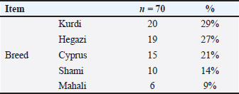
ABSTRACTBackground: In Libya, goats are considered as one of the most important livestock in which there are many breeds of goats such as Kurdi, Hegazi, Cyprus, Shami, and Mahali. A little hematological and biochemical information is known on these goat breeds raised in Libya. Aim: The main purpose was to verify the effect of breed variations on the hematological and biochemical parameters of goat breeds raised in Libya. Methods: The blood samples were collected in the winter season from 70 clinically healthy animals of different breeds for hematology and biochemical analysis. Results: Regarding the effect of breeds on blood hematology, significantly higher number (p < 0.01) of Mid cells were found in Hegazi (3.12 ± 1.30 × 10³/µl) and Cyprus (2.41 ± 1.69 × 10³/µl) when compared to other goat’s breeds including Kurdi (2.28 ± 0.95 × 10³/µl), Shami (1.90 ± 0.84 × 10³/µl), and Mahali (1.37 ± 0.88 × 10³/µl). Moreover, the percentage of Mid cells was highest in Hegazi (22.34% ± 9.40%), 11.40% ± 4.34%), followed by Kurdi (17.71% ± 8.16%), Cyprus (15.84% ± 8.33%), Shami (13.38% ± 5.76%), and lowest in Mahali (11.40% ± 4.34%). There are significant differences (p < 0. 01) in hemoglobin (Hb), red blood cells (RBCs), hematocrit (HCT), mean corpuscular hemoglobin (MCH), red cell width distribution-coefficient of variation (RDW-CV, %), and red cell distribution width-standard deviation (RDW-SD, fl) values among all different breeds of Libyan goats. The results of biochemistry displayed significant changes among the studied goats’ breeds, where the highest serum alanine aminotransferase (ALT, U/L) activity was observed in Cyprus (17.81 ± 7.95) and Shami (17.27 ± 1.15) compared with Hegazi (15.31 ± 6.13) and Mahali (14.60 ± 0.46), while Kurdi breed (11.68 ± 7.95) showed the lowest ALT activity. Moreover, significant differences (p < 0.01) in serum lactate dehydrogenase (LDH, U/l), total and direct bilirubin, glucose (GLU), creatinine, lipid profile, and electrolyte levels were recorded among different breeds used in this study. On the other hand, non-significant variations (p >0. 05) are reported in aspartate aminotransferase (U/l), alkaline phosphatase (U/l), total protein (g/dl), albumin (g/dl), urea and magnesium (Mg, mg/dl) levels. Conclusion: These results showed a significant difference between some blood parameters of goat breeds raised in Libya. This could aid veterinarians in interpreting laboratory data properly in order to improve the management and conservation of those breeds. Keywords: Goat breed, Hematology profile, Enzymes, Lipids, Electrolytes. IntroductionIn the period of 9000–7000 BC, the goat (Capra aegagrus) was one of the ancient livestock first domesticated in western Asia (Zeuner, 1963). Libya has 2.5 million heads of goats and Libyan local goats (Mahali) represent more than 90% of the total goat population (Akraim, 2012). Generally, goat breeds are named by their places of origin and classified by their uses of products. As the effect of different ecological conditions and long-term artificial selection, goat breeds with different genetic characteristics and product orientations are formed (AL-Harbi and Amer, 2012). Goats and other livestock may experience a range of behavioral, physiological, biochemical, hormonal, and molecular changes in response to various environmental stresses. These conditions include heat load and limitations on natural resources such as feed and water availability and the quality of grazing ground (Tadesse et al., 2023) which results from a total heat load (internal production and environment. The hematological and biochemical parameters are important to be determined because they provide valuable information about the breed, sex, and animal’s health status (Madan et al., 2016). Additionally, these parameters can also be used to assess the immunity status in goats (Al-Seaf and Al-Harbi, 2012). Importantly, the seasonal variations may reflect an effect on these parameters (Abdelatif et al., 2009). In the same context, some investigations have studied the hematobiochemical indicators in healthy goats in relation to breeds (Tambuwal et al., 2002). Furthermore, a Turkish study reported significant changes in red blood cells (RBC), Hb, hematocrit (HCT), and erythrocyte indices (mean corpuscular volume (MCV), MCH, mean corpuscular hemoglobin concentration (MCHC), and red cell distribution width (RDW) in Angora and Akkeçi goats (Aktaş and Pehli̇Van, 2023). Moreover, another study recorded a significant difference in most of the biochemical analytes such as alanine aminotransferase (ALT), aspartate aminotransferase (AST), and alkaline phosphatase (ALP) activities, total protein (TP), albumin (ALB), creatinine, urea, and some electrolytes among indigenous breeds of goats in Nigeria (Olaogun and Esan, 2024). However, according to the present reports, there are great alterations among goat breeds concerning their hematological and biochemical profile. Based on this data, the aim of this study was to assess the effects of differences in goat breeds on the blood hematology and biochemistry. Materials and MethodsAnimalsThe present study was approved by the Ethics Committee on Animal Care and used of the Faculty of Veterinary Medicine, University of Tripoli, Tripoli, Libya. Seventy goats of different breeds (Kurdi=20, Hegazi n=19, Cyprus n=15, Shami n=10, and Mahali n=6) and ages (14 young animals from 10 to 16 months and 56 adults above 19 months old). The animal sample included 12 males and 58 females. The animals are owned by private farms located in Tripoli, Libya. The description of the study samples is illustrated in Table 1. Blood collection and evaluation proceduresIn this study, blood samples were collected from the jugular vein into the plain tube for serum analysis and another with K3EDTA anticoagulant for whole blood analysis. The EDTA-anticoagulant tubes were analyzed using an automated hematology analyzer (Celltac α, Nihon Kohden, Tokyo, Japan). These samples were used for the determination of the following parameters: white blood cell (WBC), lymphocyte (Lymph), monocyte and eosinophil (Mid), granulocytes (Gran), hemoglobin (Hb), RBC, HCT, MCV, mean corpuscular hemoglobin (MCH), MCHC, and RDW. Moreover, the other blood samples were allowed to clot and centrifuged at 5,000 rpm for 15 minutes to obtain the serum. These samples were aliquoted in dry clean Eppendorf capped tubes and stored at –80°C for later biochemical analysis. Table 1. Description of the study samples.
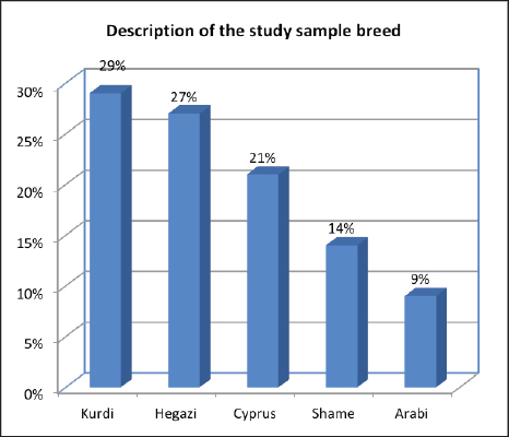
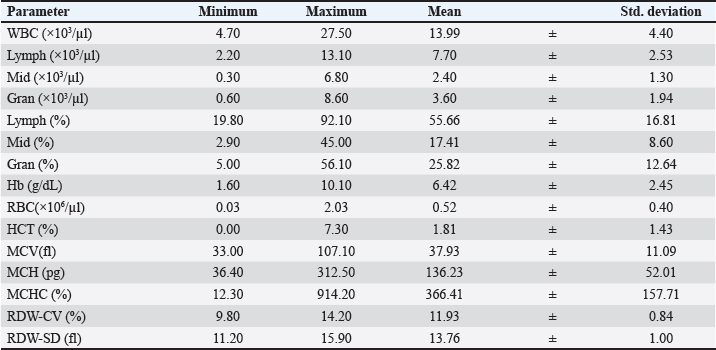
The serum alanine aminotransferase (ALAT), aspartate aminotransferase (ASAT), ALP, lactate dehydrogenase (LDH), TP, ALB, total bilirubin (T BIL), direct bilirubin (D BIL), glucose (GLU), triglycerides (TG), total cholesterol (TC), very low-density lipoprotein (VLDL), high-density lipoprotein (HDL), low-density lipoprotein (LDL), creatinine (Cr), urea, phosphorus (Phos), calcium (Ca), and magnesium (Mg) were estimated using an automatic analyzer (pz Cormay ACCENT M320). The serum concentrations of the sodium (Na), potassium (K), and chloride (Cl) electrolytes were determined by the ion-selective electrode method in the EasyLyte® Plus analyzer. All analyses were accomplished at Esraa’s and Al-shefaa’s clinical laboratories, Tripoli, Libya. Importantly, the same analyses were performed manually by investigators using 15 random blood samples. They obtained the same findings as attained from clinical laboratories. Statistical analysisThe data were analyzed using the SPSS Statistics software version 25, used to analyze the collected data; mean, standard deviation, and percentages. A non-parametric test was used because the data collected did not follow a normal distribution (Kolmogorov-Smirnov Test). To compare means, tests were used on independent two samples (Mann-Whitney test) and a one-way analysis of variances (Kruskal-Wallis test). Statistical significance happened when p < 0.05. Ethical approvalThe present study was approved by the ethics committee on animal care and use of the Faculty of Veterinary Medicine, University of Tripoli, Tripoli, Libya. ResultsThe descriptive study for the breeds is presented in Table 1 and Figure 1, while the descriptive statistics (minimum, maximum, mean, and std. deviation) for the hematological parameters in Libyan goat are presented in Table 2. The minimum and maximum values for WBC, lymphocyte, and granulocytes are 4.70–27.50 × 10³/µl, 2.20–13.10 × 10³/µl, and 0.60–8.60 × 10³/µl, respectively, while RBC, Hb, and HCT are 0.03–2.03 × 10⁶/µl, 1.60–10.10 g/dl, and 0.00%–7.30%.
Fig. 1. The descriptive study for the goat’s breeds. Table 2. Descriptive statistics for the hematological parameters.
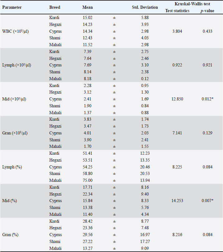
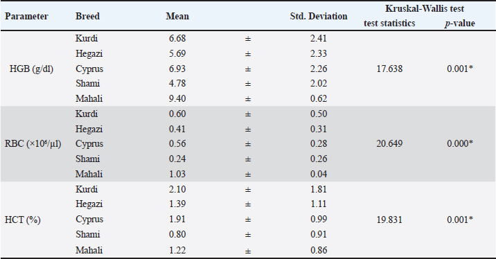
Table 3 shows the effect of different goat’s breeds on the total and differential leukocyte parameters. Higher counts of WBC, Lymph, and Gran were seen in Kurdi (15.02 ± 5.88 × 10³/µl), Mahali (8.18 ± 0.12 × 10³/µl), and Cyprus (4.01 ± 2.03 × 10³/µl), respectively, when compared with other breeds meanwhile this did not statistically significant (p > 0.129). However, Shami and Mahali goat breeds showed significantly (p < 0.01) lower mean values of mid cells (1.90 ± 0.84 × 10³/µl, 1.37 ± 0.88 × 10³/µl), and their percentages (13.38 ± 5.76 × 10³/µl, 11.40 ± 4.34 × 10³/µl), respectively, than the other breeds. There are significant differences in the Hb concentrations, RBC counts, and HCT values among all different breeds of Libya goats as illustrated in Table 4. The mean concentration value of Hb and RBC for Mahali, Kurdi, Cyprus, and Hegazi were 9.40, 6.68, 6.93, and 5.69 g/dl for Hb and 1.03 × 10⁶/µl, 0.60 × 10⁶/µl, 0.56 × 10⁶/µl, and 0.41 × 10⁶/µl for RBC, respectively. These values were significantly (p < 0.001) greater than that of Shami (4.78 g/dl, 0.24×10⁶/µl). The HCT value was significantly (p < 0.001) higher in Kurdi (2.10%), Cyprus (1.91%), Hegazi (1.39%), and Mahali (1.22%) compared with Shami (0.80%) (Table 4). Table 3. The total and differential leukocyte counts for goat’s breeds (means values± SD). n=20 Kurdi, 19 Hegazi, 15 Cyprus, 10 Shami, and 6 Mahali.
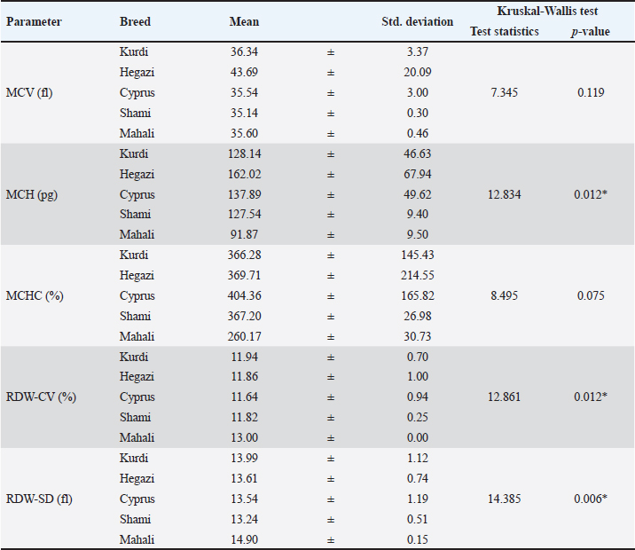
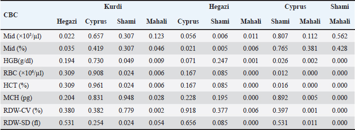
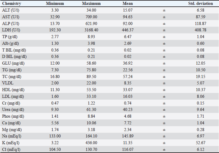
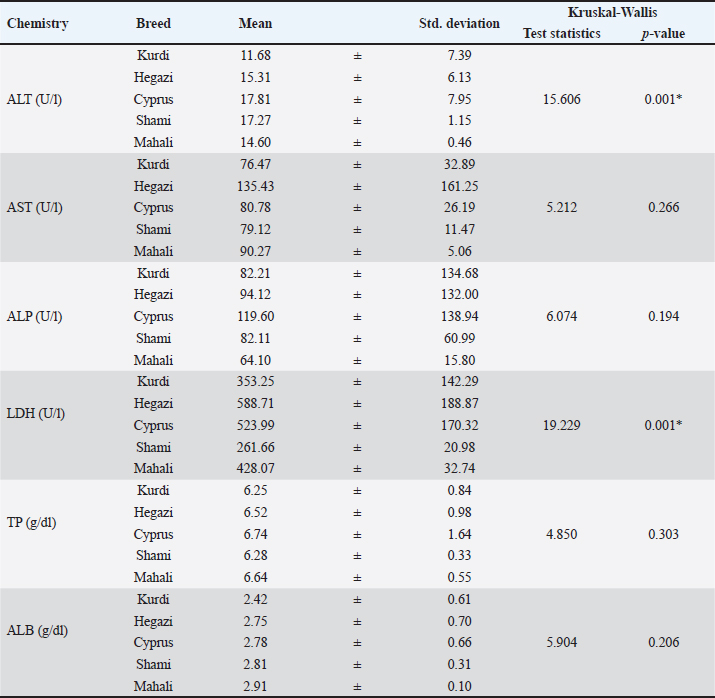
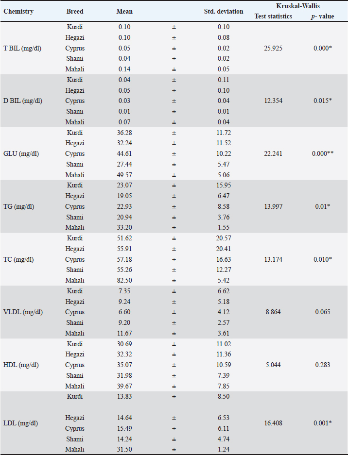
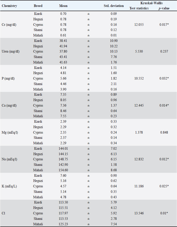
There are significant (p < 0.05) differences in the MCH, red cell width distribution coefficient of variation (RDW-CV), and red cell distribution width-standard deviation (RDW-SD) values among all different breeds of Libya goats as illustrated in Table 5. The highest significant (p < 0.01) value of MCH was recorded in Hegazi (162.02 pg), while RDW-CV (13.00%) and RDW-SD (14.90 fl) in Mahali in relation to other breeds. The lower values for MCH were detected in Mahali (91.87 pg), while RDW-CV in Cyprus (11.64%), and RDW-SD in Shami (13.24 fl) compared with others. Nevertheless, the MCV did not statistically (p > 0.1) change among all goat breeds (Table 5). The multiple comparisons between the hematological parameters (Mid, Hb, RBc, HCT, RDW-CV, and RDW-SD) which have significant differences for the different goat’s breeds were reported in Table 5. There are significant (p < 0.05) differences in the count of Mid cells between kurrdi and Hegazi, in Hb, RBc, HCT between Kurdi and Shami, in Mid, Hb, RBc, HCT, RDW-CV between kurrdi and Mahali (Table 6). Moreover, there are significant (p < 0.05) differences in all parameters between Hegazi and Mahali, as well as in all parameters except Mid between Cyprus and Mahali, and between Shami and Mahali. Regarding the effect of variations in breed on the blood parameters, descriptive statistics of chemistry in goats including minimum, maximum, mean, and standard deviation (std. deviation) were illustrated in Table 7. Table 8 compares the means of serum ALT, AST, ALP, LDH, TP, and ALB in this experiment for the different breeds of goats. The serum ALT activity showed significant differences (p < 0.001) and tended to be higher in Cyprus (17.81 ± 7.95) and Shami (17.27 ± 1.15) followed by Hegazi (15.31 ± 6.13) and Mahali (14.60 ± 0.46) compared with the lowest value in Kurdi (11.68 ± 7.39). The serum LDH activity showed a higher value (p < 0.001) in Hegazi (588.71 ± 188.87) and Cyprus (523.99 ± 170.32) when matched with Mahali (428.07 ± 32.74), Kurdi (353.25 ± 142.29) and Shami (261.66 ± 20.98). On the other hand, serum AST, ALP, TP, and ALB revealed non-significant change (p > 0.05) among the goat’s breeds. Table 9 revealed the mean values of total and D BIL, GLU, and lipid profiles in different breeds of goats. Significant differences in serum T BIL (p < 0.001), D BIL (p < 0.01), GLU ( p < 0.0001), TG (p < 0.01), TC (p < 0.01), and LDL (p < 0.001) were recorded among all used goats’ breed (Table 8). On the other hand, serum HDL, and VLDL levels revealed non-significant change (p > 0.05) among the goat’s breeds. Significant differences in serum Cr (p < 0.01), P, Ca, Na, K, and Cl among all used goats’ breeds were observed as shown in Table 10. The Mahali breed showed lower concentrations of Cr (0.61 ± 0.01 mg/dl), P (3.90 ± 0.16 mg/dl), Na (154.60 ± 8.68 mEq/l), and Cl (125.23 ± 7.54 mEq/l) than other breeds. In addition, higher levels of Ca (8.46 ± 0.64 mg/dl) and K (7.60 ± 0.90 mEq/l) were observed in Shami and Kurdi, respectively, when compared with other breeds. On the other hand, serum urea, and Mg revealed non-significant change (p > 0.05) among the goat’s breeds (Table 10). Table 4. The erythrocyte parameters in the goat’s breeds (means values± SD). n=20 Kurdi, 19 Hegazi, 15 Cyprus, 10 Shami, and 6 Mahali.
Table 5. The erythrocyte indices in the goat’s breeds (means values± SD). n=20 Kurdi, 19 Hegazi, 15 Cyprus, 10 Shami, and 6 Mahali.
Table 6. Multiple comparisons between the statistically significant hematology in the different goat’s breeds.
Table 7. Descriptive Statistics of chemistry in goats.
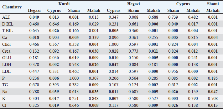
Table 11 shows the multiple comparisons between the statistically significant biochemical parameters for the different goat’s breeds. The statistically significant differences were at p < 0.05, 0.01, and 0.001. DiscussionIn order to improve the productivity of goat breeds, landowners have used special goat breeds, such as the Kurdi, Hegazi, Cyprus, Shami, and Mahali breeds, and it is important to understand the physiological characteristics of their animals, particularly the hemato-biochemical parameters, when raised in these problematic conditions. Goats can be stressed by a variety of things, including being in a restricted cage, managing their hosting, traveling, changing seasons, heat, strange places, and others (Ungerfeld et al., 2021; Rosenberger et al., 2022). After prolonged exposure to stress, goats generate physiological mechanisms that confer them the potential to tolerate stress (So-In and Sunthamala, 2023). Hematological qualities are broadly used to decide methodical connections and physiological adjustments, including evaluating the general wellbeing state of a creature (Mohammed et al., 2021). The examined breeds differed in several hematological parameters mainly related to the leukogram and erythrogram. Regarding the leukogram, higher counts of WBC, Lymph, and Gran were seen in Kurdi, and Cyprus breeds when compared with other breeds, meanwhile this was not statistically significant. The WBCs are the soldiers of the body and their high counts may also be due to the increase of the complement in the immune systems of the animals. It may also be attributed to physiological phenomena, i.e., excitement or strenuous exercise during handling (Weiss and Wardrop, 2011). However, the absolute and percentage count of mid cells were significantly (p < 0.01) lower in Shami and Mahali or Mahali goat breeds when matched with the other goats’ breed (Kurdi, Hegazi, and Cyprus). The increased values of mid cells, mainly monocytes in some goat’s breed of the present study suggested that the immune system of these breeds was well developed compared to another breeds (Shami and Mahali). These results and explanations were in agreement with those obtained by (Mohammed et al., 2021). Table 8. The means values ± SD of some biochemical parameters in goat’s breeds as Kurdi (n=20), Hegazi (n=19), Cyprus (n=15), Shami (n=10), and Mahali (n=6).
The values of the RBC, HCT, HBG, MCV, MCHC, MCH, WBC, and white blood differential count in Shami goats were reported in this study and found to be in general agreement with previously published values in healthy goats of different breeds (Mohammed et al., 2016; Al-Bulushi et al., 2017; Al-Rukibat and Ismail, 2019)concentrations for glucose ranged from 60.75 to 71.76 mg/dl, for blood urea nitrogen (BUN. Compared with other ruminants, more lymphocytes exist in circulation than neutrophils (Latimer, 2011). However, the total WBC, lymphocytes, and granulocytes did not differ in different breeds (Gorkhali et al., 2017). A study reported that the breed differences were observed in TP where Lampuchhre sheep (lowland sheep) showed a lower value than hill sheep breeds (Baruwal and Kage sheep). There was, however, some variation in a few values between our results and previously reported values that could be attributed to differences in nutritional status, geographical location, the physiologic status of the animals, and other genetic factors (Opara et al., 2010; Al-Rukibat and Ismail, 2019)WBC (X 109/l. Additionally, reported that the discrepancies in hematological parameters may be probably due to nutritional variation and breed of goats (Mohammed et al., 2021). In the present study, the mean concentration value of Hb and RBC for Mahali or Mahali, Kurdi, Cyprus, and Hegazi was significantly (p < 0.001) greater than that of Shami. These discrepancies agreed with the documentation by various researchers in different livestock (Piccione et al., 2007). These findings suggest that Mahali, Kurdi, Cyprus, and Hegazi goats have a greater propensity to transport oxygen and in situations of oxygen starvation, these breeds survive better (Okonkwo et al., 2011). Table 9. The means values± SD of some biochemical parameters in goat’s breeds as Kurdi (n=20), Hegazi (n=19), Cyprus (n=15), Shami (n=10), and Mahali (n=6).
Table 10. The means values± SD of creatinine, urea, and electrolytes in different goat’s breeds as Kurdi (n=20), Hegazi (n=19), Cyprus (n=15), Shami (n=10), and Mahali (n=6).
Table 11. Multiple comparisons between the statistically significant biochemical parameters for the different goat’s breeds.
The HCT value was significantly (p < 0.001) higher in Kurdi (2.10%), Cyprus (1.91%), Hegazi (1.39%), and Mahali (1.22%) compared with Shami (0.80%). The high HCT values in all breeds in comparison to the lowest values in the Shami breed may be attributed to either an expansion in the quantity of increasing RBC or decrease in flowing plasma volume or an increase in environmental temperature (Isidahomen et al., 2010). Furthermore, it was reported that the increased hemoglobin or HCT values might probably be a sign of healthy goats (Muayad et al., 2018). However, other authors illustrated that the high values of Hb and HCT could be related to the high altitude of the originated place of these breeds and their need for oxygen (Al-Bulushi et al., 2017). Generally, an increase in the concentration of erythrocyte parameters is associated with a greater ability to resist disease infection and a low level is an indication of disease infection and poor nutrition (Tambuwal et al., 2002). In contrast, the values of RBCs, Hb, and HCT were within the normal range that was reported formerly (Al-Bulushi et al., 2017). The highest significant (p < 0.01) value of MCH was recorded in Hegazi, while RDW-CV and RDW-SD in Mahali in relation to other breeds. However, the lower values for MCH were detected in Mahali, while RDW-CV in Cyprus and RDW-SD in Shami compared with others. Other studies showed higher values of MCV and MCH and lower values of MCHC in other goat breeds (Piccione et al., 2012; Arfuso et al., 2016). In addition, erythrocyte values were observed differently in the present study than those previously reported (Rice and Hall, 2007; Olayemi et al., 2009; Piccione et al., 2010). Finally, Mahali goat breeds showed higher mean values for RBC, HGB, RDW-CV, and RDW-SD, while HCT and MCH were significantly lower in Shami and Mahali breeds, respectively, than other goat’s breeds. These differences could be partly due to subclinical anemia caused by the higher average gastrointestinal nematode infections as reported by previous reports Gastrointestinal nematode infections in goats: Differences between strongyle faecal egg counts and specific antibody responses to Teladorsagia circumcincta in Nera di Verzasca and Alpine goats (Zanzani et al., 2020; Agradi et al., 2022). In the blood chemistry parameters of the Libyan goat breeds in this study, there are significant variations (p < 0.05) in serum ALT, LDH, total and direct BIL, GLU, TG, TC, LDL, Cr, and electrolytes (P, Ca, Na, K, and Cl) were recorded among all goats’ breed (Table 6). The Mahali breed showed higher concentrations of all previous variables with the exception of lower concentrations of Cr, P, Na, and Cl, and higher levels of Ca and K were observed in the Shami and Kurdi breeds. A previous study on Kanni goats reported that the calcium mean value (7.60 ± 0.05 mg/dl) was comparable with our finding, while the phosphorus mean value (3.58 ± 0.02 mg/dl) was lower (Ramprabhu et al., 2010). Moreover, a study carried out on West African pygmy goats found that the P mean value (8.02 ± 2.03 mg/dl) was higher than our finding (3.90–5.66mg/dl) (Tahas et al., 2012). Earlier reports stated that the serum liver enzymes and electrolytes are correlated with hormones during the different seasons and pregnancy and healthy status (Sarıbay et al., 2020). However, it was recorded that data exhibited no significant season and pregnancy status interaction on serum levels of Na, K, and iron concentrations in goats (Al-Sobaiyl, 2010). The significant dissimilarities in these parameters in the present study between different breeds were in agreement with other studies in different experimental goat breeds in Oman, Sudan, Kuwait, Nigerian, and Nepal (Oramari et al., 2014; Mohammed et al., 2016; Al-Bulushi et al., 2017; Parajuli, 2020). This could be due to differences in breed, climate, animal housing, nutrition, and subclinical diseases. Serum biochemical parameter concentrations in this study were similar to the values obtained previously in Shami goats (Al-Rukibat and Ismail, 2019)the establishment of breed-specific hematologic reference intervals (RIs. The ALT is an enzyme found in the highest amount in the liver and is typically used to detect liver injury. The values of ALT of goat breeds in this ranged from 14.60 ± 0.46–17.81 ± 7.95 U/l. These values were lower than those reported for goats in Pakistan (77.1 ± 74.2 U/l) and higher than those reported for West African Dwarf goats (8.9 ± 0.9 U/l) and the difference was highly statistically significant (Kiran et al., 2012). Another study carried out in wild goats demonstrated higher/or comparable ALT concentration (48.4 ± 52.3 U/l) to the present study (Pérez et al., 2003). Indeed, a great variation stated in ALT activity among different small ruminants was based on different geographical distributions (Kiran et al., 2012). The AST is an enzyme abundantly found in liver and heart muscles and plays an important role in amino acid metabolism (Kaneko et al., 1997). Additionally, the ALP is an enzyme produced by the liver and along with other enzymes such as ALT, AST, and GGT, can be used as an indicative of liver diseases and can also be a predictive of the health status of goats (Gwaze et al., 2012). There was no significant difference in AST and ALP levels among goats during the present study. In the study on different goat breeds in Sudan, the mean level of ALP was 104.7 IU/l with no significant difference between breeds (Elnasri, 2015). The LDH is an enzyme that catalyzes the conversion of lactate into pyruvate which is an important step in energy production in cell. The value of the LDH is higher in Hegazi (588.71 ± 188.87 U/l) than those of the Shami breed (261.66 ± 20.98 U/l) and this difference is statistically highly significant (p < 0.001). Increased activity of LDH is probably due to vascular thrombosis, hemorrhage, and tissue breakdown, especially in the liver and kidney of infected animals (Kaneko et al., 1997). The levels of T BIL, and D BIL in the present study were in the range of 0.04–014 and 0.01–0.07, respectively, in Libyan goat breeds. Higher mean values of T BIL of 0.1 mg/dl were recorded in Saanen goats and 0.22 mg/dl in Omani goat breeds (Elitok, 2012; Al-Bulushi et al., 2017). These higher values were more than those obtained in this study and could be attributed to different breeds, environmental conditions, feeding, and others. The blood GLU concentrations are indicators of energy status in animals. In domestic animals, blood GLU is regulated by the hypoglycemic and hyperglycemic hormones, but may be related to genetic predisposition (Antunović et al., 2022). A blood GLU level in the studied Libyan goat’s breed was in the range of 27.44–49.57 mg/dl and the highest level was observed in the Mahali breed (49.57 mg/dl). Several studies reported GLU level in the range of 29-35 mg/dl for breeds raised in Nigeria, 31.8 g/dl for Saanen goats raised in Turkey, 47mg/dl for Kanni goats, while higher blood GLU concentration was reported in wild goat (126.1 ± 66.0 mg/dl) than the values reported in the existing study (Pérez et al., 2003; Ramprabhu et al., 2010; Opara et al., 2010; Njidda, 2013). The difference in GLU concentration may be due to levels of nutrition and the metabolic activity of individual animals or to greater ruminal microbial propionate production because of different diets, but relatively high variability reflects the influence of many other factors (Jawasreh and Ismail, 2012; Wang et al., 2020; Belkasmi et al., 2023)non-pregnant Awassi ewes. Blood samples for hematology and blood biochemical analyses were withdrawn from 92 (30 Local Awassi; Lo-A, 34 Improved Awassi; Im-A, 28 Afec-Awassi; Af-A. Serum values of Cr and urea are used clinically to determine the integrity of urinary function, hydration, and nutritional status (Kaneko et al., 1997). The Cr is formed in skeletal muscle by the degradation of phosphocreatine to produce energy. Serum creatinine concentration has been observed to be proportional to muscle mass (Latimer, 2011). This explains the higher Cr content recorded in some breeds (Hegazi, Cyprus, and Shami) compared to others in the present study. Increases in creatinine levels are associated with a reduction of serum thyroxine levels (Yokus et al., 2006)early pregnancy (October. The blood urea is an indirect indicator of the protein composition of feed. Higher levels of urea in Shami than others although not significant are associated with greater ruminal degradation of protein with a concurrent increase in ammonia production (Kaneko et al., 1997). In the present study, the TG concentration was a very important variable probably because of differences in energy metabolism and breed. The highest serum TG level was observed in the Mahali breed but the lowest value was seen in Hegazi goat. Serum TG concentration was the most significant indicator followed by cholesterol concentration for feed restriction. Under severe feed restriction, TG concentration may increase to compensate for the maintenance energy deficit via the mobilization of fat reserve. Body condition score and body fatness under feed restriction are reported to affect TG concentration (Caldeira et al., 1999)creatinine, non-esterified fatty acids, globulins, glucose, total lipids, total protein, triglycerides, urea, β-hydroxybutyrate, and in the activity of alkaline phosphatases, aspartate aminotransferase, glucose-6-phosphate dehydrogenase, glutamate dehydrogenase, isocitrate dehydrogenase and pyruvate kinase were evaluated in the serum or plasma of ewes fed 30, 100 and 200% of theoretical maintenance energy requirements. A daily profile was observed in almost all variables, which may be of importance when interpreting data for these blood indicators. Daily variations were found even for albumin and total protein serum concentrations, showing a significant decrease of their levels after the meal. In order to maximize the diagnostic value of these indicators, the most suitable times for blood collection seem to be 16 h after the meal and (or. An increase in the level of serum TG has been reported under conditions of feed restriction because of reduced lipoprotein lipase (which transfers TG-derived fatty acids to adipose tissues for storage) activity and its mRNA expression in adipose tissues (Tadesse et al., 2023). The level of TC can differ between different animal breeds, Mahali for example was found to have a higher mean value (82.50 mg/dl) compared to Kurdi which has the lowest level of TC (51.62 mg/dl). These results disagree with the lower TC level reported in Saanen goats (45.34 mg/dl) and goat’s breeds of Northern Nigeria (2.9 ± 0.02 mmol/l), but are similar to those stated in goat breeds in Nigeria. There were no significant differences (p > 0.05) in some biochemical analytics (AST, ALP, TP, ALB, HDL, VLDL, urea, and Mg) observed among all the breeds of goats. This is in synchronization with the earlier reports which reported no significant differences in all biochemical parameters analyzed in Saanen goats breed or lame indigenous breeds of goats in Nigeria (Elitok, 2012; Olaogun and Esan, 2024). The non-significant differences in blood TP and ALB concentrations are indicators of no differences in dietary protein intake between the breed groups because serum TPs, particularly ALB, are good indicators for predicting protein status in animals (Sitaresmi et al., 2020). A high intake of grains and high temperatures were the cause of high blood TP as reported previously (Sandabe, 2000). Other reports displayed that lower protein and ALB values occurred because animals did not consume any considerable amount of grain or liver dysfunction. Similar findings reported that there was no significant difference in TP (Elnasri, 2015; Soul et al., 2019) one-year-old, of average live weight 14.13 ± 0.24 kg were assigned, in a completely randomized design, to three treatment diets. Animals were housed individually and sex was equally represented among treatments with 6 animals per treatment. Weights and blood samples were taken every fortnight. A pair of blood samples (5 ml each. ConclusionThe results obtained from this study showed a significant difference between some blood parameters of different goat breeds. This can add some knowledge to the definition of the health status of the five breeds raised in Libya. Moreover, it may serve as reference intervals for hematology and biochemistry analytes of goat breeds which could aid veterinarians to interpret laboratory data properly in order to improve the management and conservation of these breeds. AcknowledgmentNone. Authors’ contributionsConceptualization and design: F.M.B; Practical work: F.M.B, F.A.A, A.M.G, M. A. A, and A.O.B: formal analysis and interpretation of data: M.M.A and M. E.S; writing-original draft preparation: F.M.B and F.A.A; all authors revised and approved the final manuscript for publication. Conflict of interestThe authors declare that there is no conflict of interest. FundingThis research received no specific grant. Data availabilityAll data supporting the findings of this study are available within the manuscript. ReferencesAbdelatif, A.M., Ibrahim, M.Y. and Hassan, Y.Y. 2009. Seasonal variation in erythrocytic and leukocytic indices and serum proteins of female Nubian goats. Middle-East J. Sci. Res. 4, 168–174. Agradi, S., Menchetti, L., Curone, G., Faustini, M., Vigo, D., Villa, L., Zanzani, S.A., Postoli, R., Kika, T.S., Riva, F., Draghi, S., Luridiana, S., Archetti, I., Brecchia, G., Manfredi, M.T. and Gazzonis, A.L. 2022. Comparison of female verzaschese and camosciata delle alpi goats’ hematological parameters in the context of adaptation to local environmental conditions in semi-extensive systems in Italy. Animals 12, 12131703. Akraim, P. 2012. Goat production in Libya current state and production contraints. Krmiva 855, 189–194. Aktaş, R. and Pehli̇Van, E. 2023. A comparative profile of certain biochemical and hematological parameters in Angora and Akkeçi goats during the transition period. Turkish J. Vet. Anim. Sci. 47, 80–90. Al-Bulushi, S., Shawaf, T. and Al-Hasani, A. 2017. Some hematological and biochemical parameters of different goat breeds in Sultanate of Oman: a preliminary study. Vet. World 10, 461–466. AL-Harbi, M.S. and Amer, S.A.M. 2012. Preliminary comparative physiological and biochemical study of five different goat breeds inhabiting Saudi Arabia. Nat. Resour. 3, 206–212. Al-Rukibat, R. and Ismail, Z. 2019. Breed-specific reference intervals of hematologic variables in Shami goats (Capra aegagrus hircus) with the possible effects of age and sex. Vet. Clin. Pathol. 48, 762–767. Al-Seaf, A.M. and Al-Harbi, K.B. 2012. Variability of disease resistance, hematological parameters and lymphocyte proliferation in two goat breeds and their F1 and F2 crosses. Int. J. Food 2, 47–53. Al-Sobaiyl, K.A. 2010. Effect of breeding season and pregnancy status on serum progesterone, sodium, potassium, copper and iron of estrous synchronized Aradi goat does. Saudi J. Biol. Sci. 17, 259–263. Antunović, Z., Erceg, O., Šalavardić, Ž.K., Mioč, B., Đidara, M. and Novoselec, J. 2022. Hematological and biochemical parameters in the indigenous Croatian white goat in relation to age. Vet. Arh. 92, 713–722. Arfuso, F., Fazio, F., Rizzo, M., Marafioti, S., Zanghì, E., Piccione, G. 2016. Factors affecting the hematological parameters in different goat breeds from Italy. Ann. Anim. Sci. 16, 743–757. Belkasmi, F., Patra, A.K., Lourencon, R.V., Puchala, R., Dawson, L.J., dos Santos Ribeiro, L.P., Encinas, F. and Goetsch, A.L. 2023. Effects of the level and composition of concentrate supplements before breeding and in early gestation on production of different hair sheep breeds. Animals 13, 13050814. Caldeira, R.M., Almeida, M.A., Santos, C.C., Vasques, M.I. and Portugal, A.V. 1999. Daily variation in blood enzymes and metabolites in ewes under three levels of feed intake. Can. J. Anim. Sci. 79, 157–164. Elitok, B. 2012. Reference values for hematological and biochemical parameters in saanen goats breeding in Afyonkarahisar Province. Kocatepe. Vet. J. 5, 7–11. Elnasri, H. 2015. Biochemical blood parameters of different goat breeds in Sudan. Glob. J. Anim. Sci. Res. 3, 29–36. Gorkhali, A.N., Khanal, S., Sapkota, S., Prajapati, M., Shrestha, Y.K. and Khanal, D.R. 2017. Effect of breed and gender on hematological parameters and some serum biochemical profiles of apparently healthy indigenous sheep of Nepal. Nepal. Vet. J. 34, 85–94. Gwaze, F.R., Chimonyo, M. and Dzama, K. 2012. Effect of season and age on blood minerals, liver enzyme levels, and faecal egg counts in Nguni goats of South Africa. Czech J. Anim. Sci. 57, 443–453. Isidahomen, E., Ikhimioya, I., Njidda, A. and Okoruwa, M.I. 2010. Haematological parameters and Blood chemistry of different species of Ruminant animals in humid tropical environment. Niger. J. Agric. For. 3, 85–90. Jawasreh, K. and Ismail, Z.B. 2012. Normal hematology and selected serum biochemical values in different genetic lines of Awassi Ewes in Jordan. Internet J. Vet. Med. 7, 1–6. Kaneko, J., Harvey, J. and Bruss, M. 1997. Clinical biochemistry of domestic animals, 5th edition, San Diego, CA: Academy Press, pp: 22. Kiran, S., Bhutta, A.M., Khan, B.A., Durrani, S., Ali, M., Ali, M. and Iqbal, F. 2012. Effect of age and gender on some blood biochemical parameters of apparently healthy small ruminants from Southern Punjab in Pakistan. Asian Pac. J. Trop. Biomed. 2, 304–306. Latimer, K.S. 2011. Duncan and Prasse’s veterinary laboratory medicine clinical pathology, 5th edition, Chichester, UK: Wiley Blackwell. Madan, J., Sindhu, S., Gupta, M. and Kumar, S. 2016. Hematobiochemical profile and mineral status in growing beetal goat kids. J. Cell Tissue Res. 16, 5517–5522. Mohammed, M.T.A., Dhuha, J.M., Al-Bakri, S.A., Nur Hidayat, C.H.M., Amirah, S.H., Ahmed, Q.S., Basadd, H.J., Ula, H.M., Qais, N.A. and Haniza, H.M.Z. 2021. Hematological parameters of goat breeds in warm and humid weather. Indian J. Ecol. 48, 47–50. Mohammed, S.A., Razzaque, M.A., Omar, A.E., Albert, S. and Al-Gallaf, W.M. 2016. Biochemical and hematological profile of different breeds of goat maintained under intensive production system. African J. Biotechnol. 15, 1253–1257. Muayad, M.T.A., Haniza, M.Z.H., Husni, I. and Tawang, A. 2018. Haematological values of apparently healthy indigenous goats in Malaysia: a comparative study. Indian J. Anim. Res. 52, 1701–1704. Njidda, A.A. 2013. Haematological and biochemical parameters of goats of semi arid environment fed on natural Grazing Rangeland of Northern Nigeria. IOSR J. Agric. Vet. Sci. 3, 1–8. Okonkwo, J.C., Okonkwo, I.F. and Ebuh, G.U. 2011. Effect of breed, sex and source within breed on the heamatological parameter of the nigerian goats. Online J. Anim. Feed Res. 1, 8–13. Olaogun, S.C. and Esan, O.O. 2024. Blood biochemical profile and level of cortisol among lame indigenous breeds of goats in Nigeria. SVU-International J. Vet. Sci. 7, 22–38. Olayemi, F.O., Oboye, O.O., Azeez, I.O., Oyagbemi, A.A. and Soetan, K.O. 2009. Influence of management systems and sex on haematology of West African dwarf goat. African J. Agric. Res. 4, 1199–1202. Opara, M.N., Udevi, N. and Okoli, I.C. 2010. Haematological parameters and blood chemistry of apparently healthy West African Dwarf (Wad) Goats In Owerri, South Eastern. New York Sci. J. 3, 68–72. Oramari, R.A.S., Bamerny, A.O. and Zebari, H.M.H. 2014. Factors affecting some hematology and serum biochemical parameters in three indigenous sheep breeds. Adv. Life Sci. Technol. 21, 56–63. Parajuli, S. 2020. Performance of boer and their crossbreed goats in Nepal—a review. Int. J. Environ. Agric. Biotechnol. 5, 1449–1459. Pérez, J.M., González, F.J., Granados, J.E., Pérez, M.C., Fandos, P., Soriguer, R.C. and Serrano, E. 2003. Hematologic and biochemical reference intervals for Spanish ibex. J. Wildl. Dis. 39, 209–215. Piccione, G., Borruso, M., Fazio, F., Giannetto, C. and Caola, G. 2007. Physiological parameters in lambs during the first 30 days postpartum. Small Rumin. Res. 72, 57–60. Piccione, G., Casella, S., Lutri, L., Vazzana, I., Ferrantelli, V. and Caola, G. 2010. Reference values for some haematological, haematochemical, and electrophoretic parameters in the girgentana goat. Turkish J. Vet. Anim. Sci. 34, 197–204. Piccione, G., Messina, V., Vazzana, I., Dara, S., Giannetto, C. and Assenza, A. 2012. Seasonal variations of some serum electrolyte concentrations in sheep and goats. Comp. Clin. Path. 21, 911–915. Ramprabhu, R., Chellapandian, M., Balachandran, S. and Rajeswar, J.J. 2010. Influence of age and sex on blood parameters of Kanni goats in Tamil Nadu. Indian J. Small Ruminants 16, 249–251. Rice, C.G. and Hall, B. 2007. Hematologic and biochemical reference intervals for mountain goats (Oreamnos americanus): effects of capture conditions. Northwest Sci. 81, 206–214. Rosenberger, K., Simmler, M., Langbein, J., Nawroth, C. and Keil, N. 2022. Responsiveness of domesticated goats towards various stressors following long-term cognitive test exposure. Peer J. 10, 1–25. Sandabe, U. 2000. Effect of environmental temperature on some biochemical values in female sahel goats. Pak. Vet. J. 20, 10–12. Sarıbay, M.K., Naseer, Z., Doğruer, G., Özsoy, B. and Ateş, C.T. 2020. Variations in serum metabolites in response to season, cyclicity, and pregnancy in estrus-synchronized Damascus goats. Trop. Anim. Health Prod. 52, 1519–1525. Sitaresmi, P.I., Widyobroto, B.P., Bintara, S. and Widayati, D.T. 2020. Effects of body condition score and estrus phase on blood metabolites and steroid hormones in Saanen goats in the tropics. Vet. World 13, 833–839. So-In, C. and Sunthamala, N. 2023. Influence of goat management systems on hematological, oxidative stress profiles, and parasitic gastrointestinal infection. Vet. World 16, 483–490. Soul, W., Mupangwa, J., Muchenje, V. and Mpendulo, T.C. 2019. Biochemical indices and heamtological parameters of goats fed lablab purpureus and vigna unguiculata as supplements to a chloris gayana basal diet. Vet. Anim. Sci. 8, 100073. Tadesse, D., Patra, A.K., Puchala, R., Hussein, A. and Goetsch, A.L. 2023. Differentiation of hair sheep breeds based on the physiological and blood biochemical changes in response to different stressors using multivariate analysis techniques. Animals 13, 3390. Tahas, S.A., Giadinis, N.D., Kritsepi-Konstantinou, M., Papadopoulos, E., Petridou, E.J., Posantzis, D. and Dovas, C.I. 2012. Assessment of some health parameters in West African pygmy goats and cameroon dwarf sheep of a zoo in greece. J. Hell. Vet. Med. Soc. 63, 147–158. Tambuwal, F., Agale, B. and Bangana., A. 2002. Haematological and biochemical values of apparently healthy red Sokoto goats. Proceeding of 27th Annual Conference Nigerian Society of Animal Production (NSAP), pp: 50–53. Ungerfeld, R., Viera, M.N., Freitas-de-Melo, A., Giriboni, J., Casuriaga, D. and Silveira, P. 2021. Seasonality of the stress response in goat bucks when there is use of electroejaculation for semen collection. Anim. Reprod. Sci. 226, 106719. Wang, Y., Wang, Q., Dai, C., Li, J., Huang, P., Li, Y., Ding, X., Huang, J., Hussain, T. and Yang, H. 2020. Effects of dietary energy on growth performance, carcass characteristics, serum biochemical index, and meat quality of female Hu lambs. Anim. Nutr. 6, 499–506. Weiss, D.J. and Wardrop, K.J. 2011. Schalm’s veterinary hematology, 6th edition, Hoboken, NJ: John Wiley & Sons. Yokus, B., Cakir, D.U., Kanay, Z., Gulten, T. and Uysal, E. 2006. Effects of seasonal and physiological variations on the serum chemistry, vitamins and thyroid hormone concentrations in sheep. J. Vet. Med. Ser. A Physiol. Pathol. Clin. Med. 53, 271–276. Zanzani, S.A., Gazzonis, A.L., Alberti, E., Neilly, T.M., Villa, L. and Manfredi, M.T. 2020. Gastrointestinal nematode infections in goats: differences between strongyle faecal egg counts and specific antibody responses to Teladorsagia circumcincta in Nera di Verzasca and Alpine goats. Parasitol. Res. 119, 2539–2548. Zeuner, F.E. 1963. A history of domesticated animals. 1st edition. New York, NY: Harper & Row. Please provide the expansion for the Abbreviation “EDTA and SPSS.” | ||
| How to Cite this Article |
| Pubmed Style Benashour FM, Alnagar FA, Shmela ME, Buker AO, Abdunnabi MA, Gdura AM, Alssaie MA. The influence of breed's difference on the hemogram and biochemistry profile of goats raised in Libya during winter. Open Vet. J.. 2024; 14(8): 1968-1982. doi:10.5455/OVJ.2024.v14.i8.26 Web Style Benashour FM, Alnagar FA, Shmela ME, Buker AO, Abdunnabi MA, Gdura AM, Alssaie MA. The influence of breed's difference on the hemogram and biochemistry profile of goats raised in Libya during winter. https://www.openveterinaryjournal.com/?mno=202012 [Access: January 25, 2026]. doi:10.5455/OVJ.2024.v14.i8.26 AMA (American Medical Association) Style Benashour FM, Alnagar FA, Shmela ME, Buker AO, Abdunnabi MA, Gdura AM, Alssaie MA. The influence of breed's difference on the hemogram and biochemistry profile of goats raised in Libya during winter. Open Vet. J.. 2024; 14(8): 1968-1982. doi:10.5455/OVJ.2024.v14.i8.26 Vancouver/ICMJE Style Benashour FM, Alnagar FA, Shmela ME, Buker AO, Abdunnabi MA, Gdura AM, Alssaie MA. The influence of breed's difference on the hemogram and biochemistry profile of goats raised in Libya during winter. Open Vet. J.. (2024), [cited January 25, 2026]; 14(8): 1968-1982. doi:10.5455/OVJ.2024.v14.i8.26 Harvard Style Benashour, F. M., Alnagar, . F. A., Shmela, . M. E., Buker, . A. O., Abdunnabi, . M. A., Gdura, . A. M. & Alssaie, . M. A. (2024) The influence of breed's difference on the hemogram and biochemistry profile of goats raised in Libya during winter. Open Vet. J., 14 (8), 1968-1982. doi:10.5455/OVJ.2024.v14.i8.26 Turabian Style Benashour, Fathia Mahmoud, Fahima Ali Alnagar, Mansur E. Shmela, Amal O. Buker, Mouna A. Abdunnabi, Adel M. Gdura, and Mohammed A. Alssaie. 2024. The influence of breed's difference on the hemogram and biochemistry profile of goats raised in Libya during winter. Open Veterinary Journal, 14 (8), 1968-1982. doi:10.5455/OVJ.2024.v14.i8.26 Chicago Style Benashour, Fathia Mahmoud, Fahima Ali Alnagar, Mansur E. Shmela, Amal O. Buker, Mouna A. Abdunnabi, Adel M. Gdura, and Mohammed A. Alssaie. "The influence of breed's difference on the hemogram and biochemistry profile of goats raised in Libya during winter." Open Veterinary Journal 14 (2024), 1968-1982. doi:10.5455/OVJ.2024.v14.i8.26 MLA (The Modern Language Association) Style Benashour, Fathia Mahmoud, Fahima Ali Alnagar, Mansur E. Shmela, Amal O. Buker, Mouna A. Abdunnabi, Adel M. Gdura, and Mohammed A. Alssaie. "The influence of breed's difference on the hemogram and biochemistry profile of goats raised in Libya during winter." Open Veterinary Journal 14.8 (2024), 1968-1982. Print. doi:10.5455/OVJ.2024.v14.i8.26 APA (American Psychological Association) Style Benashour, F. M., Alnagar, . F. A., Shmela, . M. E., Buker, . A. O., Abdunnabi, . M. A., Gdura, . A. M. & Alssaie, . M. A. (2024) The influence of breed's difference on the hemogram and biochemistry profile of goats raised in Libya during winter. Open Veterinary Journal, 14 (8), 1968-1982. doi:10.5455/OVJ.2024.v14.i8.26 |

