


| Research Article | ||
Open Vet. J.. 2024; 14(8): 1990-1998 Open Veterinary Journal, (2024), Vol. 14(8): 1990–1998 Research Article Ameliorative effect of transfersome gel of kepok banana peel extract (Musa balbisiana) against photoaging in Wistar rat skinElly Mayangsari1,2, Arifa Mustika3*, Nurdiana Nurdiana2 and Nozlena Abdul Samad41Doctoral Program of Medical Science, Faculty of Medicine, Universitas Airlangga, Surabaya, Indonesia 2Department of Pharmacology, Faculty of Medicine, Universitas Brawijaya, Malang, Indonesia 3Department of Anatomy Histology and Pharmacology, Faculty of Medicine, Universitas Airlangga, Surabaya, Indonesia 4Department of Toxicology, Advanced Medical and Dental Institute USM, Kepala Batas, Malaysia *Corresponding Author: Arifa Mustika. Department of Anatomy Histology and Pharmacology, Faculty of Medicine, Universitas Airlangga, Surabaya, Indonesia. Email: fat.ashour [at] uot.edu.ly; arifa-m [at] fk.unair.ac.id Submitted: 24/05/2024 Accepted: 27/07/2024 Published: 31/08/2024 © 2024 Open Veterinary Journal
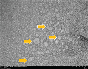
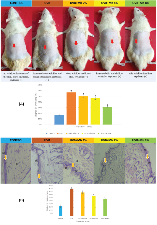
ABSTRACTBackground: Repeated acute exposure to ultraviolet B (UVB) rays can cause photoaging. Musa balbisiana peel contains flavonoid compounds which act as antioxidants. However, the physicochemicals of flavonoids are unstable, have high molecular weight, and are easily oxidized, causing their use is still limited and transdermal delivery to be inefficient. Aim: To investigate the ameliorative effect of transfersome gel of M. balbisiana peels against photoaging in Wistar rat skin. Methods: Transfersome gel was characterized by transmission electron microscopy (TEM). In vivo research was used to determine the ameliorative effects of M. balbisiana peel. The composition of transfersome consists of ethanol extracts of M. balbisiana peel, soybean phosphatidylcholine, and tween 80. The gel was applied three times a week for 4 weeks with a total UVB radiation dose of 840 mJ/cm2. To evaluate the repair mechanism by measuring the degree of wrinkles, epidermal thickening, dermal thinning, collagen fiber irregularity, matrix metalloproteinase 1 (MMP-1), and transforming growth factor-β (TGF-β) expression, malondialdehyde (MDA) and tumor necrosis factor-α (TNFα) levels. Results: TEM results show that gel transfersome M. balbisiana peel has a round morphology with a diameter of ±50 nm and no aggregation, which are defined as nanoparticles. Transfersome gel ameliorated the degree of wrinkle, epidermal thickening, dermal thinning, and irregularity of collagen fibers caused by UVB exposure, suppresses lipid peroxidation by decreasing MDA and TNFα level, also collagen imbalance by inhibiting MMP-1 expression and activating TGF-β expression, which was found statistically significantly different from non-transfersome gel group. Conclusion: Transfersome gel of M. balbisiana peel can act as an alternative medicine to ameliorate clinical photoaging due to exposure to UVB. Keywords: Medicine, Musa balbisiana peel, Photoaging, Transfersome gel, Ultraviolet B radiation. IntroductionPhotoaging can cause skin aging caused by ultraviolet (UV) radiation. Photoaging causes photodamage to the skin. Skin aging is a complex biological process that affects the appearance of the skin. Skin aging cannot be avoided due to the degenerative process of normal skin function (Ahmad, 2017; Zahruddin and Damayanti, 2018). There are two types of skin aging. First, intrinsic aging that occurs with age. Second, extrinsic aging is due to UV radiation and can accelerate the skin aging process, known as photoaging. The majority of causes of facial skin aging are related to UV rays (Muslim et al., 2021). Acute exposure to ultraviolet B (UVB) rays can help the body’s synthesis of vitamin D, but repeated exposure can cause symptoms of photoaging such as redness, wrinkles, loss of elasticity, skin laxity, and precancerous lesions. Photoaging symptoms have an impact on an individual’s social life because these symptoms affect their appearance when interacting with other people (Mayangsari et al., 2024). UVB radiation can directly damage DNA, increase reactive oxygen species (ROS), and decrease antioxidant enzyme activity. Oxidative stress due to ROS can trigger lipid peroxidation which is characterized by increasing malondialdehyde (MDA), activating activator protein-1 (AP-1), and nuclear factor kappa B which stimulates the production of tumor necrosis factor-α (TNF-α), increasing matrix metalloproteinase 1 (MMP-1) and decreasing transforming growth factor-β (TGF-β). This mechanism causes irregularity of collagen fibers (Respati et al., 2022; Mayangsari et al., 2024). Therefore, natural plants are needed that have antioxidant activity and can be used as an alternative for developing anti-aging skin products, one of which is Musa balbisiana peel, which has not been widely used as medicine. The banana plant can be said to be a plant with many benefits, which can be used from the roots, stems, tubers, leaves, and fruit. The parts used are generally fruit for consumption and leaves as a means of wrapping food. Apart from that, bananas are also used as a traditional medicine for various diseases, including diarrhea, diabetes, intestinal lesions, and dysentery. Literatures such as DOI: 10.24198/jf.v16i2.17789.g8492. Another part that has not been widely utilized is banana peel, even though the potential of banana peels is very large for skin health. Banana peels contain flavonoids, catecholamines, carotenoids, eugenol which are effective for eliminating acne, brightening the skin, reducing swelling, moisturizing the skin, and disguising acne scars. Banana peels contain flavonoid compounds that have potential as antioxidants (Jami’ah et al., 2018). In other research, it has been proven that banana peels contain flavonoids which have the potential as antioxidants and wound healing with strong antioxidant activity of 3,601.11 ± 0.46 mg GAEAC/kg (Rahmi et al., 2019; Rita et al., 2020). In Rosida’s research, the gel of banana peel extract showed the spreadability, stickiness, and pH test of the extracted gel which had significant differences compared to bioplacenton and base gel, but the concentration given was still quite high, namely 12.5%, 25%, and 50% (Rosida et al., 2014). Musa balbisiana peel contains flavonoid compounds that act as anti-inflammatories and antioxidants (Mayangsari et al., 2023). However, the physicochemical properties of flavonoids are unstable, have a high molecular weight, and are easily oxidized, so their use is less effective, and transdermal delivery becomes inefficient (Rosida et al., 2014). Therefore, in this study, we tried to develop a transdermal preparation in the form of a transfersome gel of M. balbisiana peel as an alternative preparation to increase permeation into the skin, so that it is hoped that its use will be more efficient with minimal concentration. Transfersome is a carrier molecule consisting of lipids in the form of vesicles that absorb drugs or active compounds into them. Transfersomes are a development of liposomes and are more effective in delivering active substances through the skin because of their flexible ability to change shape when passing through small pores, without destroying the initial size and shape, thus facilitating the penetration of drugs into the skin and increasing the absorption of active ingredients (Cheng et al., 2020). Transfersome formulations can be applied in semisolid dosage forms, such as transdermal gel. The transdermal gel is easy to apply and has better absorption percutaneously because it has a high water content so it can moisturize the skin and spread easily when applied (Rajabi and Mousa, 2016; Cheng et al., 2020). In this study, we hypothesized that gel transfersome M. balbisiana peel with topical administration can act synergistically to prevent UVB-induced photoaging effects. We investigated the effects of gel transfersome M. balbisiana peel on the degree of wrinkles, epidermal thickening, dermal thinning, and irregularity of collagen fibers. To understand the repair mechanism by measuring the expression of MMP-1 and TGF-β, the level of MDA and TNFα on UVB-induced photoaging in Wistar rat skin. Materials and MethodsPreparation of extract M. balbisiana peelThe sample in this study was kepok banana peel (Musa balbisiana) and it was obtained from Batu City, East Java Province, Indonesia. Musa balbisiana peel is dried, and then blended into a fine powder. Musa balbisiana peel powder was macerated with 70% ethanol and evaporated until it became a thick extract (Mayangsari et al., 2023). Transfersomes formulationThe thin film hydration method was used to make a transfersome formulation of M. balbisiana peel. Ethanol extracts of M. balbisiana peel, soybean phosphatidylcholine (SPC), and tween 80 (Sigma-Aldrich, Indonesia) were put in a flask and dissolved in ethanol. SPC is used as the main component to form lipid vesicles. Tween 80 is used as a surfactant/penetration enhancer, which is used as an edge activator to soften and deform phospholipid membranes. Ethanol was used as an organic solvent and PBS pH 7.4 as a hydration medium to form a transfersome suspension. The mixture was evaporated at 37°C, speed 150 rpm, for 70 minutes until a thin layer formed on the flask. Then the phosphate buffer saline (PBS, pH 7.4) hydration process was carried out and evaporated again at a temperature of 37°C, for 60 minutes and speed of 100 rpm. The resulting dispersion was sonicated to reduce the particle size for 10 minutes and then stored at 4°C (Khotimah et al., 2022). Gel formulationThe gel preparation consists of a transfersome of M. balbisiana peel, triethanolamine, carbopol 940, propylene glycol (Sigma-Aldrich, Indonesia), and distilled water. The formulation begins with carbopol 940 dissolved in aqqquadest and left at room temperature for 24 hours. The carbopol solution was homogenized at a speed of 1,500 rpm, then carbopol base gel was formed. After that, the carbopol gel base was added with TEA and propylene glycol. Then, add transfersome of ethanol extract M. balbisiana peel until it becomes a suspension. This formula was homogenized for 15 minutes at a speed of 500 rpm (Khotimah et al., 2022). Transmission electron microscopy (TEM)TEM is a tool for imaging the detailed internal structure of samples in high resolution. The TEM HT7700 has a maximum acceleration voltage of 120 kV. The maximum magnification of the TEM HT7700 is 600,000 times. TEM samples must be able to be penetrated by the electron beam, namely with a maximum sample thickness of 100 nm. The TEM examination procedure is that the sample has been dispersed in the solution, then 15 ul is dropped on to the TEM grid and then left for about 60 minutes until the sample dries and leaves the sample attached to the grid (Rachman et al, 2017). Animal experimentalThis study used 10-week-old male Wistar rats (n=30, average weight 150–250 g). Rats were obtained from the Pharmacology Laboratory of Universitas Brawijaya Indonesia and maintained in standard environmental conditions. Rats were divided into five groups randomly, with six rats in each group. i) Group 1: Control (no UVB radiation + no based gel) ii) Group 2: UVB (UVB radiation + based gel) iii) Group 3: UVB + Mb 2% (UVB radiation + Musa balbisiana peel transfersome gel 2%) iv) Group 4: UVB + Mb 4% (UVB radiation + Musa balbisiana peel transfersome gel 4%) v) Group 5: UVB + Mb 8% (UVB radiation + Musa balbisiana peel transfersome gel 8%) The skin surface of the rat backs was shaved with dimensions of length 5 cm and width 5 cm, and kept hairless during the experimental period. The UVB frequency was set three times a week for 4 weeks, using a Phillips UVB lamp that emits radiation with a wavelength of 290–320 nm. The total UVB dose reached 840 mJ/cm2. The dorsal skin of the rat was smeared with a transfersome gel of M. balbisiana peel evenly three times a week for 4 weeks. The dorsal skin of the rat was photographed using a camera. The degree of wrinkling was evaluated and measured via ImageJ 1.53e software. The rats were sacrificed using cervical dislocation after 4 weeks, and skin tissue was collected for further analysis. Histological examinationPreparation and staining of histology slides using Hematoxylin and eosin (HE) and Masson staining. HE staining is used to observe epidermal and dermal thickness. Masson’s trichrome staining was used to observe collagen fiber density. Histology slides were observed and photographed randomly at five locations using an Olympus light microscope (40× and 100× magnification). Results were measured using ImageJ 1.53e software (Khotimah et al., 2022; Mayangsari et al., 2024). Immunohistochemistry analysisImmunohistochemical staining to assess the expression of MMP-1 and TGF-β in skin tissue, with using antibodies anti-TGF-β and anti-MMP-1 (Sigma-Aldrich, Indonesia). The slides were observed under an Olympus light microscope. The images were photographed using the camera. The result was analyzed using ImageJ 1.53e software (Khotimah et al., 2022). MDA analysisSkin tissue was homogenized (10,000 rpm, 20 seconds) with a 1:9 volume of water for deionization (1.8 ml), then centrifuged at 12,000 rpm for 5 minutes. The supernatant was collected to measure MDA levels in Wistar rat skin. MDA levels were measured using the thiobarbituric acid test (Mayangsari et al., 2023). TNF-α analysisThe examination of TNF α level in skin tissue using the ELISA technique. The skin tissue must be homogenized, then make a Trizma Base Solution solution from Trizma Base 50 mM, NaCl 0.2 M, and distilled water 1,000 ml with pH 7.4, then centrifuge and measure the pH. The scouring results were sonicated with a long sonicator with a frequency of 400 Hz, for 10 minutes so that the tissue was homogeneous (Mayangsari et al., 2023). Statistical analysisResults are presented as mean ± SD. Differences between groups were analyzed by one-way ANOVA and considered statistically significant when the p-value <0.05. Then, analyzed with Tukey’s post hoc test using SPSS software (SPSS, Version 24.0). Ethical approvalAnimals were maintained according to guidelines for animal care. All efforts were taken to minimize any pain or discomfort. Research procedures were approved by the Health Research Ethics Committee of the Faculty of Medicine, Universitas Brawijaya, Indonesia (No. 227/EC/KEPK/10/2022). ResultsCharacterization of gel transfersome M. balbisiana peel using TEMThe use of transfersome preparations for transdermal drug delivery makes the active ingredients easier to penetrate into the skin, where they usually have an average particle size of between 1 and 100 nm. Characterization of gel transfersome M. balbisiana peels using a TEM HT7700 tool. TEM results show that gel transfersome M. balbisiana peel has a round morphology with a diameter of ±50 nm and no aggregation (Fig. 1). These results are in accordance with the characteristics of nanoparticles which are defined as particles that have a diameter between 1 nm to less than 100 nm. There is no aggregation which means the nanoparticles are stable (Rachman et al., 2017). Transfersome gel of M. balbisiana peel ameliorating visual appearance and prevents epidermal thicknessThe degree of wrinkles was assessed using the rating scale from Surini et al. (2020). Macroscopic assessment of the degree of skin wrinkling of mice was evaluated in the last week of the study (Fig. 2A). The UVB + Mb8% group showed thin wrinkles/fine lines erythema (+), which was 32% ± 1.8% (p < 0.05).
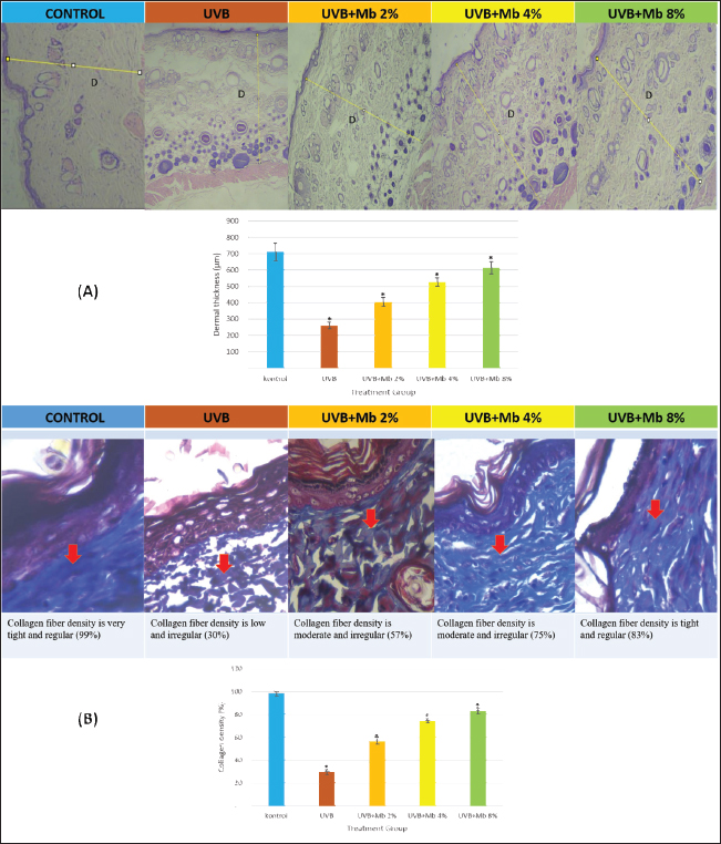
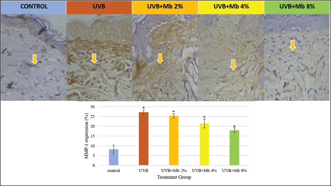
Fig. 1. Characterization of gel transfersome M. balbisiana peel using a TEM HT7700. The scale bar is 50 μm. Yellow arrow : morphology of transfersome gel of M. balbisiana peel. Assessment of epidermis thickness to assess skin damage due to UVB, using HE staining (Fig. 2B). Epidermis thickness is measured by measuring from five different locations. The UVB + Mb8% group showed epidermal thickness with a mean value of 27.72 ± 1.0 µm (p < 0.05). Transfersome gel of M. balbisiana peel ameliorating dermal thickness and replenished collagen degradationAssessment of dermis thickness by HE staining showed the UVB + Mb8% group with a mean value of 615 ± 39 µm (p < 0.05) (Fig. 3A). Clinical manifestations due to UVB exposure can be seen from the irregularity of collagen fibers in the dermis (Fig. 3B). In the UVB + Mb8% group, showed the collagen density in the dermis with a mean value of 83% ± 2.2% (p < 0.05). Transfersome gel of M. balbisiana peel inhibits and decreases MMP-1 expressionMMP-1 expression may explain the molecular mechanism of gel transfersome M. balbisiana peels to prevent UVB-induced collagen degradation. The UVB group increased significantly in MMP-1 expression by 27.11% ± 1.19%. MMP-1 expression decreased significantly in the UVB + Mb2%, UVB + Mb4%, and UVB + Mb8% groups, showing estimates of 25.29% ± 1.16%, 21.40% ± 2.19% and 17.96% ± 1.14% (Fig. 4). Therefore, transfersome gel of M. balbisiana peel could significantly inhibit MMP-1 expression due to UVB exposure through synergistic action. Transfersome gel of M. balbisiana peel activates and upregulates TGFβ expressionTGF-β expression decreased significantly by 7.35% ± 2.08% after exposure to UVB. TGF-β expression increased significantly in the UVB + Mb2%, UVB + Mb4%, and UVB + Mb8% groups, showing estimates of 18.06% ± 2.11%, 22.31% ± 1.43% and 24.71% ± 2.60% (Fig. 5). These results indicate that transfersome gel of M. balbisiana peel has antioxidant effects that can improve collagen density. Transfersome gel of M. balbisiana peel reduced oxidative stressMDA can be used as a parameter to assess oxidative stress and be a byproduct of lipid peroxidation. MDA level in the UVB group was significantly increased (2.89 ± 0.06 ng/ml). The transfersome gel of M. balbisiana peel has a great effect in reducing oxidative stress on the skin due to UVB exposure., with MDA levels of UVB + Mb2%, UVB + Mb4%, and UVB + Mb8% groups, showing an approximate 1.85 ± 0.05 ng/ml; 1.40 ± 0.07 ng/ml; 0.78 ± 0.04 ng/ml (p < 0.05). Transfersome gel of M. balbisiana peel decreased the TNFα levelExposure to UVB light increases the expression of the inflammatory cytokine TNFα in skin tissue. TNFα level in the UVB group was significantly increased (52.14 ± 2.38 ng/ml). The transfersome gel of M. balbisiana peel has an effect on reducing erythema triggered by TNFα secretion in the skin, which is caused by UVB radiation, with TNFα levels of UVB + Mb2%, UVB + Mb4%, and UVB + Mb8% groups, showing an approximate 43.18 ± 2.12 ng/ml; 39.16 ± 0.38 ng/ml; 34.56 ± 2.33 ng/ml (p < 0.05). DiscussionCharacterization using TEM HT 7700 proves that the transfersome gel of M. balbisiana peel has achieved the preparation of nanoparticles. These TEM results are in line with the characteristics of nanoparticles which are defined as particles that have a diameter between 1 nm and less than 100 nm (Rachman et al., 2017). Manifestations of the aging process due to direct exposure to UV light include rough skin, wrinkles, and sagging. In this study, we demonstrated the effect of transfersome gel of M. balbisiana peel and UVB radiation. Macroscopically, administration of transfersome gel of M. balbisiana peel shows improvement in wrinkles on the skin surface. This finding is in line with other research which proves that bananas have high antioxidant activity because they contain high levels of flavonoid compounds, so they are effective in preventing skin damage due to UVB (Handayani et al., 2019; Khan et al., 2020; Mayangsari et al., 2023). The assessment of epidermis thickness showed that the transfersome gel of M. balbisiana peel was able to reduce the epidermal thickness of the skin due to UVB exposure. These findings are in line with previous research which shows that the histological parameter that plays a role in the formation of wrinkles and the level of skin damage due to exposure to UVB rays is epidermal thickness (Khotimah et al., 2022). Meanwhile, transfersome gel of M. balbisiana peel can act as an anti-inflammatory and antioxidant in photoaging because it contains flavonoids (Mayangsari et al., 2023). The assessment of dermal thickness shows that transfersome gel of M. balbisiana peel can correct the irregularities of the dermis layer. These results are in accordance with previous which show that flavonoid compounds in bananas can be useful as antioxidants and can be used to reduce symptoms of photoaging (Rachmi et al., 2019; Mayangsari et al., 2023).
Fig. 2. Effect of transfersome gel of M. balbisiana peel. (A) Wrinkle formation on rat skin exposed to UVB (red arrow), p value <. (B) Assessment of epidermal thickness with HE staining; microscope magnification 100×; scale bar is 50 μm. SC: stratum corneum; EP: epidermis; D: dermis; yellow arrow: epidermal thickness. Data are presented as mean ± SD (n=5). Statistical analysis was done by oneway ANOVA followed by post hoc test analysis using the SPSS software. Data with notation (*) implied a significant difference (p < 0.05).
Fig. 3. Effect of transfersome gel of M. balbisiana peel. (A) Assessment of dermal thickness with HE staining (yellow line). (B) Assessment of collagen density with Masson trichrome staining (red arrow); microscope magnification 40×; scale bar is 50 μm. Data are presented as mean ± SD (n=5). Statistical analysis was done by oneway ANOVA followed by post hoc test analysis using the SPSS software. Data with notation (*) implied a significant difference (p < 0.05). Extrinsic aging of the skin caused by UVB exposure can trigger an imbalance between collagen synthesis and degradation, resulting in a decrease in the amount of collagen. Collagen can be directly degraded by exposure to UVB rays and cause wrinkles. Collagen density assessment showed that the transfersome gel of M. balbisiana peel could influence collagen regularity and increase the amount of collagen in the dermis layer. These results are in accordance with previous research which shows that photoaging caused by UVB exposure shows a decrease in collagen density (Zhang et al., 2020).
Fig. 4. Effect of transfersome gel of M. balbisiana peel on MMP-1 expressions. Immunohistochemistry sections of MMP-1; microscope magnification 100×; scale bar is 50 μm; brown staining indicates positive cells (yellow arrow). Quantitative analysis of expression intensity was performed using ImageJ 1.53e software. Data are presented as mean ± SD (n=5). Statistical analysis was done by oneway ANOVA followed by post hoc test analysis using the SPSS software. Data with notation (*) implied a significant difference (p < 0.05).
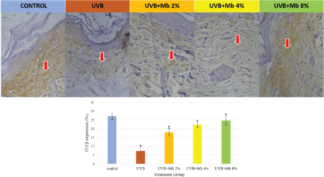
Fig. 5. Effect of transfersome gel of M. balbisiana peel on TGF-β expressions. Immunohistochemistry sections of TGF-β; microscope magnification 100×; scale bar is 50 μm; brown staining indicates positive cells (red arrow). Quantitative analysis of expression intensity was performed by ImageJ 1.53e software. Data are presented as mean ± SD (n=5). Statistical analysis was done by oneway ANOVA followed by post hoc test analysis using the SPSS software. Data with notation (*) implied a significant difference (p < 0.05). MMPs contribute to the degradation of collagen. ROS activates mitogen activated protein kinase and leads to an increase in AP-1. AP-1 induces the synthesis and expression of MMP-1 and causes collagen degradation in skin tissue (Khotimah et al., 2022). This study shows that transfersome gel of M. balbisiana peel significantly suppresses the increase in MMP-1 expression. These results were correlated with macroscopic and histological examination, in which transfersome gel of M. balbisiana peel significantly increased collagen density, inhibited epidermal thickness, and reduced wrinkle formation and erythema. Collagen synthesis is increased through the activation of TGF-β via TGF-β /Smad pathway. UVB exposure can interfere with TGF-β thereby reducing procollagen type 1 synthesis and causing a decrease in the amount of collagen on the dermis layer (Zhang et al., 2020; Khotimah et al., 2022). This study also showed that UVB exposure reduced TGF-β expression, and administration of transfersome gel of M. balbisiana peel increased TGF-β expression. The pathogenesis of photoaging due to UVB rays is associated with increased oxidative stress. MDA is an important biomarker of oxidative stress to evaluate lipid preoxidation. Furthermore, this process can increase the expression of the inflammatory cytokine TNF-α in skin tissue. In this study, transfersome gel of M. balbisiana significantly reduced MDA and TNF-α levels due to UVB exposure. These results are in accordance with previous studies showing that UVB exposure in mice causes an increase in MDA activity (Khotimah et al., 2022; Mayangsari et al., 2023). Other research also shows that TNF-α released after UVB exposure induces inflammatory cells that secrete collagenase (MMP-1) and cause skin aging (Ho et al., 2023). ConclusionAdministration of transfersome gel of M. balbisiana peel can significantly reduce the formation of wrinkles, improve epidermis and dermis thickness, increase the density of collagen fibers, can reduce MMP-1 expression and increase TGF-β expression, reduce MDA and TNF-α levels which overall can inhibit clinical photoaging due to exposure UVB rays. The most optimal concentration is a concentration of 8%. AcknowledgmentsThe author thanks the Center for Higher Education Fund (Balai Pembiayaan Pendidikan Tinggi) or Center or Education Service (Pusat Layanan Pendidikan-Puslapdik) and Indonesia Endowment Fund for Education (Lembaga Pengelola Dana Pendidikan—LPDP) Ministry of Education, Culture, Research and Technology of the Republic of Indonesia (Kementerian Pendidikan, Kebudayaan, Riset dan Teknologi Indonesia) with BPI ID Number: 202101121613; the Doctoral Program of Medical Science, Faculty of Medicine, Universitas Airlangga Indonesia and Faculty of Medicine Universitas Brawijaya Indonesia; Research Collaboration Universiti Sains Malaysia (represented by its Advanced Medical and Dental Institute) and Universitas Airlangga Indonesia (represented by its Faculty of Medicine) which have all supported this research. Conflict of interestThe authors declare they have no conflicts of interest. FundingThis study was supported by the Center for Higher Education Fund (Balai Pembiayaan Pendidikan Tinggi) or Center or Education Service (Pusat Layanan Pendidikan-Puslapdik) and Indonesia Endowment Fund for Education (Lembaga Pengelola Dana Pendidikan—LPDP) Ministry Education, Culture, Research, and Technology of the Republic of Indonesia (Kementerian Pendidikan, Kebudayaan, Riset dan Teknologi Indonesia), Research Collaboration Universiti Sains Malaysia (represented by its Advanced Medical and Dental Institute), and Universitas Airlangga Indonesia (represented by its Faculty of Medicine). Authors’ contributionsEM: conceptualized the study, also conducted the experiment and laboratory examination, then analyzed the data, wrote and revised the manuscript; AM, ND, NAS: supervised the study, and wrote and reviewed the manuscripts. Data availabilityAll data are provided in the manuscript. ReferencesAhmad, S.I. 2017. Ultraviolet light in human health, diseases and environment. Adv. Exp. Med. Biol. 996, 1–360. Cheng, Y.C., Li, T.S., Su, H.L., Lee, P.C. and Wang, H.D. 2020. Transdermal delivery systems of natural products applied to skin therapy and care. Molecules (Basel, Switzerland) 25(21), 5051. Handayani, R., Fans, K., Mastuti, T.S. and Rosa, D. 2019. Comparison study of antioxidant activity from three banana leaves extracts. Bali-Indonesia J. Teknol. Dan Industri Pangan. 32(1), 92–97. Ho, H., Hong, K., Kim, S., Kim, B., Hyung, S. and Suh, J. 2023. Effects of fish collagen on hairless mice skin photoaging induced by ultraviolet irradiation via regulation of the TGF- β signaling pathway anti-photoaging effect of fish collagen in UVB-induced hairless mice. J. Funct. Foods.105, 105554. Jami’ah, S.R., Ifaya, M., Pusmarani, J. and Nurhikma, E. 2018. Uji Aktivitas Antioksidan Ekstrak Metanol Kulit Pisang Raja (Musa paradisiaca sapientum) Dengan Metode DPPH (2,2-Difenil-1-Pikrilhidrazil). Jurnal Mandala Pharmacon Indonesia 4(1), 33–38. Khan, A., Bai, H., Khan, A. and Bai, Z. 2020. Neferine prevents ultraviolet radiationinduced skin photoaging. Exp. Ther. Med. 31, 89–96. Khotimah, H., Dewi Lestari Ismail, D., Widasmara, D., Riawan, W., Retnaningtyas, E. and Weka Nugraheni, R. 2022. Ameliorative effect of gel combination of Centella asiatica extract transfersomes and rosemary essential oil nanoemulsion against UVB-induced skin aging in Balb/C mice. F1000 Res. 11, 288. Mayangsari, E., Mustika, A., Nurdiana, N. and Ardhayudicva, S. 2023. Potency antiinflammatory of ethanol extract gel of Kepok banana peel (Musa balbisiana). Med. J. Malaysia 78(4), 488–490. Mayangsari, E., Mustika, A., Nurdiana, N. and Samad, N.A. 2024. Comparison of UVA vs UVB photoaging rat models in short-term exposure. Med. Arch. 78(2), 88–91. Muslim, M., Jusuf, N.K. and Putra, I.B. 2021. The dermoscopic features of facial aging among diverse ethnicity in Medan, Indonesia. Bali Med. J. 10(2), 904–909. Rachman, H.A.A., Aritonang, H. and Koleangan, H. 2017. Sintesis Dan Karakterisasi Nanopartikel Platina (Pt) Dari Larutan Kalium Tetrakloroplatinat (II) (K2PtCl4). Chem. Prog. 10(2), 50–55. Rahmi, D., Purnomo, D., Films, B.P., Chodijah, S. and Husaini, A. 2019. Microemulgel formulation of Kepok banana peel extract (Musa paradisiaca L) as an antioxidant. J. Phys. Conf. Ser. 1402, 055090 Rajabi, M. and Mousa, S.A. 2016. Lipid nanoparticles and their application in nanomedicine. Curr. Pharma. Biotechnol. 17(8), 662–672. Respati, R.A., Yusharyahya, S.N., Wibawa, L.P. and Widaty, S. 2022. The dermoscopic features of photoaging and its association with Sun Index Score in the coastal population at Cilincing, Jakarta: a cross-sectional study. Clin. Cosmet. Investing. Dermatol. 15, 939–946. Rita, W., Swantara, I.M., Astiti A.I. and Puspawati, N. 2020. Antibacterial activity and antioxidant capacity of selected local banana peel (Musa Sp.) methanol extracts cultivated in Bali. Int. J. Agric. Environ. Biores. 5, 242–251. Rosida, A., Sukardiman, B. and Khotib, J. 2014. The increasing of vegf expression and re-epithelialization on dermal wound healing process after treatment of banana peel extract (Musa acuminata colla). Int. J. Pharm. Pharma. Sci. 6(11), 427–430. Surini, S., Leonyza, A. and Suh, C.W. 2020. Formulation and in vitro penetration study of recombinant human epidermal growth factor-loaded transfersomal Emulgel. Adv. Pharm. Bull. 10(4), 586–594. Zahruddin, A. and Damayanti, D. 2018. Penuaan Kulit: Patofisiologi dan Manifestasi Klinis. Berkala Ilmu Kesehatan Kulit Dan Kelamin, 30(3), 208–215. Zhang, Z., Zhu, H., Zheng, Y., Zhang, L., Wang, X., Luo, Z., Tang, J., Lin, L., Du, Z. and Dong, C. 2020. The effects and mechanism of collagen peptide and elastin peptide on skin aging induced by D-galactose combined with ultraviolet radiation. J. Photochem. Photobiol. Biol. 210, 111964. | ||
| How to Cite this Article |
| Pubmed Style Mayangsari E, Mustika A, Nurdiana N, Samad NA. Ameliorative effect of transfersome gel of kepok banana peel extract (Musa balbisiana) against photoaging in Wistar rat skin. Open Vet. J.. 2024; 14(8): 1990-1998. doi:10.5455/OVJ.2024.v14.i8.28 Web Style Mayangsari E, Mustika A, Nurdiana N, Samad NA. Ameliorative effect of transfersome gel of kepok banana peel extract (Musa balbisiana) against photoaging in Wistar rat skin. https://www.openveterinaryjournal.com/?mno=202227 [Access: January 25, 2026]. doi:10.5455/OVJ.2024.v14.i8.28 AMA (American Medical Association) Style Mayangsari E, Mustika A, Nurdiana N, Samad NA. Ameliorative effect of transfersome gel of kepok banana peel extract (Musa balbisiana) against photoaging in Wistar rat skin. Open Vet. J.. 2024; 14(8): 1990-1998. doi:10.5455/OVJ.2024.v14.i8.28 Vancouver/ICMJE Style Mayangsari E, Mustika A, Nurdiana N, Samad NA. Ameliorative effect of transfersome gel of kepok banana peel extract (Musa balbisiana) against photoaging in Wistar rat skin. Open Vet. J.. (2024), [cited January 25, 2026]; 14(8): 1990-1998. doi:10.5455/OVJ.2024.v14.i8.28 Harvard Style Mayangsari, E., Mustika, . A., Nurdiana, . N. & Samad, . N. A. (2024) Ameliorative effect of transfersome gel of kepok banana peel extract (Musa balbisiana) against photoaging in Wistar rat skin. Open Vet. J., 14 (8), 1990-1998. doi:10.5455/OVJ.2024.v14.i8.28 Turabian Style Mayangsari, Elly, Arifa Mustika, Nurdiana Nurdiana, and Nozlena Abdul Samad. 2024. Ameliorative effect of transfersome gel of kepok banana peel extract (Musa balbisiana) against photoaging in Wistar rat skin. Open Veterinary Journal, 14 (8), 1990-1998. doi:10.5455/OVJ.2024.v14.i8.28 Chicago Style Mayangsari, Elly, Arifa Mustika, Nurdiana Nurdiana, and Nozlena Abdul Samad. "Ameliorative effect of transfersome gel of kepok banana peel extract (Musa balbisiana) against photoaging in Wistar rat skin." Open Veterinary Journal 14 (2024), 1990-1998. doi:10.5455/OVJ.2024.v14.i8.28 MLA (The Modern Language Association) Style Mayangsari, Elly, Arifa Mustika, Nurdiana Nurdiana, and Nozlena Abdul Samad. "Ameliorative effect of transfersome gel of kepok banana peel extract (Musa balbisiana) against photoaging in Wistar rat skin." Open Veterinary Journal 14.8 (2024), 1990-1998. Print. doi:10.5455/OVJ.2024.v14.i8.28 APA (American Psychological Association) Style Mayangsari, E., Mustika, . A., Nurdiana, . N. & Samad, . N. A. (2024) Ameliorative effect of transfersome gel of kepok banana peel extract (Musa balbisiana) against photoaging in Wistar rat skin. Open Veterinary Journal, 14 (8), 1990-1998. doi:10.5455/OVJ.2024.v14.i8.28 |

