


| Review Article | ||
Open Vet. J.. 2024; 14(8): 1751-1760 Open Veterinary Journal, (2024), Vol. 14(8): 1751–1760 Review Article Ultrasonographic evaluation of thoracic and abdominal neoplasia in domestic ruminants: A systemic reviewMohamed Tharwat1, El-Sayed El-Shafaey2,3* and Abdulrahman A. Alkheraif41Department of Clinical Sciences, College of Veterinary Medicine, Qassim University, Buraidah, Saudi Arabia 2Department of Surgery, Anesthesiology and Radiology, Faculty of Veterinary Medicine, Mansoura University, Mansoura-City, Egypt 3Department of Veterinary Surgery, Salam Veterinary Group, Buraydah, Saudi Arabia 4Department of Pathology and Laboratory Diagnosis, College of Veterinary Medicine, Qassim University, Buraydah, Saudi Arabia *Corresponding Author: El-Sayed El-Shafaey. Department of Surgery, Anesthesiology and Radiology, Faculty of Veterinary Medicine, Mansoura University, Mansoura-City, Egypt. Email: eelshafay [at] mans.edu.eg Submitted: 07/06/2024 Accepted: 20/07/2024 Published: 31/08/2024 © 2024 Open Veterinary Journal
ABSTRACTThere is a lack of literature on the usefulness of ultrasonography in the diagnosis and prognosis of thoracic and abdominal neoplasia in domestic ruminants, such as cattle, camels, sheep, and goats. This review aims to shed light on the current applications and prospects of ultrasonography in the diagnosis of thoracic and abdominal neoplasia in domestic ruminants. The scientific literature on ultrasonographic evaluation of the thoracic and abdominal neoplasia in domestic ruminants has been systematically reviewed to verify the potential role of ultrasonography in diagnosing such neoplasia. Based on the literature results, cattle (71.03%) were the most affected animals by thoracic and abdominal neoplasia, followed by sheep (11.86%), goats (11.57%), and camels (5.54%). In all included species, the abdominal neoplasia was more frequent (6.18%) than the thoracic neoplasia (2.97%), and the most frequent neoplasms were forestomach neoplasia. It is concluded that ultrasonography is not widely used in diagnosing thoracic and abdominal neoplasia in ruminant practice. Using ultrasonography in conjunction with guided needle aspiration and biopsy can offer veterinarians the opportunity for more precise diagnosis and treatment decisions guidance of thoracic and abdominal neoplasia in domesticated ruminants. Keywords: Animals, Diagnosis, Ruminant, Tumors, Ultrasound. IntroductionDomestic ruminants are an important source of meat and milk production in the world. The increase in consumption, along with increased herds and production, can induce stress and disease, particularly neoplasms (FAO, 2021). Diagnosis of neoplasia in ruminants represents a challenge for veterinarians due to its nonspecific lesions and the limited role of clinical and laboratory diagnosis. Neoplasms are life-threatening diseases, and most of them are detected in slaughterhouses or post-mortem examinations (Moharram et al., 2019). Thus, the need for an accurate diagnosis of such affection is very important for animal survival and productivity maintenance. In veterinary practice, ultrasonography is an integral part of contemporary ruminant medicine. The procedure is used routinely as a simple, safe, and cost-effective noninvasive diagnostic tool for the investigation of different ruminant disorders (Mohamed and Buczinski, 2011; Buczinski et al., 2012; Buczinski et al., 2013; Tharwat, 2020a; Tharwat, 2020b; Tharwat, 2020c, Tharwat, 2021a, Sadan et al., 2023; Tharwat et al., 2023; Tharwat, 2024; Tharwat and Al-Hawas, 2024a,b,c; Tharwat et al., 2024a,b,c,d). Ultrasound technology permits clinicians to get real-time information on various body systems. In certain cases, it can also evaluate the dynamic functions of organs (Braun et al., 2022). Ultrasound allows performing a detailed repeatedly, non-invasively imaged study of the thoracic and abdominal wall’s layers, thickness, and surrounding organs without discomfort or radiation exposure for the animal (Mohamed, 2010; Tharwat and Oikawa, 2011). Ultrasonography can be performed at any location and tolerated in un-sedated animals without the need for specific safety precautions, making serial scanning techniques to monitor the progression of the condition possible (Tharwat and Al-Sobayil, 2016). The incidence of tumors in cattle is increasing, which reduces their productivity. Cattle rank second to dogs in tumor incidence (Shruthi et al., 2018). Thoracic and abdominal neoplasia in domestic ruminants is resulting in economic losses of meat, milk, and productivity. The disease has non-specific clinical presentation and laboratory abnormalities, making it challenging to identify, characterize, and stage. In routine veterinary practice, imaging techniques are crucial for diagnosing diseases (Tharwat and Al-Sobayil, 2017). Ultrasound is a powerful diagnostic tool that can be used to evaluate not only thoracic and abdominal neoplasia but also other mass lesions. Additional techniques such as color Doppler imaging, contrast-enhanced ultrasound, and ultrasound-guided biopsy can be used to accurately differentiate unclear mass lesions with low risk (Tharwat, 2021b). However, the diagnostic applications of ultrasonography in ruminant practice were mostly directed at imaging the reproductive system (Ribadu and Nakao, 1999). This review aims to discuss the use of ultrasonography in evaluating abdominal and thoracic tumors in domestic ruminants, including cattle, camels, sheep, and goats. Thoracic NeoplasiaUltrasonography of cardiovascular neoplasmsIn cattle, diagnosis of heart disease involves using a variety of additional tests, with echocardiography being the most reliable tool due to its high sensitivity and specificity. The prognosis depends on various factors, including the disease stage, diagnosis, cow’s value, and withdrawal losses caused by therapeutic efforts (Galofaro et al., 2005). In all reported cases of cardiac neoplasms, the outcome is invariably fatal. Detecting an illness in its early stages is, therefore, crucial to avoid unnecessary treatment and minimize the suffering of animals (Tharwat et al., 2011). The main heart neoplasia in ruminants includes lymphoma, fibrosarcoma, and hemangiosarcoma (Tharwat et al., 2012). Lymphoma is a type of tumor that can affect multiple organs in cattle. However, the most prevalent kind of heart tumor in cattle is the lymphoma. This condition is often characterized by the infiltration of the right atrium and pericardial effusion. It is important to note that treating cardiovascular disturbance alone may not be enough to improve the animal’s condition, since many organ systems can be affected simultaneously in most cases of lymphoma (Van Biervliet et al., 2006). During an ultrasonographic examination of a six-year-old female White Alpine sheep, the following symptoms of right-sided cardiac insufficiency were observed: tachycardia, jugular vein engorgement, brisket edema, and ascites. Ultrasound examination revealed an echogenic mass in the right atrium, which may be either a tumor or a thrombus. Abdominal ultrasonography showed persistent liver congestion and significant ascites, which may be related to right-sided cardiac insufficiency. At the post-mortem examination, the clinical and sonographic results were later verified. A pedunculated fibrosarcoma was recognized as the mass found in the right atrium (Braun et al., 1995). Ultrasonography of respiratory neoplasmsUltrasonography is a useful tool for examining and identifying different lesions of the pleura and lungs. It is highly accurate and can be used in combination with radiography to allow see the lesion or to guide aspiration. The degree and severity of pulmonary changes affecting the pleura can be accurately assessed using ultrasonography. However, lower lesions in the lung tissue are not evident (Babkine and Blond, 2009). Thoracic ultrasonography is effective in visualizing intrathoracic masses and pleural effusion. At the same time, imaging alone may not be sufficient to diagnose neoplasia (Davis and Rush, 2013). In cattle, small pulmonary nodules measuring about 1 cm in diameter may appear in cases of bronchopneumonia (Floeck, 2004). These tiny nodules could be metastases, tiny abscesses, inflammatory masses, or necrotic areas. Ultrasonography can guide the aspiration of these nodules for accurate diagnosis. Main thoracic neoplasms in ruminants include adenocarcinoma, lymphosarcoma, osteosarcoma, thymomas, and mesothelioma (Backer and Smith, 2008). When the healthy lungs are examined with ultrasonography, the visceral pleura appears as a bright white line close to the transducer, along with many reverberation artifacts visible below it in aerated parenchyma. If there are any areas consistent with ovine pulmonary adenocarcinoma (OPA), these can be identified by a loss of the bright white line. The areas where the line is missing are known as lung consolidation areas, which are hypoechoic and well-demarcated. Additionally, echogenic areas of various sizes that correspond to neoplastic nodules may also be noticed. Using transthoracic ultrasonography, small tumors (i.e., those with a diameter of more than 1 cm) can be detected even before the onset of clinical symptoms (Quintas et al., 2018). Moreover, the use of ultrasound scanning enables the follow-up progress of small superficial lung lesions, which are shown at necropsy to be OPA (Scott et al., 2018). Ultrasonography of thymic lymphoma in a 2-year-old Holstein cow revealed a significant amount of subcutaneous fluid and a highly echogenic mass at the thoracic inlet, medial to the left and right jugular veins. The jugular veins above the thoracic inlet had a diameter of 3.8 cm and looked significantly enlarged with brisket edema (Alexander et al., 1996). In another case, a 16-year-old goat underwent an ultrasound examination of the thorax and heart. The sonogram identified a minor pleural and pericardial effusion, along with an 11 cm diameter multi-chambered pericardial mass. The goat was euthanized and necropsied, and a mediastinal or pleural neoplasia was tentatively diagnosed. A histological examination of the mass confirms the diagnosis of thoracic extra-skeletal osteosarcoma in a goat (Braun et al., 2011). Thymomas are tumors that develop from thymic epithelial cells and typically, show mixed cortical and/or medullary differentiation (Tharwat and Abd El-Rahim, 2012). When goats have thymomas, ultrasonography can detect a well-encapsulated mass with mixed echogenicity, often containing distinct anechoic fluid pockets. These masses are consistently found near the carotid arteries and jugular veins, just cranial to the thoracic inlet (Hill et al., 2017). In a six-year-old Holstein cow, ultrasonography showed an irregular pericardial surface and an intrapleural effusion, which are signs of pleural mesothelioma. On ultrasonography, pleural mesothelioma manifests as several tiny nodules on a thick serosal surface (Davis and Rush, 2013). Thus, ultrasonography is a valuable tool for early diagnosis and prognosis of mesothelioma in cows, and it can help rule out alternative causes of non-inflammatory ascites (Tharwat et al., 2012). Abdominal NeoplasiaUltrasonography of gastrointestinal neoplasmsAbdominal ultrasonography is a highly effective method used by veterinarians to diagnose and predict outcomes of abdominal diseases in farm animals. It helps veterinarians determine whether an animal should receive medical or surgical treatment or if it is better to be slaughtered. Ultrasonography is particularly useful in investigating gastrointestinal neoplasia in ruminants. This includes tumors such as granulosa cell tumors, mesothelioma, leiomyoma, squamous cell carcinoma, adenocarcinoma, and stromal tumors (Braun, 2003). During an ultrasound examination, a two-year-old Swiss Braunvieh cow was diagnosed with metastases of a granulosa cell tumor in the abdomen. The ultrasound showed a significant accumulation of hypoechoic fluid in the abdomen and thorax. Abdominal and thoracentesis procedures were performed, which yielded red fluid indicating bleeding in the abdominal and thoracic cavities. The ultrasound examination revealed multiple nodular lesions in the omentum, liver, spleen, and lungs. As well as the left ovary was noticeably enlarged and had nodules. Further examination of the lesions confirmed the presence of a granulosa cell tumor in the left ovary, which had spread to the omentum, liver, spleen, and lungs (Trösch et al., 2015). Mesotheliomas are malignant tumors that develop from the single layer of cells known as the mesothelium. This layer lines the pleura, pericardial, and peritoneum (Dobromylskyj et al., 2011). Mesotheliomas are very rare in cattle (Tharwat et al., 2012). During an ultrasound examination of a 6-year-old Holstein cow with mesothelioma, a significant volume of hypoechogenic fluid was found in the abdominal cavity. There were several masses with mixed echogenicity located in the abdomen, which were attached to the abdominal wall in different areas. Additionally, smaller masses were found next to the dorsal part of the spleen and on the mesentery’s surface. The large amount of abdominal fluid, along with the tumor masses, could be visualized. Therefore, ultrasonography is an important technique for rapid diagnosis and prognosis of mesothelioma in cattle, and it can help rule out other causes of non-inflammatory ascites such as right-sided heart failure, caudal vena cava thrombosis, or portal hypertension (Fig. 1) (Tharwat et al., 2012). The most prevalent neoplasms in domesticated female animals are leiomyomas. Gastrointestinal leiomyomas are often detected in the small intestine of cattle (Mohamed et al., 2004; Sadan et al., 2024). Ultrasonographic examination of a 6-year-old, 534-kg cow, revealed the presence of a mass within the omasum, which is causing intermittent fever, anorexia, depression, and ataxia. The mass’s edges were asymmetric, and it was echogenic with a lack of echotexture consistency. Necropsy findings showed an 18 × 14 cm mass within the omasum lumen. Histopathologically, interlacing bundles of well-differentiated smooth muscle cells with abundant cytoplasm and irregular fusiform nuclei with vesiculated chromatin were detected (Fig. 2) (Mohamed et al., 2004). Ultrasound is a useful non-invasive diagnostic technique for identifying neoplasia in the gastrointestinal tract of bovines. Other ultrasonographic findings that may indicate the presence of tumors include uneven reticular shape, deposits of echogenic material on the reticular wall, and non-audible biphasic reticular contractions. A hypoechogenic fluid may cavitate these deposits. These tumors have a histologic appearance that supports a leiomyoma diagnosis (Mohamed et al., 2004; Sadan et al., 2024). On ultrasonographic examination of an 8.8-year-old Simmental cow with squamous cell carcinoma, it was observed that the liver had multiple, poorly defined, and heterogeneous echogenic changes. A biopsy of the affected areas confirmed that it was indeed squamous cell carcinoma. The cow was euthanized and further examination revealed that the reticular mass, which had entered the liver through the portal vein, was the primary tumor (Braun et al., 2012). Gastric adenocarcinoma is a type of cancer that affects both humans and pets. It ranks third globally in terms of tumor-related fatalities and is the fifth most frequent type of tumor. However, this type of tumor is not often diagnosed in large ruminants. It has been documented in cattle abomasum (Ritchey et al., 1996). A 15-year-old female dromedary camel was admitted due to a history of decreased appetite, weight loss, and colic over the past 6 months. The transabdominal ultrasonographic examination showed heterogeneous contents and a significantly thicker, corrugated omaso-abomasum wall, with suspicion of neoplasia. During the postmortem examination, it was found that the abomasum had enlarged and weighed around 10.5 kg. Its wall was hardened, and filled with foul-smelling, bloody material. Additionally, there was thickening in the parietal peritoneum and congestion, thickening, and ulceration in the abomasal and omasal folds. Further examination through histopathology confirmed that there was a well-differentiated invasive adenocarcinoma present in the walls of both the abomasum and omasum (Fig. 3) (Tharwat et al., 2018).
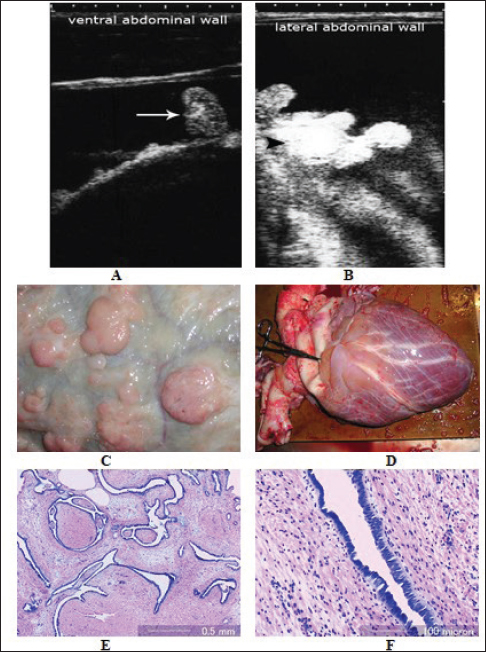
Fig. 1. Ultrasonograms in a Holstein cow with mesothelioma (3.5 MHz). Image (A) shows the ultrasonographic appearance of the greater omentum viewed at the left ventral abdomen. The greater omentum is surrounded by hypoechoic ascitic fluid and contains multiple nodules (arrow). Image (B) shows the ultrasonographic appearance of the lateral aspect of the abdomen taken at the right 11th intercostal space where multiple coalesced nodules (arrowhead) were scanned floating in hypoechogenic fluid. Image (C) shows multiple nodules in the greater omentum, varying in size from one to ten cm. Image (D) shows numerous masses adherent to the epicardium. Image (E) shows tubular structures lined by neoplastic cells with basophilic cytoplasm and supported by connective tissue; magnification (HE, x40). Image (F) shows the lumina of the tubular structures are lined by solitary columnar neoplastic mesothelial cells with basophilic cytoplasm; magnification (HE, ×200) (Tharwat et al., 2012). A nine-year-old castrated pygmy goat buck that had respiratory stertor, fever, and off-food was diagnosed with a gastrointestinal stromal tumor in the rumen with liver metastasis. A diagnostic work-up for respiratory issues showed slight enlargement of the arytenoids, but no other abnormalities were found. Despite a month of symptomatic treatment with little improvement and deteriorated general health, it was eventually euthanized. During necropsy, fibrinous and fibrous adhesions were found to attach the diaphragm, forestomachs, liver, spleen, and ventral and lateral sides of the cranial third of the abdominal cavity wall. The liver had several hard, white lumps that measured about 2 cm in diameter. Moreover, a sizable sessile mass from the rumen wall protruded into its lumen (Pesato et al., 2018).
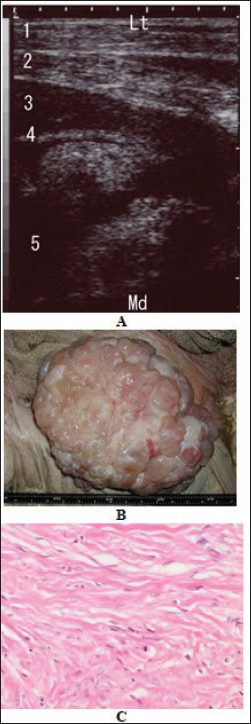
Fig. 2. Longitudinal sonogram of an omasal leiomyoma imaged at the seventh intercostal space in a 6-year-old Holstein cow. Note the asymmetric, non-homogenous, and echogenic patterns of the tumor (A). The mass appeared to project into the omasal lumen. Image (B) shows necropsy findings in the same cow showing an 18×14 cm mass in the omasum. Image (C) shows a photomicrograph of a section of the mass revealing interlacing bundles of well-differentiated smooth muscle cells with abundant cytoplasm and irregular fusiform nuclei with vesiculated chromatin (Hematoxylin and eosin ×40). Lt, lateral; Md, medial, 1, right ventral thoracic wall; 2, sternal part of the diaphragm and peritoneum; 3, liver; 4, omasal wall, 5, omasal lumen (Mohamed et al., 2004). Ultrasonography of hepatic neoplasmsUltrasonography is a useful diagnostic means for diagnosing liver disorders. It allows imaging, aspiration, and biopsy of isolated or diffuse liver lesions under visual guidance. Aspirating bile from the gallbladder with this imaging technique can also be utilized to diagnose liver flukes (Braun, 2009). In ruminants, hepatic tumors are not commonly found. These are typically metastatic tumors that travel from the gastrointestinal tract through the portal vein or the hepatic artery from the lungs to the liver. However, the tumor may in a few uncommon cases; have originated in the liver itself (Mohajeri et al., 2008). Hepatic neoplasia in ruminants includes carcinoma, adenoma, and melanoma (Braun, 2009). On ultrasound, hepatic tumors appear as circumscribed structures, either singular or numerous (Braun et al., 2005). This includes hepatocellular adenoma, adenocarcinoma, and bile duct tumors. Ultrasound characteristics of liver tumors show modifications to the liver’s structure and texture as well as the displacement of bile ducts and arteries. Both homogeneous and heterogeneous neoplastic alterations may be observed (Braun et al., 2005). The hepatic contour may appear to swell due to tumors that are situated on the surface of the liver. The appearance of most metastases on ultrasound scans differs from that of the liver tissue. Certain metastases may give a bulging appearance in the liver and have the same echogenicity as the liver. The vascularity and growth rate of metastases can have a significant impact on their echogenic pattern. Hypo-echogenicity is common in fast-growing metastases that contain predominantly tumor cells since these cells have fewer acoustic surfaces. While, slow-growing metastases are typically more vascularized and connective tissue-rich, giving them a high echogenic appearance. Echogenic thrombi can be visible when a tumor bursts into a vessel. Sometimes, there can be a decrease in hepatic perfusion, which causes the portal vein to become congested (Braun et al., 2005). In most cases, percutaneous ultrasound-guided liver biopsy can identify the kind of tumor and is advised for verifying a preliminary diagnosis of neoplasia (Braun, 2009). Typically isolated, hepatocellular carcinoma might have intrahepatic metastases all around it. Additionally, the tumors can spread to the rumen, spleen, and portal vein, leading to portal hypertension. Most cases of cholangiocellular carcinomas are diffuse or multifocal, with solitary tumors being rare. Animals that are afflicted often have normal livers (Stalker and Hayes, 2007). During an ultrasonographic examination of a 10-year-old cow, an 8-month-old heifer, and a 3-year-old sheep with polycythemia, it was observed that they had a huge liver with a focal echogenic lesion. The cow and heifer were diagnosed with multifocal liver neoplasms, specifically hepatocellular carcinoma and cholangiocellular carcinoma. The sheep, on the other hand, was diagnosed with cholangiocellular carcinoma. These diagnoses were confirmed through histopathological investigation (Braun et al., 1997). In two cases involving miniature goats, an ultrasonographic examination was performed due to in case 1, abdominal distension resulted in a pear-shaped belly, and in case 2, stranguria and pollakiuria. Several circular echoic structures that were partially encircled by a hypoechoic zone were noticed throughout the study. Based on the ultrasonographic findings, a liver tumor was tentatively diagnosed. However, post-mortem examination confirmed the final diagnosis of intrahepatic metastatic bile duct carcinoma (Trösch et al., 2015).
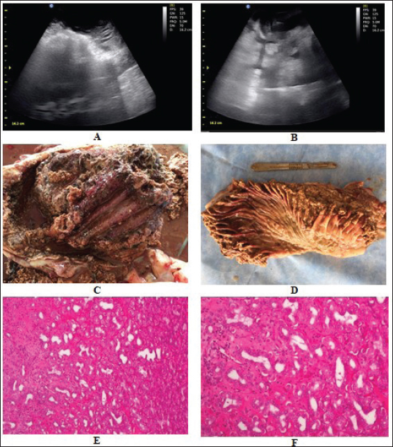
Fig. 3. Intraoperative ultrasonographic findings of the abomasum in a female camel with inappetence and colic during the past 6 months. In image (A), the abomasal wall appeared thickened, vascularized, and hyperechoic and the contents appeared heterogeneous (B). At necropsy, the abomasal (C) and omasal (D) folds were thickened, congested, and ulcerated. The tumor consisted of variable-sized acini lined by malignant epithelial cells with moderate atypia separated by desmoplastic stroma with dense mixed inflammatory cell infiltrate. Extensive areas of necrosis with suppuration were also seen (HE: (E) ×200; (F) ×400) (Tharwat et al., 2018).
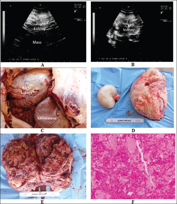
Fig. 4. Transrectal ultrasonographic findings in a female camel with renal cell carcinoma of the right kidney. Image (A) shows a hypoechoic mass involving the right renal parenchyma while image (B) shows the normal left kidney. Postmortem findings showed that the right kidney occluded almost the pelvic cavity (C) and a severely enlarged right kidney (18 Kg) compared to a normal, 1.5 Kg, left kidney (D). Image (E) shows cross section through the right kidney mass with a hemorrhagic, irregular shaped, and cavitated tumor. Image (F) shows histopathological results of the tumor revealing tubular differentiation with malignant epithelial lining and nuclear anaplasia (HE ×400) (Tharwat et al., 2017). On an ultrasound scan of an 8-year-old female Saanen goat with metastatic melanoma, several ovoid non-encapsulated structures with varied echogenicity were found in the liver. These structures contained multifocal to coalescent hypoechoic spots with about 12 × 11.65 cm in diameter. Additionally, in the mammary parenchyma, a multilobulated non-encapsulated round structure was identified in the dorsal area of the udder. This structure had hyperechoic cores encircled by hypoechogenic tissue with variant echogenicity (Conceição et al., 2021). Ultrasonography of urinary neoplasiaNumerous ruminants who suffer from urinary tract disorders may have a medical history that suggests the presence of other illnesses. Despite having abnormal urine constituents, this often goes unnoticed. Even though there may be substantial parenchymal defects. Additionally, serum biochemical profiles are not frequently performed on cattle because of the costs involved. Urinary tract abnormalities can be identified with the help of ultrasonography. However, a comprehensive and meticulous ultrasound evaluation scan of the whole urinary tract is necessary to ensure an accurate diagnosis of urinary tract disease since only one kidney might be impacted. The limits are diffuse or tiny parenchymal changes that are invisible due to the low-frequency transducers’ poor resolution, and the investigation of the entire urinary tract is time-consuming (Floeck, 2009). Urinary bladder tumors can cause hematuria, anemia, weight loss, stranguria, and secondary cystitis, and they are frequently linked to bracken poisoning (Radostits et al., 2007). Increased bladder wall thickness and irregular masses spreading into the bladder lumen, can be identified with ultrasound (Hoque et al., 2002). A 13-year-old female dromedary camel was admitted for clinical investigation because of emaciation, colic, and voiding of bloody urea for thirty days. On the trans-rectal ultrasound scan, a massive, irregularly shaped, hypoechoic, and cavitated mass was found protruding caudally and involving the right renal parenchyma. The left kidney, however, seemed to be functioning normally as stated. On necropsy, the right kidney harbored a hemorrhagic, atypically shaped, and cavitated tumor. The right kidney largely occupies the pelvic cavity and weighs eighteen kilograms in comparison to the left’s weight. Based on the histopathological investigation of the renal samples, renal cell carcinoma with tubular differentiation, malignant epithelial lining, and nuclear anaplasia, were identified. There was no evidence of metastases in the left kidney or other organs (Fig. 4) (Tharwat et al., 2017). ConclusionThe use of ultrasonography in conjunction with guided needle aspiration and biopsy can offer veterinarians the opportunity for more precise diagnosis and treatment decisions guidance of thoracic and abdominal neoplasia in domesticated ruminants. Further studies are required in abattoirs and farms to learn more about the total number of animals, the impact of neoplasms on carcass production and condemnation, and the related risk factors. AcknowledgmentNone. Conflict of interestThe authors declare that there is no conflict of interest. FundingThis research received no specific grant. Authors’ contributionsMT: conceptualization, data collection, and writing of manuscript draft. ES and AA: rereview and editing. All authors approved the final manuscript for publications. Data availabilityAll data supporting the findings of this study are available within the manuscript and no additional data sources are required. ReferencesAlexander, A.N., Constable, P.D., Meier, W.A., French, R.A., Morin, D.E., Lowry, J.E. and Hoffman, W.E. 1996. Clinical and immunohistochemical characterization of thymic lymphosarcoma in a heifer. J. Vet. Intern. Med. 10, 275–278. Babkine, M. and Blond, L. 2009. Ultrasonography of the bovine respiratory system and its practical application. Vet. Clin. North Am. Food Anim. Pract. 25, 633–649. Backer, J.C. and Smith, J.A. 2008. Miscellaneous conditions. In Large animal internal medicine. Ed., Smith BP, editor. 4th edition. St. Louis, MO: Mosby, pp: 666. Braun U., Caplazi, P., Linggi, T. and Graf, F. 1997. Polycythemia caused by liver carcinoma in cattle and sheep. Schweiz Arch Tierheilkd 139, 165–171. Braun, U. 2009. Ultrasonography of the liver in cattle. Vet. Clin. North Am. Food Anim. Pract. 25, 591–609. Braun, U., Nuss, K., Reif, S., Hilbe, M. and Gerspach, C. 2022. Left and right displaced abomasum and abomasal volvulus: comparison of clinical, laboratory and ultrasonographic findings in 1982 dairy cows. Acta Vet. Scand. 64, 40–57. Braun, U., Nuss, K., Soldati, G. and Ossent, P. 2005. Clinical and ultrasonographic findings in four cows with liver tumours. Vet. Record 157, 482–484. Braun, U. 2003. Ultrasonography in gastrointestinal disease in cattle. Vet J. 166, 112–124. Braun, U., Hagen, A., Pusterla, N. and Caplazi, P. 1995. Echocardiographic diagnosis of a cardiac fibrosarcoma in the right atrium of a sheep. Schweiz Arch Tierheilkd 137, 187–192. Braun, U., Schwarzwald, C.C., Forster, E., Becker-Birck, M., Borel, N. and Ohlerth S. 2011. Extraskeletal osteosarcoma of the thorax in a goat: case report. BMC Vet. Res. 7, 55–60. Braun, U., Trösch, L., Koschwanez, M., Rütten, M. and Hilbe, M. 2012. Squamous cell carcinoma of the reticulum and liver in a Simmental cow. Schweiz Arch Tierheilkd. 154, 331–335. Buczinski, S., Tolouei, M., Rezakhani, A. and Tharwat M. 2013. Echocardiographic measurement of cardiac valvular thickness in healthy cows, cows with bacterial endocarditis, and cows with cardiorespiratory diseases. J. Vet. Cardiol. 15, 253–261. Buczinski, S., Tsuka, T. and Tharwat, M. 2012. The diagnostic criteria used in bovine bacterial endocarditis: a meta-analysis of 460 published cases from 1973 to 2011. Vet. J. 193, 349–357. Conceição, A.I., Cajueiro, J.F.P., Mendonça, C.L., Souza, M.I., Afonso1, J.A.B., Oliveira, J.B.S., Santos, R.L. and Arenales, A. 2021. Metastatic melanoma in a Saanen goat: clinical, ultrasonographic and anatomopathological aspects – case report. Arq. Bras. Med. Vet. Zootec. 73, 827–833. Davis, E.G. and Rush, B.R. 2013. Diagnostic challenges: equine thoracic neoplasia. Equine Vet. Educ. 25, 96–107. Dobromylskyj, M.J., Copas, V., Durham, A., Hughes, T.K. and Patterson-Kane, J.C. 2011. Disseminated lipid-rich peritoneal mesothelioma in a horse. J. Vet. Diagn. Invest. 23, 615–618. FAO. 2021. Meat market review: emerging trends and outlook, Rome, Italy: FAO. Floeck, M. 2004. Diagnostic ultrasonography in cattle with thoracic disease. Vet J 167, 272–280. Floeck, M. 2009. Ultrasonography of bovine urinary tract disorders. Vet. Clin. North Am. Food Anim. Pract. 25, 651–667. Galofaro, V., Rapisarda, G., Ferrara, G. and Marino, F. 2005. Pericardial angiolipoma in a young bull. Vet. Record 157, 812–813. Hill, J.A., Fubini, S.L. and Hackett, R.P. 2017. Clinical features, treatment, and outcome in goats with thymomas: 13 cases (1990–2014). JAVMA. 251, 829–834. Hoque, M., Somvanshi, R., Singh, G.R. and Mogha, I.V. 2002. Ultrasonographic evaluation of urinary bladder in normal, fern fed and enzootic bovine haematuria-affected cattle. J. Vet. Med. A 49, 403–407. Mohajeri, D., Rezaie, A. and Mousavi, G.H. 2008. An abattoir study on hepatic tumors in sheep. Pakistan J. Bio.l Sci. 11, 1477–1481. Mohamed, T. 2010. Clinicopathological and ultrasonographic findings in 40 water buffaloes (Bubalus bubalis) with traumatic pericarditis. Vet. Rec. 167, 819–824. Mohamed, T. and Buczinski, S. 2011. Clinicopathological findings and echocardiographic prediction of the localisation of bovine endocarditis. Vet. Record 169, 180–185. Mohamed, T., Oikawa, S., Koiwa, M., Sato, H. and Kurosawa, T. 2004. Ultrasonographic diagnosis of omasal leiomyoma in a cow. Vet. Rec. 155, 530–531. Moharram, I., Awadin, W.F., Hamed, M.F., Salem, M.G. and Mosbah, E.A. 2019. Survey of tumors affecting cattle, buffaloes and sheep, in El-Dakahlyia Governorate. Mans. Vet. Med. J. 20, 37–45. Pesato, M., Boyle, A., Fecteau, M., Hamberg, A. and Smith, B. 2018. Gastrointestinal spindle cell tumor of the rumen with metastasis to the liver in a goat. J. Vet. Diagn. Invest. 30, 451–454. Quintas, H., Pires, I., Garcês, A., Prada, J., Silva, F. and Alegria, N. 2018. The diagnostic challenges of ovine pulmonary adenocarcinoma. Ruminants 1, 8–71. Radostits, O.M., Gay, C.C., and Hinchcliff, K.W. 2007. Diseases of the urinary system. In A textbook of the diseases of cattle, horses, sheep, pigs, and goats. 10th edition, Edinburgh, UK: Saunders Elsevier, pp: 543–573. Ribadu, A. and Nakao, T. 1999. Bovine reproductive ultrasonography: a review. J. Reprod. Dev. 45, 13–28. Ritchey, J.W., Marshall, C., David, C. and Brown, T.T. 1996. Mucinous adenocarcinoma in the abomasum of a cow. Vet. Pathol. 33, 454–456. Sadan, M., Tharwat, M. and El-Deeb, W. 2023. Deep swellings in sheep and goats: clinical, ultrasonographic and post-mortem findings. Int. J. Vet. Sci. 12, 793–801. Sadan, M., Tharwat, M., Alkhedhairi, S., Refaai, W., Moghazy, H.M.E.L., Khodier, M.M., Alkhamiss, A.S. and Ghallab, A. 2024. Abdominal pedunculated leiomyoma in a male dromedary camel: clinical, hematobiochemical, ultrasonographic and pathologic findings. Int. J. Vet. Sci. 13, 458–462. Scott, P.R., Dagleish, M.P. and Cousens, C. 2018. Development of superficial lung lesions monitored on farm by serial ultrasonographic examination in sheep with lesions confirmed as ovine pulmonary adenocarcinoma at necropsy. Ir. Vet. J. 71, 23–31. Shruthi, P.J., Sujatha, K., Srilatha, C.H. and Rayulu, V.C. 2018. Incidence of different tumors in bovines. Open Access J. Sci. 2, 220‒222. Stalker, M.J. and Hayes, M.A. 2007. Liver and biliary system. In Jubb, Kennedy, and Palmer’s pathology of domestic animals. Eds., Grant Maxie M., 5th edition, Edinburgh, UK: Saunders Elsevier, pp: 297‒388. Tharwat M, Al-Sobayil F, Ali A, Derar D, Khodeir M. 2017. Renal cell carcinoma in a female aranian camel: clinical, hematobiochemical, ultrasonographic and pathologic findings. J. Camel Pract. Res. 24, 61–66. Tharwat, M. 2020a. Ultrasonography of the abdomen in healthy and diseased camels (Camelus dromedaries)—a review. J. Appl. Anim. Res. 48, 300–312. Tharwat, M. 2020b. Ultrasonography of the kidneys in healthy and diseased camels (Camelus dromedaries). Vet. Med. Intern. 2020, 7814927. Tharwat, M. 2020c. Ultrasonography of the liver in healthy and diseased camels (Camelus dromedaries). J. Vet. Med. Sci. 82, 399–407. Tharwat, M. 2021a. Clinical, ultrasonographic, and postmortem findings in sheep and goats with urinary tract disorders, Vet. World 14, 1879–1887. Tharwat, M. 2021b. Obstructive urolithiasis in dromedary camels: clinical, ultrasonographic and postmortem findings. J. Camel Pract. Res. 28, 85–93. Tharwat, M. 2024. Fundamentals of diagnostic ultrasound in dromedary camel medicine. Int. J. Vet. Sci. 13, 1–6. Tharwat, M. and Al-Hawas, A. 2024a. Liver diseases in sheep and goats: parallel sonographic and pathologic findings. Int. J. Vet. Sci.13, 284-290. Tharwat, M. and Al-Hawas, A. 2024b. Suppurative pyelonephritis in a caprine buck: Clinical, laboratory, ultrasonographic and pathologic findings. Int. J. Vet. Sci. 13, 479–483. Tharwat, M., Al-Hawas, A. 2024c. The emerging topic of cosmetic medicine in dromedary camels. Int. J. Vet. Sci. 13, 139–146. Tharwat, M. and Al-Sobayil, F. 2017. Diagnostic ultrasonography in goats with contagious caprine pleuropneumonia caused by Mycoplasma capricolum subsp. Capripneumoniae. BMC Vet. Res. 13, 263–270. Tharwat, M. and Oikawa, S. 2011. Ultrasonographic evaluation of cattle and buffaloes with respiratory disorders. Trop. Anim. Health Prod. 43, 803–810. Tharwat, M. and Abd El-Rahim, I. 2012. Clicopathological and biochemical studies on thymic lymphoma in cattle. Assiut Vet. Med. J. 58, 27–32. Tharwat, M. and Al-Sobayil, F. 2016. Ultrasonographic findings in camels (Camelus dromedarius) with different urinary affections. J. Camel Pract. Res. 23, 301–308. Tharwat, M., Al-Sobayil, F., Ali, A. and Buczinski, S. 2012. Ultrasonographic evaluation of abdominal distension in 52 camels (Camelus dromedarius). Res. Vet. Sci. 93, 448–456. Tharwat, M., El Moghazy, H.M. and Oikawa, S. 2023. Ultrasonographic verification of hepatic hydatidosis in a female dromedary camel: a case report. J. Vet. Med. Sci. 85, 1286–1290. Tharwat, M., El-Ghareeb, W.R. and Almundarij, T.I. 2024a. Depraved appetite in dromedary camels: clinical, ultrasonographic, and postmortem findings. Open Vet. J. 14, 652–663. Tharwat, M., Hegazy, Y. and Alkheraif, A.A. 2024b. Discolored urine in sheep and goats: Clinical, etiological, hematobiochemical, sonographic and postmortem findings. Open Vet. J. 14, 1059–1071. Tharwat, M., Sadan, M. and Al-Hawas, A. 2024c. The emerging topic of injected cosmetic fillers in the perinasal region of dromedary camels: ultrasonographic and radiographic verification. Open Vet. J. 14, 840–845. Tharwat, M., Haridy, M., Elmoghazy, H.M.M., Elnahas, A. and Alkheraif, A.A. 2024d. Abdominal fat necrosis in a female dromedary camel: clinical, hematobiochemical, sonographic and pathologic findings. Open Vet. J. 14(7), 1726. Tharwat, M., El-Shafaey, E., Sadan, M., Ali, A., Al-Sobayil, F. and Al-Hawas, A. 2018. Omaso-abomasal adenocarcinoma in a female Arabian camel (Camelus dromedarius). J. Appl. Anim. Res. 46, 1268–1271. Tharwat, M., Oikawa, S. and Buczinski, S. 2011. Ultrasonographic prediction of hepatic fat content in dairy cows during the transition period. J. Veterinar. Sci. Technolo. 3, 111–115. Trösch, L., Müller, K., Brosinski, K. and Braun U. 2015. Haemoabdomen and haemothorax in a cow with metastatic granulosa cell tumor. Schweiz Arch Tierheilkd 157, 339–343. Van Biervliet, J., Kraus, M., Woodie, B., Divers, T.J., Gelzer, A. and Ainsworth, D. 2006. Thoracoscopic pericardiotomy as a palliative treatment in a cow with pericardial lymphoma. J. Vet. Cardiol. 8, 69–73.37. | ||
| How to Cite this Article |
| Pubmed Style Tharwat M, El-shafaey E, Alkheraif AA. Ultrasonographic evaluation of thoracic and abdominal neoplasia in domestic ruminants: A systemic review. Open Vet. J.. 2024; 14(8): 1751-1760. doi:10.5455/OVJ.2024.v14.i8.2 Web Style Tharwat M, El-shafaey E, Alkheraif AA. Ultrasonographic evaluation of thoracic and abdominal neoplasia in domestic ruminants: A systemic review. https://www.openveterinaryjournal.com/?mno=204787 [Access: January 25, 2026]. doi:10.5455/OVJ.2024.v14.i8.2 AMA (American Medical Association) Style Tharwat M, El-shafaey E, Alkheraif AA. Ultrasonographic evaluation of thoracic and abdominal neoplasia in domestic ruminants: A systemic review. Open Vet. J.. 2024; 14(8): 1751-1760. doi:10.5455/OVJ.2024.v14.i8.2 Vancouver/ICMJE Style Tharwat M, El-shafaey E, Alkheraif AA. Ultrasonographic evaluation of thoracic and abdominal neoplasia in domestic ruminants: A systemic review. Open Vet. J.. (2024), [cited January 25, 2026]; 14(8): 1751-1760. doi:10.5455/OVJ.2024.v14.i8.2 Harvard Style Tharwat, M., El-shafaey, . E. & Alkheraif, . A. A. (2024) Ultrasonographic evaluation of thoracic and abdominal neoplasia in domestic ruminants: A systemic review. Open Vet. J., 14 (8), 1751-1760. doi:10.5455/OVJ.2024.v14.i8.2 Turabian Style Tharwat, Mohamed, El-sayed El-shafaey, and Abdulrahman A. Alkheraif. 2024. Ultrasonographic evaluation of thoracic and abdominal neoplasia in domestic ruminants: A systemic review. Open Veterinary Journal, 14 (8), 1751-1760. doi:10.5455/OVJ.2024.v14.i8.2 Chicago Style Tharwat, Mohamed, El-sayed El-shafaey, and Abdulrahman A. Alkheraif. "Ultrasonographic evaluation of thoracic and abdominal neoplasia in domestic ruminants: A systemic review." Open Veterinary Journal 14 (2024), 1751-1760. doi:10.5455/OVJ.2024.v14.i8.2 MLA (The Modern Language Association) Style Tharwat, Mohamed, El-sayed El-shafaey, and Abdulrahman A. Alkheraif. "Ultrasonographic evaluation of thoracic and abdominal neoplasia in domestic ruminants: A systemic review." Open Veterinary Journal 14.8 (2024), 1751-1760. Print. doi:10.5455/OVJ.2024.v14.i8.2 APA (American Psychological Association) Style Tharwat, M., El-shafaey, . E. & Alkheraif, . A. A. (2024) Ultrasonographic evaluation of thoracic and abdominal neoplasia in domestic ruminants: A systemic review. Open Veterinary Journal, 14 (8), 1751-1760. doi:10.5455/OVJ.2024.v14.i8.2 |

