| Short Communication | ||
Open Vet. J.. 2024; 14(12): 3630-3639 Open Veterinary Journal, (2024), Vol. 14(12): 3630-3639 Short Communication Effects of a blend extract of Sauropus androgynus, Moringa oleifera, and Coleus amboinicus on milk production in lactating ratsPutri Reno Intan1,2, Sukmayati Alegantina3, Ani Isnawati3, Nanang Yunarto4, Fitrine Ekawasti5, Ratih Rinendyaputri2, Sunarno Sunarno2, Yulvian Sani5, Sela Septima Mariya2 and Ekowati Handharyani6*1Animal Biomedical Study Program, IPB Postgraduate School, School of Veterinary Medicine and Biomedicine, IPB University, Bogor, Indonesia 2Center for Biomedical Research, Research Organization for Health, National Research and Innovation Agency (BRIN), Cibinong Science Center, Bogor, Indonesia 3Research Center for Pharmaceutical Ingredients and Traditional Medicine, Research Organization for Health, National Research and Innovation Agency (BRIN), Cibinong Science Center,—Bogor, Indonesia 4Department of Pharmacy, STIKES Widya Dharma Husada, South Tangerang, Indonesia 5Research Centre for Veterinary Science, Research Organization for Health, National Research and Innovation Agency, Cibinong, Indonesia 6Division of Pathology, School of Veterinary Medicine and Biomedical Sciences, IPB University, Bogor, Indonesia *Corresponding Author: Ekowati Handharyani. Division of Pathology, School of Veterinary Medicine and Biomedical Sciences, IPB University, Bogor, Indonesia. Email: ekowatieko [at] apps.ipb.ac.id Submitted: 03/09/2024 Accepted: 05/11/2024 Published: 31/12/2024 © 2024 Open Veterinary Journal
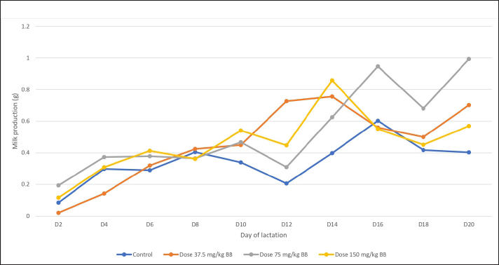
AbstractBackground: Breastfeeding is vital for infant health, providing essential nutrients and protection against infections. Despite the benefits, many postpartum women face challenges in breastfeeding due to hypogalactia caused by stress, anxiety, or maternal illness. While medications, such as metoclopramide and domperidone, are sometimes prescribed to increase milk supply, they are limited due to safety concerns. As a result, there is a growing interest in alternative nonpharmacological interventions, including herbal galactagogues. Plants, such as Sauropus androgynus, Moringa oleifera, and Coleus amboinicus, have shown potential in enhancing milk production due to their unique lactogenic properties. Aim: This study aimed to investigate the influence of a combination of S. androgynus, M. oleifera, and C. amboinicus extracts on milk production while assessing whether these extracts possess galactagogue properties. Methods: Twenty-four Sprague-Dawley rats were randomly divided into four groups at parturition. Each group, comprising six dams, was assigned a specific dose of 37.5, 75, and 150 mg/kg of the blended extract orally for 20 days postpartum, with one group serving as the control. Mammary glands were harvested and assayed for prolactin expression by immunohistochemistry. Results: The results showed that while the rate of milk production did not increase linearly with the duration of lactation, rats treated with the blended extract at a dose of 75 mg/kg exhibited higher milk production than the control group. Immunohistochemical analysis revealed increased prolactin expression in the mammary glands of rats treated with blend extract at 37.5 mg/kg (p < 0.05), suggesting its potential as a galactagogue. Conclusion: The blended extract of S. androgynus, M. oleifera, and C. amboinicus may stimulate milk production by modulating prolactin expression and mammary gland morphology. Keywords: C. amboinicus, Immunohistochemistry, Mammary glands, M. oleifera, S. androgynus. IntroductionBreast milk is widely regarded as the most suitable food for infants during the first 6 months of life. Breast milk has been associated with improved infant health, including improved neurodevelopment and gastrointestinal microbiota function, enhanced immunity, and reduced risk of gastrointestinal diseases and mortality (Campoy et al., 2018; Lyons et al., 2020). The World Health Organization (WHO) recommends that newborns should be breastfed for at least 6 months, and the preference is for continued breastfeeding for 1 year or more. This recommendation is based on the numerous benefits that breastfeeding provides to both infants and mothers. Breastfeeding offers ideal infant nutrition, which protects newborns from infections and provides nutrients necessary for healthy growth and development. Furthermore, breastfeeding strengthens the emotional bond between the mother and child and positively impacts mothers’ mental health. The WHO recommendation is grounded in extensive research and evidence-based practices and is widely regarded as the gold standard for infant feeding (Vesel et al., 2010). However, many postpartum women with hypogalactia face difficulties in breastfeeding because of stress, anxiety, or maternal illness (Mustofa et al., 2020). Medications, such as metoclopramide and domperidone, are sometimes used to increase milk supply but are limited because of safety concerns (Doggrell and Hancox, 2014; Liu et al., 2015; Grzeskowiak et al., 2018). Alternative options include herbal, synthetic, and plant-derived galactagogues. These remedies are commonly used as nonpharmacological interventions to increase breast milk production. Herbal galactagogues, such as Sauropus androgynus, Moringa oleifera, and Coleus amboinicus, have been shown to enhance milk production in postpartum mothers because of their specific interactions (Iwansyah et al., 2016; Indrayani et al., 2020; Fungtammasan and Phupong, 2021). This study used a blend of three plant combinations known for their galactagogue properties. The rationale behind this mixture harnesses synergistic effects, expands the range of activity, minimizes the risk of plant-specific resistance, reduces adverse reactions, incorporates complex active ingredients, and aligns with traditional medicinal practices. Coleus amboinicus, recognized for its milk-enhancing qualities and rich flavonoid content, exhibits lower antioxidant activity than other plants. Consequently, pairing it with M. oleifera, a plant with higher antioxidant content could amplify its effects (Lutfiani and Nasrulloh, 2023; Zulkarnain et al., 2024). Although M. oleifera boasts abundant nutrients and potent antioxidant and anti-inflammatory properties, its standalone galactagogue efficacy is less pronounced than S. androgynus, demonstrating significant lactogenic capabilities. However, studies have suggested that excessive consumption of S. androgynus may lead to adverse effects. The anti-inflammatory characteristics of M. oleifera can help mitigate potential side effects and enhance its overall effectiveness. Combining these three extracts can achieve a more potent and synergistic effect, leveraging the unique strengths of each plant to boost breast milk production and promote the overall well-being of nursing mothers. This study aimed to investigate the influence of a combination of S. androgynus, M. oleifera, and C. amboinicus extracts on milk production while assessing whether these extracts possess galactagogue properties. Materials and MethodsPlant collection, identification, and extractionThe Breynia androgyna (L.) Chakrab. & N. P. Balakr., M. oleifera Lam., and C. amboinicus Lour were assigned by Dr. Atik Retnowati, SP., M.Sc., Head of the Botany Division, Research Center for Biology—Indonesian Institute of Sciences, West Java, Indonesia; these simplicia were confirmed as authentic by Document No. 856/IPH.1.01/If.07/VIII/2020. The three simplicia were collected and supplied from a horticultural garden in Wonosobo Regency, Central Java, Indonesia (GPS location: 7°21’32.2″ S, 109°54’16.8″ E). The extraction process for the simplicia utilized the percolation at 60°C for 90 minutes with pharmaceutical-grade ethanol 70% solvent. The three-leaf combinations were combined in equal proportions, each contributing one part. The resulting liquid extract mixture was dried and thickened using spray drying and evaporation. AnimalsThe procedure for animal research was systematically evaluated and compared to that of animal research. Twenty-four female Sprague-Dawley rats weighing 200–250 g were provided by the Indonesian Food and Drug Authority (BPOM Indonesia). Before the commencement of the study, the animals were housed in a regulated animal care facility that maintained a 12-hour day–night cycle, standard feed, unrestricted access to water, temperature ranging between 20°C and 25°C, and humidity levels between 40% and 70% for 1 week to enable them to become acclimated to their environment. After parturition, the rats were randomly divided into four groups comprising six dams and five pups per dam. Dam rats and their offspring were housed in cages. The four lactating groups were classified as Group 1, which served as the normal control and were treated orally with distilled water; Groups 2–4 were orally administered the blended extracts at 37.5, 75, and 150 mg/kg, respectively. The dams began receiving the extracts 2 days after parturition until day 20. Milk production of rat damsThe body weight of the pups was assessed three times daily. Initial weighing (W1) was performed at 8:00 a.m., after which pups were separated from their dams for 4 hours. The second weighing (W2) was performed at 12:00 p.m. The pups were reunited with their dams and allowed to nurse for 1 hour. Finally, the pups were weighed (W3) at 1:00 p.m. and allowed to remain with their dams for the remainder of the day. Daily milk production of the rat dams was determined during the entire lactation period. It was calculated as the difference between W3 and W2, plus the weight loss due to metabolic processes in the offspring during the suckling period, divided by four (W2—W1)/4. Therefore, milk production could be expressed as (W3 − W2) + [(W2 − W1)/4] (Cai et al., 2015), (De Los Ríos et al., 2018). Sample collectionMammary gland tissue samples were obtained from all the dams on the 21st day of lactation. Rats were anesthetized using a combination of ketamine (80-mg/kg body weight) and xylazine (10-mg/kg body weight) administered intraperitoneally, followed by dissection to remove mammary glands. The collected samples were placed in labeled container tubes containing 10% formalin and stored at room temperature (TitiMutiara, K, 2013). Histopathology of the mammary glandMammary gland tissues were processed using 10% neutral buffered formalin and embedded in paraffin. Subsequently, sections measuring 5 mm in thickness were utilized with hematoxylin and eosin (H&E). Microscopic observations were captured and analyzed using an Olympus BX53 microscope equipped with an Olympus DP25 camera and Stream program at a magnification of 400×. Immunohistochemical analysisSeveral steps were taken to prepare the mammary glands for analysis, including saline washing, 10% formalin fixation, ethanol concentration in progressive dehydration, xylene clearing, and paraffin embedding. Tissue slices were cut using a microtome and stained using immunohistochemistry to evaluate prolactin expression. Rabbit monoclonal anti-prolactin/PRL antibody (ERP18018-31) at 1:2000 (Abcam) was used as the primary antibody, and Starr Trek Universal HRP Detection System (Biocare Medical) was used as the secondary antibody. Staining pattern analysis aims to elucidate prolactin distribution (Taylor and Levenson, 2006) in specific tissues. This method helps determine the precise location of prolactin within glands or tissues and identifies any shifts in its localization due to particular treatments or conditions. Such information can shed light on the mechanisms underlying breast milk production and tissue responses to various plant extracts. Staining intensity measurements are used to quantify prolactin expression levels (Kim et al., 2016). A more intense staining indicated a higher concentration of prolactin in the tissue. By comparing staining intensities across different treatment groups, researchers can ascertain whether specific combinations of plant extracts significantly affect prolactin production. Lumen morphometric evaluation focuses on examining structural alterations in glandular ducts (Jolicoeur, 2005) such as changes in lumen size or shape associated with breast milk secretion activity. This analysis helps to determine whether galactagogue extracts influence lumen dimensions or capacity, providing evidence for enhanced breast milk production and secretion. Understanding the relationship between lumen size and prolactin production can help clarify how plant-derived bioactive compounds boost breast milk production. The interplay between the staining pattern, staining intensity, and lumen morphometry parameters, when correlated with breast milk production volume, offers a comprehensive view of plant extract efficacy. These parameters are interconnected, as increased prolactin expression, as measured by immunohistochemistry, is expected to correlate with elevated breast milk production and the corresponding morphological changes in the gland that support this secretion. A semi-qualitative evaluation of a positive or negative reaction and the staining pattern, which was classified as membranous when the positive expression was localized to the apical membrane and cytoplasmic when it was intracytoplasmic, was used to assess prolactin expression. Using morphometric analysis, the number of positive and negative cells in the luminal epithelial cells was determined in ten fields of view (400×) from six blocks of each group. Prolactin protein levels were measured as the percentage of positive cells relative to the total alveolar cells per field of view, and the intensity of the brown color in the alveolar lumen was assessed as weak, medium, and vigorous. The results are expressed as the mean ± SD using an Olympus BX53 microscope with a camera device (Olympus DP25) and Stream program. Statistical analysisMilk production data are presented as mean ± standard deviation (SD). Statistical differences between groups were determined using Kruskal–Wallis and Mann–Whitney U post hoc tests with SPSS (version 29.0; p < 0.05). Using a light microscope, immunohistochemical data were analyzed using a semi-quantitative method based on immune reactions. Ethical approvalThe experimental procedures and protocols used in this study were approved by the Health Research Ethics Commission, Health Research and Development Agency (KEPK-BPPK), Ministry of Health of the Republic of Indonesia (Approval no: LB.02.01/2/KE.090/2021). Results and DiscussionMilk production of rat damsThe effects of the blended extracts of S. androgynus, M. oleifera, and C. amboinicus on milk production were assessed by calculating the milk production. Milk production in female rats was measured daily after administration of the blended extract, and the findings revealed that milk production was not proportional to the number of lactation days (Fig. 1). There were intervals between the high and low increases in milk production. Notably, milk production increased at intervals of D6–D10 and D16–D18 when lactating rats were treated with 75- and 150-mg/kg blend extract, respectively. Milk production in the 75-mg/kg BW group was higher than in other groups. However, decreased production was observed on days D12 and D18. Peak lactation is typically defined as the maximum milk secretion yield. Throughout lactation, milk secretion gradually peaks and then decreases. Research has categorized lactation into three phases: the early phase (from birth to day 6), the middle phase (from day 7 to day 14), where milk production peaks, and the final phase (from day 15 until the end of lactation), where milk production declines (Knight et al., 1986; Bautista et al., 2019). Fluctuations in milk production during these phases are influenced by changes in metabolic needs, hormone levels, and nursing stimuli from pups.
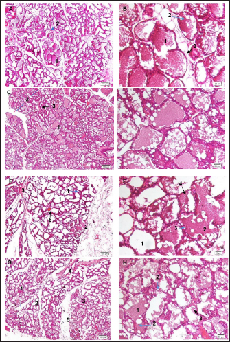
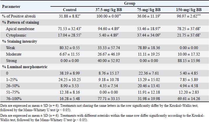
Fig. 1. Effects of blend extracts on milk production for 20 days. Data are expressed as mean ± SD (n=6). Statistical analysis using the Kruskal–Wallis test. Administration of a combination of herbal extracts showed a positive effect on milk production, with a noticeable increase in volume in lactating rats administered a dose of 75 mg/kg compared to the control group. The larger alveolar images in the extract group indicate that these extracts enhanced the tissue structure that supports lactation. This suggests that the extracts may contain substances that stimulate milk production by regulating lactation hormones or repair defects in the metabolism that affect lactation(Liu et al., 2015; Mustofa et al., 2020) (Liu et al., 2015; Mustofa et al., 2020). To ensure consistent results, the number of rat pups in each experimental group was standardized to five pups per group. Thus, the observed differences in milk production and the weight of the pups can be more accurately attributed to the effects of extract administration rather than differences in litter size. Nevertheless, a larger litter size can generally affect the quality and quantity of milk produced by the mother, which ultimately determines the weight gain of the pups (Xavier et al., 2019). Several studies have measured the weight and weight gain of pups (Sampson and Jansen, 1984; Mahmood et al., 2012; Liu et al., 2015; Mustofa et al., 2020). This is believed to reflect the milk production during lactation. This is because the daily litter weight gain is regarded as an approximate estimate of milk production in rats (Mahmood et al., 2012). The results did not reveal any substantial variation in milk production between the control group, which received distilled water, and the group administered blended extracts. Although lactating rats were administered the blended extracts at various dosage levels, they produced more milk than the control group. The results revealed that the 75-mg/kg group displayed the highest milk production compared with the other groups. These findings imply that the ethanol extract obtained from a blend of S. androgynus, M. oleifera, and C. amboinicus, or its herbal components, could contain chemical compounds that might stimulate lactation. Milk production in rats was not proportional to the duration of lactation, which was also observed when evaluating the galactagogue activity of Musa × paradisiacal leaf extracts in rats (Mahmood et al., 2012). Repeated cell growth cycles and programmed cell elimination in mammary glands during lactation influence milk production and lactation length. Consequently, the rate of milk synthesis varies depending on the lactation stage. After reaching the peak of milk production on day 16, the equilibrium shifted, causing reduced milk production on days 12 and 18 in most experimental groups. However, the pups gained weight during the study, indicating that the milk produced by lactating rats was good quality. Based on the findings of Nosrati et al. (2021) this conclusion suggests that the weight gain of rat pups indicates milk production in female rats. The study conducted by Nosrati et al. (2021) concluded that this finding was consistent with the mechanism of induced galactagogue activity by examining its expression in lactating rats treated with the blended extract. The results revealed extract-induced PRL expression (Lompo-Ouedraogo et al., 2004). Histopathology of the mammary glandDuring lactation, alveoli undergo significant changes, including increased alveolar lumen size and enhanced proliferation of epithelial cells, which indicate active milk production (Salahshoor et al., 2018; Utary et al., 2019). Histological analysis revealed that compared to the normal control group, mammary glands consisted of small lobules dispersed within adipose tissue (Fig. 2A, B), and the groups treated with blended extract had increased lobular and alveolar volume, expanded alveoli filled with breast milk, along with a reduction in adipose tissue (Fig. 2C–H). These observations indicate that the blended extracts may influence the functional aspects of alveolar development and milk synthesis. In the 75-mg/kg group (Fig. 2D), each lobule contained a marginally larger alveolus than that in the 37.5, 150 mg/kg, and control groups, indicating a positive effect of the extract on the alveolar size. During pregnancy, glandular tissue develops into secretory alveoli surrounded by contractile myoepithelial cells. Expansion of the alveolar lumen, driven by epithelial cell growth, enhances milk production capacity. Our results showed that lactating rats treated with the 75-mg/kg extract had notably prominent alveolar lumens, suggesting that the extract stimulated the proliferation of myoepithelial cells, leading to more alveoli and potentially higher milk production (Utary et al., 2019). In addition, the increase in lobular size and reduction in adipose tissue in treated groups indicate that the extracts promote glandular tissue expansion and optimize conditions for lactation. Immunohistochemical studyA blend containing extracts of Sauropus, Moringa, and Coleus may have galactagogue effects since it can increase the production of prolactin. Mammary gland samples from breastfeeding rats were administered the extract daily and were subjected to histological examination. The findings revealed that the 37.5-mg/kg bw blend extract dose significantly impacted the percentage of positive alveoli and increased apical membrane staining, followed by doses of 150-mg/kg BW and 75-mg/kg BW, as demonstrated in Table 1. Specifically, at 150 mg/kg, the blended extract exhibited a vigorous intensity of prolactin compared to the other groups and the control group, particularly in the mammary ductules compared to the alveolar membrane (Fig. 3D). Various hormones, including prolactin, have been shown to influence milk secretion in rats. Immunohistochemistry was used to identify these hormones in the mammary gland tissue of rats. Using prolactin-specific antibodies, they determined variations in prolactin concentration in the cytoplasm, apical epithelial cells, and alveolar lumen/ductus. The results of this study indicated that prolactin was only present during the late gestation and lactation periods, as evidenced by the brown color in these areas (Naylor et al., 2003). Lactation during and after childbirth is mainly managed by the continuous secretion of prolactin (Neville et al., 2001). Prolactin stimulates the growth of receptors on the surface of alveolar cells, thereby promoting the influx of water and nutrients necessary for lactation (Liu et al., 2015). Consuming an adequate amount of plants rich in antioxidants, particularly flavonoids and phenolic compounds, may slow the aging process of mammary alveolar cells. (Jalili et al., 2015). An increase in the number of alveolar cells can potentially increase milk production (Lompo-Ouedraogo et al.,2004). The ability of the extract to activate prolactin receptors on lactotrophs, which produce prolactin, was thought to be the cause of the results observed in the group that received the extract. Activation of prolactin receptors stimulates the release of neurohormones, which in turn trigger the secretion of prolactin-releasing factor. The hypothesis suggests that this group’s observed elevated prolactin levels and vivid color intensity were caused by phytosterols present in S. androgynus. These sterols function similar to cholesterol, which is involved in steroidogenesis. Steroidogenesis is a biosynthetic pathway that produces steroid hormones, including estrogen, which stimulates prolactin synthesis in the pituitary gland. According to lactation theory, increased milk secretion during lactation is closely associated with elevated prolactin levels in the blood (Miharti et al., 2018). The function of the active components in S. androgynus leaves in milk production in secretory glands involves two pathways. First, hormonal action is involved, and S. androgynus leaves can influence lactogenic hormones directly or indirectly. Directly, prostaglandins and steroid hormones are affected by the action of S. androgynus leaves. At the same time, stimulation of pituitary gland cells indirectly triggers the release of hormones, such as prolactin and oxytocin. Second, metabolic activity occurs through the hydrolysis of active compounds in S. androgynus leaves, which can then participate in the metabolism of carbohydrates, proteins, and fats. Beta-carotene, present in S. androgynus, plays an active role and is transformed into vitamin A in the body. Vitamin A is essential for protein synthesis, growth, and development of secretory cells in mammary glands (Suprayogi, 2000). The significance of phytosterols in the hormonal pathways of Moringa, particularly in increasing prolactin levels, cannot be overstated. Raising prolactin levels directly stimulates the protoplasmic activity of secretory cells in mammary glands, thereby increasing milk production. Furthermore, it encourages the discharge of nerve endings into mammary glands, thereby enhancing milk secretion. In addition, phytosterols in Moringa stimulate prolactin production, which acts on alveolar epithelial cells to promote milk production (Farah Rizki, 2013; Raguindin et al., 2014). Moringa oleifera has gained extensive recognition for enhancing glucose metabolism, particularly lactose synthesis, which leads to increased milk production through metabolic pathways (Sari. I.P, 2003).
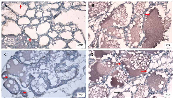
Fig. 2. Effect of the blended extract of S. androgynus, M. oleifera, and C. amboinicus on mammary gland tissue. A and B control group: A) 1. alveolar-containing milk and 2. cuboidal epithelial lining cells (blue arrow) HE ×100; B) 1. alveolar-containing milk, 2. fat globules (blue arrows), and 3. cuboidal epithelial lining cells (black arrows). HE ×400; C-D Group 2 with 37.5 a blended extract: C) 1. alveolar-containing milk, 2. lobules (blue arrows), and 3. interlobular duct (black arrow) HE ×100; D) 1. alveolar-containing milk, 2. fat globules (blue arrows), 3. cuboidal epithelial lining cells (black arrows), and 4. secretory ducts. HE ×400; E and F. Group 3: 75 mg/kg of the blended extract: E) 1. alveolar, 2. alveolar containing milk, 3. connective tissues, 4. duct (blue arrow), and 5. blood vessels (black arrow) HE ×100; F) 1. alveolar, 2. alveolar containing milk, 3. fat globules (blue arrows), and 4. cuboidal epithelial lining cells (black arrows). HE ×400; G and H group 4 treated with 150 mg/kg of the blended extract: G) 1. lobules of the mammary glands (blue arrow), 2. alveolar, 3. alveolar-containing milk, 4. interlobular duct, and 5. interseptum. HE ×100; H) 1. alveolar-containing milk, 2. fat globules (blue arrows), and 3. cuboidal epithelial lining cells (black arrows). HE ×400. The effects of the phytoestrogen content of C. amboinicus on prolactin levels in the hormonal pathway were minimal. Nevertheless, its bioactive components influence metabolic pathways by regulating casein secretion and lactose synthesis in the mammary epithelial cells (Iwansyah et al., 2019). The effects of plant-derived galactagogues are related to their bioactive compounds that promote lactation by boosting milk production. Blended extracts have been shown to contain various chemical components, such as quercetin, which is linked to increased mammary gland development, lactation volume, and protein expression in milk production (Saxena VK, 1998; Mapuskar et al., 2019). Quercetin has also been reported to stimulate prolactin secretion (Lin et al., 2018). The three investigated plants exhibited similar characteristics; however, they all demonstrated the same effect of increasing milk production. S. androgynus, M. oleifera, and C. amboinicus are all rich in lactogague, a chemical compound that promotes milk production. The presence of saponins, flavonoids, and alkaloids in C. amboinicus and M. oleifera may stimulate lactation hormones such as prolactin and oxytocin. Sauropus contains carotenoids, an active ingredient that acts as a source of vitamin A and can increase prolactin stimulation. Scientific research has suggested that the bioactive compounds in this extract possess the potential to elevate serum prolactin levels, which plays a critical role in milk production. Moreover, existing literature indicates that specific herbs classified as galactagogues can enhance milk production in lactating rats by stimulating cell division within the mammary glands. These herbs are believed to affect the function of the cells responsible for milk secretion, and their effect on these cells can serve as a marker of secretory cell activity during milk production (Lompo-Ouedraogo et al., , 2004; Okasha et al., 2008; Damanik et al., 2017; Fikri and Purnama, 2020; Fatmawati et al., 2022). These findings suggest a plausible relationship between prolactin, proteins related to milk production, and the function of the blended extract. Further experiments are necessary to determine how the blended extract enhances milk production, which could stimulate prolactin secretion. Nonetheless, the observed increase in milk production and known milk production-related components in the blend extract lend credence to its galactagogue effect. Previous studies investigated the effects of herbal galactagogues on milk production-related proteins (Liu et al., 2015; Mustofa et al., 2020). The findings of this investigation demonstrated that the alveolar lumen was more spacious in the treatment group administered a blend extract of S. androgynus, M. oleifera, and C. amboinicus (Fig. 3B, C, and D) than that in the control group (Fig. 3A). Furthermore, the lumen was filled with milk in the milk group. Treatment with the extract at 150-mg/kg BW (Fig. 3D) led to this outcome. Table 1. Expression of prolactin of mammary tissue using immunohistochemical staining.
Fig. 3. Histological sections of the mammary glands of lactating rats were treated with a blend extract of S. androgynus, M. oleifera, and C. amboinicus. The image shows the following samples. A. Control, positive prolactin reaction (PRL) in apical membrane epithelial cells (red arrow). B. Dose of 37.5-mg/kg BW, positive prolactin reaction (PRL) in the ductus (red arrow. C. Dose of 75-mg/kg BW, positive prolactin reaction (PRL) in the cytoplasm (red arrow). D. Dose of 150-mg/kg BW, intracellular lipid droplets (red arrow), and strong positive prolactin reaction (PRL) in the ductus (black arrow). All groups were subjected to prolactin antibody immunohistochemical staining at 400× magnification. This study indicated that the alveolar lumen in the treatment group that received S. androgynus, M. oleifera, and C. amboinicus extracts were more expansive and filled with milk. This is because, during the lactation period, the mammary parenchyma includes alveoli with large lumens filled with milk and flattened epithelium owing to the accumulation of milk in the lumen (Delpal et al., 2013). ConclusionIn conclusion, this study highlights the potential of a blended extract of S. androgynus, M. oleifera, and C. amboinicus to enhance milk production in lactating rats. The increased milk production and prolactin expression in mammary glands indicated the galactagogue properties of the blended extract. Further research is warranted to elucidate the mechanisms underlying its action and to explore its potential application in lactation support for postpartum women. AcknowledgmentsThe authors acknowledge the Center for Research and Development of Biomedical and Basic Health Technology at the National Institute of Health Research and Development, Ministry of Health, Republic of Indonesia, for providing fund support to complete this study. Conflict of interestsThe authors declared that there is no conflict of interest. FundingThe authors acknowledge the Center for Research and Development of Biomedical and Basic Health Technology at the National Institute of Health Research and Development, Ministry of Health, Republic of Indonesia, for providing fund support to complete this study. Authors’ contributionsPRI, SA, AI, NY, and EH developed the concept and design. PRI, SA, AI, NY, FE, RR, SS, YS, SSM, and EH contributed to data analysis/interpretation. PRI, SA, AI, NY, FE, RR, SS, YS, SSM, and EH contributed to drafting the manuscript. PRI, SA, AI, NY, FE, RR, SS, YS, SSM, and EH contributed to the critical revision of the manuscript. PRI, FE, and SSM contributed to statistical analysis. PRI, SA, AI, NY, FE, RR, and SS contributed to admin and technical or material support. YS, SSM, and EH contributed to supervision. PRI, SA, AI, NY, FE, RR, SS, YS, SSM, and EH contributed to the final approval. Data availabilityAll data are provided in the manuscript. ReferencesBautista, C.J., Bautista, R.J., Montaño, S., Reyes-Castro, L.A., Rodriguez-Peña, O.N., Ibáñez, C.A., Nathanielsz, P.W. and Zambrano, E. 2019. Effects of maternal protein restriction during pregnancy and lactation on milk composition and offspring development. Br. J. Nutr. 122(2), 141–151. Cai, B., Chen, H., Sun, H., Sun, H., Wan, P., Chen, D. and Pan, J. 2015. Lactogenic activity of an enzymatic hydrolysate from Octopus vulgaris and Carica papaya in SD rats. J. Med. Food 18(11), 1262–1269. Campoy, C., Campos, D., Cerdó, T., Diéguez, E. and Garciá-Santos, J.A. 2018. Complementary feeding in developed countries: The 3 Ws (When, what, and why?). Ann. Nutr. Metab. 73(suppl 1), 27–36. Damanik, R.M., Kustiyah, L., Hanafi, M. and Iwansyah, A.C. 2017. Evaluation lactogenic activity of ethyl acetate fraction of torbangun (Coleus amboinicus L.) leaves. IOP Conf. Ser. Earth Environ. Sci. 101(1), 012007. Delpal, S., Pauloin, A., Hue-Beauvais, C., Berthelot, V., Schmidely, P. and Ollivier-Bousquet, M. 2013. Effects of dietary fish oil and corn oil on rat mammary tissue. Cell Tissue Res. 351(3), 453–464. Doggrell, S.A. and Hancox, J.C. 2014. Cardiac safety concerns for domperidone, an antiemetic and prokinetic, and galactogogue medicine. Expert Opin. Drug Saf. 13(1), 131–138. Farah Rizki, S. 2013. The miracle of vegetables. Jakarta, Indonesia: Agro Media. Fatmawati, A., Sucianingsih, D., Kurniawati, R. and Abdurrahman, M. 2022. Microscopic identification and determination of total flavonoid content of Moringa leaves extract and ethyl acetate fraction (Moringa oleifera L.). Indones. J. Pharm. Sci. Technol. 1(1), 66. Fikri, F. and Purnama, M.T.E. 2020. Pharmacology and phytochemistry overview on sauropus androgynous. Syst. Rev. Pharm. 11(6), 124–128. Fungtammasan, S. and Phupong, V. 2021. The effect of Moringa oleifera capsule in increasing breastmilk volume in early postpartum patients: a double-blind, randomized controlled trial. PLoS One 16(4), 1–7. Grzeskowiak, L.E., Smithers, L.G., Amir, L.H. and Grivell, R.M. 2018. Domperidone for increasing breast milk volume in mothers expressing breast milk for their preterm infants: a systematic review and meta-analysis. BJOG An Int. J. Obstet. Gynaecol. 125(11), 1371–1378. Indrayani, D., Shahib, M.N. and Husin, F. 2020. The effect of katuk (Sauropus androgunus (L) Merr) leaf biscuit on increasing prolactine levels of breastfeeding mother. J. Kesehat. Masy. 16(1), 1–7. Iwansyah, A.C., Damanik, R.M., Kustiyah, L. and Hanafi, M. 2016. Relationship between antioxidant properties and nutritional composition of some galactopoietics herbs used in indonesia: a comparative study. Int. J. Pharm. Pharm. Sci. 8(12), 236–243. Iwansyah, A.C., Damanik, R.M., Kustiyah, L. and Hanafi, M. 2019. The ethyl acetate fraction of torbangun ( Coleus amboinicus L .) leaves increasing milk production with up-regulated genes expression of prolactin receptor. J. Trop. Life Sci. 9(2), 147–154. Jalili, C., Salahshoor, M.R., Yousefi, D., Khazaei, M., Shabanizadeh Darehdori, A. and Mokhtari, T. 2015. Morphometric and hormonal study of the effect of Utrica diocia extract on mammary glands in rats. Int. J. Morphol. 33(3), 983–987. Jolicoeur, F. 2005. Intrauterine breast development and the mammary myoepithelial lineage. J. Mammary Gland Biol. Neoplasia 10(3), 199–210. Kim, S.W., Roh, J. and Park, C.S. 2016. Immunohistochemistry for pathologists: protocols, pitfalls, and tips. J. Pathol. Transl. Med. 50(6), 411–418. Knight, C.H., Maltz, E. and Docherty, A.H. 1986. Milk yield and composition in mice : effects of litter size and lactation. Comp. Biochem. Physiol. A Comp. Physiol. 84, 127–133. Lin, M., Wang, N., Yao, B., Zhong, Y., Lin, Y. and You, T. 2018. Quercetin improves postpartum hypogalactia in milk-deficient mice via stimulating prolactin production in pituitary gland. Phyther. Res. 32(8), 1511–1520. Liu, H., Hua, Y., Luo, H., Shen, Z., Tao, X. and Zhu, X. 2015. An herbal galactagogue mixture increases milk production and aquaporin protein expression in the mammary glands of lactating rats. Evidence-based Complement. Altern. Med. 2015(1), 760585. Lompo-Ouedraogo, Z., van der Heide, D., van der Beek, E.M., Swarts, H.J., Mattheij, J.A. and Sawadogo, L. 2004. Effect of aqueous extract of Acacia nilotica ssp adansonii on milk production and prolactin release in the rat. J. Endocrinol. 182(2), 257–266. De Los Ríos, E.A., Ruiz-Herrera, X., Tinoco-Pantoja, V., Ĺopez-Barrera, F., De La Escalera, G.M., Clapp, C. and Macotela, Y. 2018. Impaired prolactin actions mediate altered offspring metabolism induced by maternal high-fat feeding during lactation. FASEB J. 32(6), 3457–3470. Lutfiani, L. and Nasrulloh, N. 2023. Total flavonoid and antioxidant activity of food bar torbangun – katuk on the effectiveness of breast milk production. Amerta Nutr. 7(1), 88–97. Lyons, K.E., Ryan, C.A., Dempsey, E.M., Ross, R.P. and Stanton, C. 2020. Breast milk, a source of beneficial microbes and associated benefits for infant health. Nutrients 12(4), 1–30. Mahmood, A., Omar, M.N. and Ngah, N. 2012. Galactagogue effects of Musa x paradisiaca flower extract on lactating rats. Asian Pac. J. Trop. Med. 5(11), 882–886. Mapuskar, K.A., Wen, H., Holanda, D.G., Rastogi, P., Steinbach, E., Han, R., Coleman, M.C., Attanasio, M., Riley, D.P., Spitz, D.R., Allen, B.G. and Zepeda-Orozco, D. 2019. Persistent increase in mitochondrial superoxide mediates cisplatin-induced chronic kidney disease. Redox Biol. 20, 98–106. Miharti, S.I., Oenzil, F. and Syarif, I. 2018. Pengaruh ekstrak etanol daun katuk terhadap kadar hormon prolaktin tikus putih menyusui. J. Iptek Terap. 12(3), 202–211. Mustofa, Yuliani, F.S., Purwono, S., Sadewa, A.H., Damayanti, E. and Heriyanto, D.S. 2020. Polyherbal formula (ASILACT®) induces milk production in lactating rats through upregulation of α-Lactalbumin and aquaporin expression. BMC Complement. Med. Ther. 20(1), 1–8. Naylor, M.J., Lockefeer, J.A., Horseman, N.D. and Ormandy, C.O. 2003. Proliferation via autocrine / Paracrine mechanism. Endocrine 20(1), 111–114. Neville, M.C., Morton, J. and Umemura, S. 2001. Lactogenesis: the transition from pregnancy to lactation. Pediatr. Clin. North Am. 48(1), 35–52. Nosrati, H., Hamzepoor, M., Sohrabi, M., Saidijam, M., Assari, M.J., Shabab, N., Gholami Mahmoudian, Z. and Alizadeh, Z. 2021. The potential renal toxicity of silver nanoparticles after repeated oral exposure and its underlying mechanisms. BMC Nephrol. 22(1), 228. Okasha, M.A.M., Abubakar, M.S. and Bako, I.G. 2008. Study of the effect of aqueous hibiscus sabdariffa linn seed extract on serum prolactin level of lactating female albino rats. Eur. J. Sci. Res. 22(4), 575–583. Raguindin, P.F.N., Dans, L.F. and King, J.F. 2014. Moringa oleifera as a galactagogue. Breastfeed. Med. 9(6), 323–324. Salahshoor, M.R., Mohammadi, M.M., Roshankhah, S. and Jalili, C. 2018. Effect of Falcaria Vulgaris on milk production parameters in female rats’ mammary glands. J. Fam. Reprod. Heal. 12(4), 177–183. Sampson, D.A. and Jansen, G.R. 1984. Measurement of milk yield in the lactating rat from pup weight and weight gain. J. Pediatr. Gastroenterol. Nutr. 613–617. Sari. I.P. 2003. Daya Laktagogum Jamu Uyup-uyup dan Ekstrak Daun Katu (Sauropus androgynus Merr.) pada Glandula Ingluvica Merpati: Lactagogue effect of uyup-uyup (traditional medicine) and Sauropus androgynus Merr extract on pigeon inglu. Maj. Farm. Indones. 14, 2003. Saxena, V.K. and Singal, M. 1998. Genistein 4’-O-α-L-rhamnopyranoside from Pithecellobium dulce. Fitoter. 69(4), 305–306. Suprayogi, A. 2000. Studies on the biological effects of Sauropus androgynus (L.) Merr: effects on milk production and the possibilities of induced pulmonary disorder in lactating sheep. Cuvilier. 140. Taylor, C.R. and Levenson, R.M. 2006. Quantification of immunohistochemistry—issues concerning methods, utility and semiquantitative assessment II. Histopathology 49(4), 411–424. TitiMutiara, K, Titi, E.S. and Estiasih, W. 2013. Effect Lactagogue Moringa leaves ( Moringa oleifera Lam ) Powder in Rats. J. Basic. Appl. Sci. Res. 3(4), 430–434. Utary, N., Murti, K. and Septadina, I.S. 2019. Effects of Moringa (Moringa oleifera) leaf extract on alveolar diameter of breastfeeding and weight of infant Wistar rats. J. Phys. Conf. Ser. 1246(1), Vesel, L., Bahl, R., Martines, J., Penny, M., Bhandari, N. and Kirkwood, B.R. 2010. Use of new World Health Organization child growth standards to assess how infant malnutrition relates to breastfeeding and mortality. Bull. World Health Organ. 88(1), 39–48. Xavier, J.L.P., Scomparin, D.X., Pontes, C.C., Ribeiro, P.R., Cordeiro, M.M., Marcondes, J.A., Mendonça, F.O., Silva, M.T.D., Oliveira, F.B.D., Franco, G.C. and Grassiolli, S. 2019. Litter size reduction induces metabolic and histological adjustments in dams throughout lactation with early effects on offspring. An. Acad. Bras. Cienc. 91(1), 1–17. Zulkarnain, Z., Fitriani, U., Ardiyanto, D., Saryanto, Wijayanti, E., Triyono, A. and Novianto, F. 2024. Galactagogue activity of poly-herbal decoction from Indonesia: a randomized open label controlled trial. J. Complement. Integr. Med. 21(4), 532–539. | ||
| How to Cite this Article |
| Pubmed Style Intan PR, Alegantina S, Isnawati A, Yunarto N, Ekawasti F, Rinendyaputri R, Sunarno S, Sani Y, Mariya SS, Handharyani E. Effects of a blend extract of Sauropus androgynus, Moringa oleifera, and Coleus amboinicus on milk production in lactating rats. Open Vet. J.. 2024; 14(12): 3630-3639. doi:10.5455/OVJ.2024.v14.i12.44 Web Style Intan PR, Alegantina S, Isnawati A, Yunarto N, Ekawasti F, Rinendyaputri R, Sunarno S, Sani Y, Mariya SS, Handharyani E. Effects of a blend extract of Sauropus androgynus, Moringa oleifera, and Coleus amboinicus on milk production in lactating rats. https://www.openveterinaryjournal.com/?mno=218447 [Access: January 11, 2026]. doi:10.5455/OVJ.2024.v14.i12.44 AMA (American Medical Association) Style Intan PR, Alegantina S, Isnawati A, Yunarto N, Ekawasti F, Rinendyaputri R, Sunarno S, Sani Y, Mariya SS, Handharyani E. Effects of a blend extract of Sauropus androgynus, Moringa oleifera, and Coleus amboinicus on milk production in lactating rats. Open Vet. J.. 2024; 14(12): 3630-3639. doi:10.5455/OVJ.2024.v14.i12.44 Vancouver/ICMJE Style Intan PR, Alegantina S, Isnawati A, Yunarto N, Ekawasti F, Rinendyaputri R, Sunarno S, Sani Y, Mariya SS, Handharyani E. Effects of a blend extract of Sauropus androgynus, Moringa oleifera, and Coleus amboinicus on milk production in lactating rats. Open Vet. J.. (2024), [cited January 11, 2026]; 14(12): 3630-3639. doi:10.5455/OVJ.2024.v14.i12.44 Harvard Style Intan, P. R., Alegantina, . S., Isnawati, . A., Yunarto, . N., Ekawasti, . F., Rinendyaputri, . R., Sunarno, . S., Sani, . Y., Mariya, . S. S. & Handharyani, . E. (2024) Effects of a blend extract of Sauropus androgynus, Moringa oleifera, and Coleus amboinicus on milk production in lactating rats. Open Vet. J., 14 (12), 3630-3639. doi:10.5455/OVJ.2024.v14.i12.44 Turabian Style Intan, Putri Reno, Sukmayati Alegantina, Ani Isnawati, Nanang Yunarto, Fitrine Ekawasti, Ratih Rinendyaputri, Sunarno Sunarno, Yulvian Sani, Sela Septima Mariya, and Ekowati Handharyani. 2024. Effects of a blend extract of Sauropus androgynus, Moringa oleifera, and Coleus amboinicus on milk production in lactating rats. Open Veterinary Journal, 14 (12), 3630-3639. doi:10.5455/OVJ.2024.v14.i12.44 Chicago Style Intan, Putri Reno, Sukmayati Alegantina, Ani Isnawati, Nanang Yunarto, Fitrine Ekawasti, Ratih Rinendyaputri, Sunarno Sunarno, Yulvian Sani, Sela Septima Mariya, and Ekowati Handharyani. "Effects of a blend extract of Sauropus androgynus, Moringa oleifera, and Coleus amboinicus on milk production in lactating rats." Open Veterinary Journal 14 (2024), 3630-3639. doi:10.5455/OVJ.2024.v14.i12.44 MLA (The Modern Language Association) Style Intan, Putri Reno, Sukmayati Alegantina, Ani Isnawati, Nanang Yunarto, Fitrine Ekawasti, Ratih Rinendyaputri, Sunarno Sunarno, Yulvian Sani, Sela Septima Mariya, and Ekowati Handharyani. "Effects of a blend extract of Sauropus androgynus, Moringa oleifera, and Coleus amboinicus on milk production in lactating rats." Open Veterinary Journal 14.12 (2024), 3630-3639. Print. doi:10.5455/OVJ.2024.v14.i12.44 APA (American Psychological Association) Style Intan, P. R., Alegantina, . S., Isnawati, . A., Yunarto, . N., Ekawasti, . F., Rinendyaputri, . R., Sunarno, . S., Sani, . Y., Mariya, . S. S. & Handharyani, . E. (2024) Effects of a blend extract of Sauropus androgynus, Moringa oleifera, and Coleus amboinicus on milk production in lactating rats. Open Veterinary Journal, 14 (12), 3630-3639. doi:10.5455/OVJ.2024.v14.i12.44 |


