| Research Article | ||
Open Vet. J.. 2025; 15(2): 1009-1023 Open Veterinary Journal, (2025), Vol. 15(2): 1009-1023 Research Article Bibliometric analysis of the use of pathological techniques in camel researchAhmed Aljazzar*Department of Pathology, College of Veterinary Medicine, King Faisal University, Al Hofuf, Saudi Arabia *Corresponding Author: Ahmed Aljazzar. Department of Pathology, College of Veterinary Medicine, King Faisal University, Al Hofuf, Saudi Arabia. Email: ajazzar [at] kfu.edu.sa Submitted: 31/12/2024 Accepted: 07/02/2025 Published: 28/02/2025 © 2025 Open Veterinary Journal
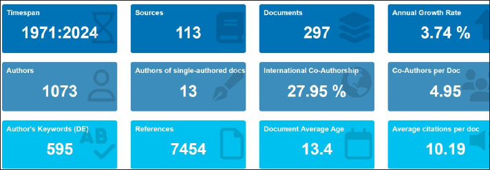
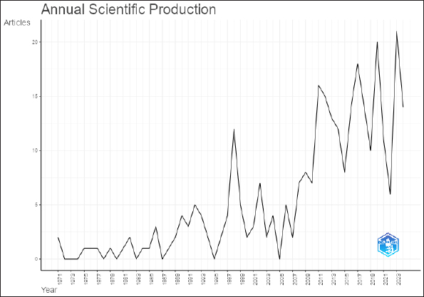
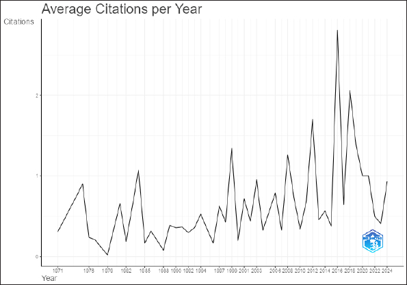
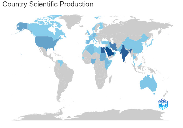
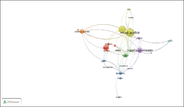
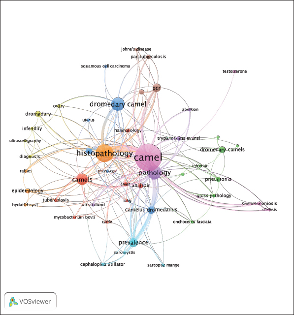
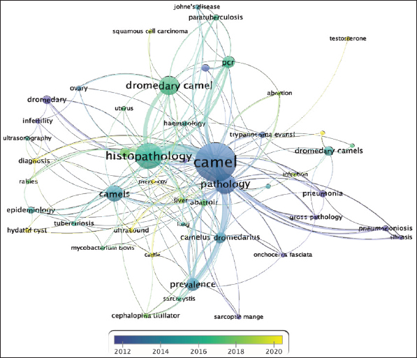
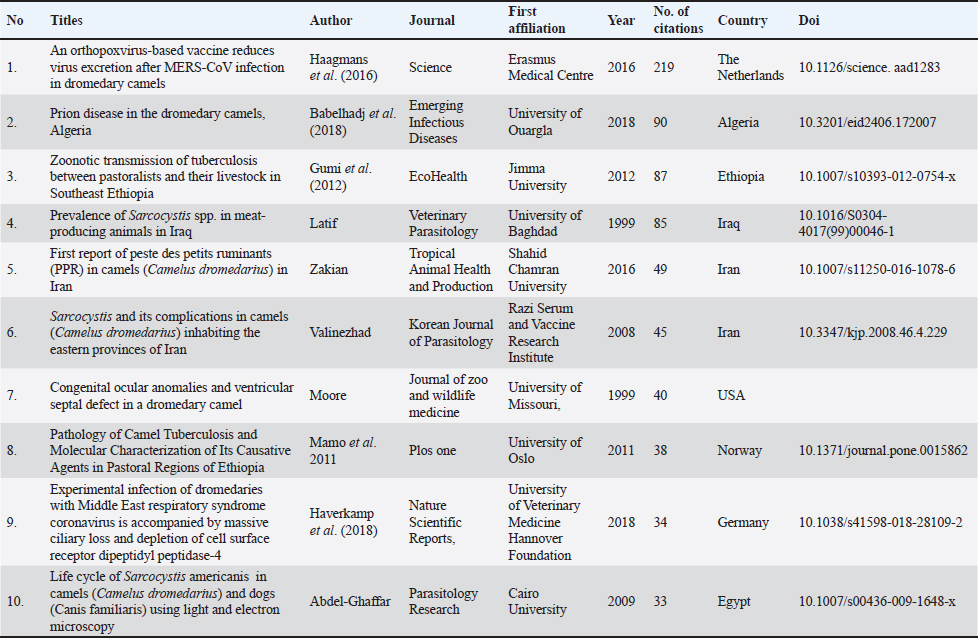
AbstractBackground: Camel pathology research has evolved significantly over the past five decades, reflecting the growing recognition of camels’ importance in agriculture, zoonotic disease control, and economic contributions. However, there has been limited evaluation of research trends, thematic advancements, and international collaborations in this field. Aim: This study aimed to conduct a bibliometric analysis of camel pathology research published between 1971 and 2024. The study identified key research themes, assessed collaboration patterns, and highlighted gaps to guide future research efforts in camel pathology. Results: The analysis included 297 research articles retrieved from Scopus. A steady increase in publications was observed, with a notable surge after 2010 due to advancements in molecular techniques and rising global interest in camel health. Thematic analysis identified well-established motor themes, such as camel histopathology and tuberculosis, while basic themes, such as infertility and postmortem studies, were found to be underexplored. Collaboration analysis revealed the dominance of Saudi Arabia and Egypt, emphasizing their pivotal role in camel research. However, under-represented regions, such as Sub-Saharan Africa, showed limited participation in collaborative networks. Conclusion: This study highlights the transition from traditional pathological techniques to advanced molecular and epidemiological approaches, enabling better disease detection, diagnosis, and management in camels. Despite significant progress, the slower adoption of cutting-edge technologies in certain regions indicates the need for targeted capacity-building initiatives. By addressing these gaps and fostering international collaborations, camel pathology research can contribute to sustainable development, improved public health, and improved global food security. Keywords: Bibliometric, Camel, Histopathology, Pathology, Trend. IntroductionCamels (Camelus dromedarius) are uniquely adapted to arid environments and play a crucial role in the livelihoods, food security, and economies of many regions, particularly in the Middle East, Africa, and parts of Asia (Gupta et al., 2015; Masebo et al., 2023). Over the years, camel research has expanded, focusing on camels’ use as a source of milk, meat, and other products, as well as their role in zoonotic diseases (Nagy et al., 2022; Faye et al., 2023; Seifu, 2023). Furthermore, studies have explored the unique biological and physiological traits of camels, such as their immune system, nanobodies, and adaptations to extreme environments (Ali et al., 2019; Hussen and Schuberth, 2021; Al-Numair et al., 2022). Despite these advancements, the application of pathological techniques in camel research remains underexplored, compared to other livestock species. Pathological studies are critical for understanding disease mechanisms, diagnosing health issues, and improving camel productivity and welfare (Levy et al., 2008; Hardy, 2012; Schulze et al., 2016; Rossi and Salguero, 2024). The emerging challenges of zoonotic diseases, such as MERS-CoV, and the increasing global interest in camels, such as sustainable livestock species, highlight the importance of advancing pathology research in this field (Widagdo et al., 2016; Alnaeem et al., 2018; Faye, 2018; Haverkamp et al., 2018; Zhu et al., 2019; Khalafalla, 2023; Faye et al., 2023). This study aimed to perform a comprehensive bibliometric analysis of camel pathology research, with a particular focus on the use of pathological techniques. By analyzing the trends, themes, and collaborations in the field, this study seeks to identify research gaps, highlight emerging areas of interest, and provide insights to guide future research efforts. Materials and MethodsData collection and search strategyThe data used in this study were collected from the Scopus database, a well-known source for a wide range of scholarly publications. The strategy and keywords were used as follows: (camel) AND (pathology OR histopathology OR histology OR microscopic OR lesion OR lesions) [(camel) AND (pathology OR histopathology OR histology OR microscopic OR lesion OR lesions)] AND [LIMIT-TO (DOCTYPE, “ar”)] AND [EXCLUDE (AFFILCOUNTRY, “Undefined”)] AND [LIMIT-TO (LANGUAGE, “English”)]. The search was limited to titles and abstracts in English. Research articles with no clear affiliation were also excluded. The search began on October 16, 2024, for publications from 1971 and later. The total number of articles was 1282. Publication screening processTo ensure that all retrieved research articles used pathological techniques, the retrieved data were exported as CSV and Excel files. Titles, abstracts, and articles were reviewed based on the following: 1. These articles used pathological techniques. 2. The humped camel (Camelus dromedarius) was the main animal or one of the studied animals. 3. Articles addressing two humped camels (Camelus bactrianus) were excluded. 4. Duplicate records were eliminated. 5. Articles unrelated to camel pathology were excluded. Bibliometric analysisVOS viewer and Bibliometrix packages in R The VOS viewer and Bibliometrix R packages [13–15] were used for the analysis. Visual thematic clusters, maps, and keyword co-occurrence were generated using VOSviewer. The Bibilometrix R package was used to perform statistical analyses of the annual growth rate, citation impact, emerging themes, and annual scientific production. ResultsCamel pathological growth and statusThis study covers the period from 1971 to 2024 and retrieved 297 research articles from 113 sources and 595 different keywords. This suggests that the topics in camel pathology research are multidisciplinary and have a broad scope of the topics explored. The growth rate of the use of pathology in camel research was 3.74%, indicating a growing interest in the use of pathological techniques in camel research. The research work appears to be of a collaborative nature since the average number of authors per document is 4.95, with only 13 documents produced by a single author. The average age of the research articles was 13.4 years, indicating a gap in the continuous advancement and application of pathological techniques in camel research. However, the impact of the research was evident, with an average of 10.19 citations per document and 7454 referenced articles across all publications (Fig. 1). Annual research article production in camel pathologyThere have been many changes in the contribution of pathology to camel research between 1971 and 2024. From 1971 to 1997, the number of publications was between one and four, suggesting a very low contribution of pathology during these 36 years. From 1998 to 2010, the contribution of pathology increased, ranging from as low as 2 to 12 publications per year, with an average of 4.9 papers per year. However, in 2011 and later, there was a boom in camel research in general (Kandeel et al., 2023), and this was reflected in the involvement of pathology in these research articles, with an average of 13.69 publications per year (Fig. 2). In addition, the research work has had more impact during the last 13 years, which is evident from the average number of citations per publication (Fig. 3). Moreover, the top five cited countries are Saudi Arabia, India, Egypt, UAE, and Iran (Fig. 4). International collaborationThe analysis of the collaboration data revealed that Saudi Arabia was the initiating country for many of the international collaborations related to camel pathology, with a total of 58 articles international collaboration; 33 of 58 articles are in collaboration with Egypt. Other countries with lesser collaboration between 1 and 4 documents include the United States, Sudan, Oman, Canada, Libya, UAE, Belgium, China, Finland, France, Germany, Pakistan, Senegal, and the United Kingdom (Fig. 5). After Saudi Arabia, Egypt, UAE, Germany, Sudan, and Oman, they produced 48, 29, 16, 13, and 12 documents, respectively.
Fig. 1. Summarization of research related to pathology in camels.
Fig. 2. Annual research outgrowths for pathological techniques in camel from 1971 to 2023. This graph shows the increasing interest and use of pathological techniques in camel research.
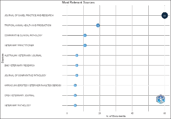
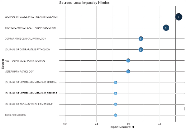
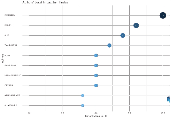
Fig. 3. Average citations for pathological techniques used in camel research from 1971 to 2024. The top publishing journals and their impactsConsidering the number of research articles published by journals, the Journal of Camel Practice and Research has the lead of 60 published articles related to camel pathology. The Journal of Tropical Animal Health and Production ranks second with 19 published articles, which is a large gap compared with the number of published articles in the Journal of Camel Practice and Research. The comparative clinical pathology and veterinary practitioner journals ranked third, with 11 published articles in both journals. The Journals of the Australian Veterinary Journal, BMC Veterinary Research, and Comparative Pathology have six articles each (Fig. 6). Kafas University Veterinary Fakulltesi Dergisi, Open Veterinary Journal, and Veterinary Pathology had five articles each (Fig. 5). The impact of the research on camel pathology is also dominated by the Journal of Camel Practice and Research, which has an H-index of 9 (Fig. 7). The Journals of Tropical Animal Health and Production and Comparative Clinical Pathology came in second and third with H-indexes of 6 each. However, although the Journal of Comparative Pathology has only 6 published papers, the impact of these publications was significant, with an H-index of 6. This made this journal comparable with Journals of Tropical Animal Health and Production and Comparative Clinical Pathology (Fig. 7). Journals such as Australian Veterinary Journal and Veterinary Pathology have an H-index of 5. The Journal of Veterinary Medicine Series A, Veterinary Medicine Series B, and the Journal of Wildlife and Zoo Medicine and Theriogenology had an H-index of 4 (Fig. 7). The most relevant authors are listed as followsThe highest research impact in camel research using pathological techniques among the top 10 authors in this sector was made by Wernery U, with an H-index of 10, suggesting that he has published at least 10 research articles, and each of these articles has been cited 10 times at a minimum. Another significant contribution was made by Kinne J, Ali A, and Tharwat M with an H-index of 8, 7, and 6, respectively. The results show that Ali M, Gameel AA, Narnaware SD, and Oryan A have an H-index of 5. Finally, Abu-samra MT and Al-hawas A had an H-index of 4 (Fig. 8). Co-occurrence of keywords and classical and emerging topics in camel pathologyCamel research related to pathology, or the use of pathological techniques, can be grouped into 9 clusters according to the authors’ keywords (Fig. 9). Red cluster This cluster was related to camel research and included words such as Tuberculosis, Mycobacterium bovis, cattle, liver, lung, abattoir, postmortem, and hematology. Obviously, this suggests research-related tuberculosis, which is caused mainly by M. bovis, and the use of postmortem examinations and examination of meat carcasses in abattoirs (Mamo et al., 2011; Ahmed et al., 2017; Jibril, 2019). In addition, most lesions observed postmortem or in the abattoir will be seen in the liver and lungs of the infected animals (Infantes-Lorenzo et al., 2020). This procedure also involves the use of histopathological and use of Ziehl-Neelsen stain.
Fig. 4. Word map with a blue gradient showing the countries with the highest cited countries in articles using pathological techniques in camel research. The dark blue countries are the highest, indicating that Saudi Arabia, India, Egypt, and Iran have the highest number of citations, respectively.
Fig. 5. VOSviewer international collaboration network. This figure illustrates which countries are highest in the number of collaborations defined by the size of the circles, and which countries participated in the collaboration defined by the lines between the countries. The thicker the link, the stronger the collaboration. Therefore, it is clear that most of the collaboration was initiated by Saudi Arabia and Egypt, and the link between the two countries is thick, suggesting a strong collaborative relationship. Next comes Iran, the UAE, and the United States.
Fig. 6. Most relevant journals in relation to articles using pathological techniques in camel research.
Fig. 7. Illustrated impact of journals according to their H-index.
Fig. 8. Visual illumination of impact by different authors based on their H-index.
Fig. 9. The network visualization generated by VOSviewer illustrates thematic clusters in camel research using pathological techniques, with each cluster representing a specific area of study. The node size reflects the frequency of a research topic, and the thickness of the connecting lines represents the strength of the relationship between different topics. Green cluster Research in this cluster involves topics such as infection, pneumonia, gross pathology, Onchocerca fasciata, and dehydration and rehydration. In this cluster, the research is connected to the use of gross pathology for the diagnosis of many infections, including pneumonia and O. fasciata (Tafti et al., 2015; Dinaol et al., 2015; Asopa et al., 2022) Blue cluster Topics in the blue cluster include MERS-CoV, immunohistochemistry, squamous cell carcinoma, and uterine cancer. Therefore, it is clear that it is related to the group of research that used immunohistochemistry for the diagnosis of many diseases, such as MERS-CoV, squamous cell carcinoma, and uterine infections and conditions (Ghoneim et al., 2013; Alnaeem et al., 2018; Abdelazim et al., 2023; Jaber et al., 2023; Ibrahim et al., 2023). Yellow cluster The yellow cluster has research topics related to infertility, ultrasonography, ovaries, and diagnosis. This indicates the use of histopathological techniques in the diagnosis of reproductive problems in camels. Such topics might include genital and reproductive disorders and causes of infertility (Waheed et al., 2009; Ali et al., 2010; Ali et al., 2017; Hussein and Saad, 2017; Al-Afaleq et al., 2022). Purple cluster This cluster is positioned around pathology, with keywords such as abortion, ultrasound, and Trypanosoma evansi (Narnaware et al., 2017; Tharwat, 2020; Jaber et al., 2023). Cyan cluster Pathology has always been a significant part of parasitic disease prevalence investigation, and this cluster is arranged around the use of pathological techniques in this area. This includes topics such as Cephalopina titillator, Sarcocystis, and sarcoptic mange (Abu El Ezz et al., 2018; Ahmed et al., 2020a; Shamsi et al., 2023; Narnaware et al., 2024; Wieser et al., 2024). Orang cluster The orange cluster indicates research areas connected to histopathology, epidemiology, hydatid cysts, and rabies (Singh et al., 2017; Sosango et al., 2019; Ahmed et al., 2020a; Ahmed et al., 2020b; Ranjan, 2023; Andadari et al., 2024). Brown cluster In this cluster, PCR is the focus, and topics are arranged around it. In the mentioned topics, Johne’s disease, paratuberculosis, and camelpox, and these diseases have pathological techniques heavily involved in the research and understanding of the disease (Alharbi et al., 2012; Hereba et al. 2015; Narnaware et al., 2018; Joseph et al., 2021; Hussein, 2021; Narnaware et al., 2021; El Tigani-Asil et al., 2023). Pink cluster This cluster deals with research related to pneumonia, silicosis, and testosterone and how pathological techniques are used to diagnose and research these conditions (Derar et al., 2017; Ismail, 2017; Ali et al., 2024; Hassaneen et al., 2024). During the years 2012–2020, there were significant changes in research interests and the evolution of research techniques, which influenced the nature of camel pathology research. From 2012 to 2016, topics such as pneumonia, silicosis, gross pathology, infertility, camelpox, O. fasciata, and sarcoptic mange were areas of primary focus, relying mainly on traditional pathological techniques. However, from 2016 to 2020, there was a noticeable shift toward better integration of pathological methods with advanced molecular approaches. This latter period introduced keywords such as PCR, Trypanosoma evansi, paratuberculosis, MERS-CoV, ovary, uterus, prevalence, Sarcocystis, Cephalopina titillator, M. bovis, tuberculosis, epidemiology, rabies, diagnosis, hydatid cyst, immunohistochemistry, and testosterone. These trends reflect a transition from traditional pathology to molecular techniques, which are increasingly employed for disease detection, diagnosis, and understanding (Fig. 10). The visualization in Figure 10 further illustrates this evolution through a keyword co-occurrence network. Earlier years (2012–2015) focused on traditional pathological terms, such as gross pathology, and conditions, such as sarcoptic mange, represented by cooler shades (purple and blue). As time progressed (2016–2020), advanced molecular techniques and disease-related studies emerged with warmer shades (green and yellow). This transition underscores the growing integration of molecular and epidemiological approaches into camel pathology. The increasing connectivity among keywords reflects a multidisciplinary approach that links traditional pathology with advanced diagnostic tools. This shift is consistent with global trends in veterinary medicine, where molecular techniques enhance the understanding, detection, and management of diseases. Ultimately, this evolution broadens the scope of camel pathology research and demonstrates the adoption of innovative methodologies to address camel health challenges. The highly cited publicationsThe use of pathology in camel research has been important for understanding and preventing diseases and protecting camels as animals and as food sources. For instance, a 2016 publication by Haagmans et al. (2016) showed that a vaccine designed for camels made from an orthopox virus could reduce the shading of the MERS-coV virus from infected camels and therefore reduce the transmission of the infection to humans (Haagmans et al., 2016). Furthermore, Babelhadj et al. (2018) reported for the first time a prion disease camel. In his study, he examined camels showing neurological signs during an antemortem inspection at a slaughterhouse, which demonstrated the importance of the study. Examination of the brains of ill animals at the microscopic level revealed pathognomonic lesions of prion disease (Babelhadj et al., 2018). In addition, in Ethiopia in 2012, Gumi et al. (2012) used pathological techniques to diagnose tuberculosis in infected camels. Then, after isolation of M. bovis from both infected camel and human patients, he showed the evidence of tuberculosis transmission to camel workers (Gumi et al., 2012). Other studies included the characterization of causative agents of tuberculosis in infected camels, calculation of the prevalence of sarcocystosis, and the report of paste des petits ruminants in camels for the first time. These studies all emphasize one health concept because all the articles investigated zoonotic diseases, animal diseases, or meat meant for human consumption (Table 1).
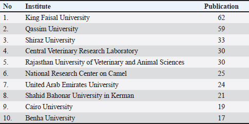
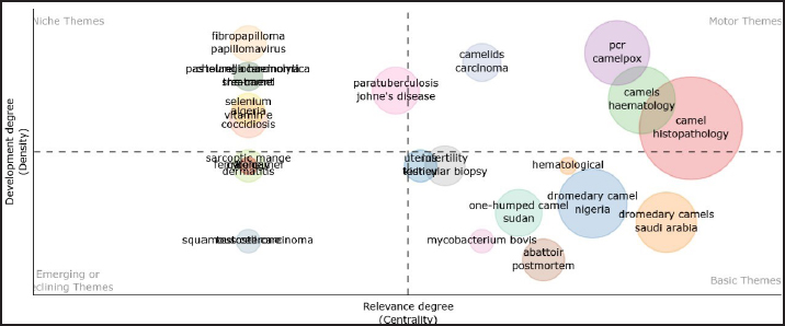
Fig. 10. VOSviewer overlay visualization highlights trends in camel pathology research, with classical topics represented in blue and more recent topics depicted in yellow. The most active institutionsThe use of pathological techniques in camel research is led by Saudi Arabia, where King Faisal University and Qassim University produced a total of 121 publications. This project was led by King Faisal University, which produced 62 publications. Subsequently, it was followed by Qassim University, which produced 59 publications. Then, Shiraz University from Iran with 33 publications. Next came the Central Veterinary Research Laboratory in the United Arab Emirates and the Rajasthan University of Veterinary and Animal Science in India with 30 publications each. The National Research Center on Camel in India has 25 publications. This was followed by United Arab Emirates University, Shahid Bahonar University of Kerman from Iran, Cairo University, and Benha University from Egypt with 25, 24, 21, and 19 publications, respectively (Table 2). Thematic map of camel pathologyTo understand the themes of research in which pathological techniques were used. Thematic analysis of the research that used histopathological techniques revealed four quadrants. In the first quadrant, motor themes, camel histopathology, camelpox PCR, camel hematology, and camelid carcinomas are well-developed and central themes that drive which are used as drivers for other research themes. In niche themes, areas such as fibropapilloma/papilloma virus, Pasteurella haemolytica, she-camel, selenium Algeria, vitamin E, and coccidiosis represent well-developed topics not central to camel pathology research. The basic quadrant represents topics central to camel pathology research but not well-developed. This quadrant provides driving material for other quadrants and contains topics such as dromedary camels in Nigeria, Sudan, and Saudi Arabia, infertility, testicular biopsy, uterus and kidney, M. bovis, abattoir, and postmortem findings. Finally, the emerging or declining theme represents topics that are less central and less developed. Included topics such as squamous cell carcinoma, sarcoptic mange, dermatitis, and she-camel may either be growing or declining (Fig. 11). Strengths and limitations of this study and future directionsBibliometric analysis of camel pathology research offers much strength that contributes to the understating of the status of pathology in camel research. Statistical analysis, annual scientific production, most cited journal and impact, authors H-index international collaboration, research area, most cited country, top cited papers, trends, most active institutes, and themes of research. One of the limitations is the use of key words to retrieve data, which is a powerful tool; however, at the same time, it could limit the keyword used in the search which might exclude another research not included in the search. Moreover, the search was limited to publications in the English language, leaving out research work on other languages. Table 1. Prominent highly cited studies in camel pathology research and collaborative efforts with other countries.
Table 2. Leading institutions conducting camel pathology research.
Fig. 11. The thematic map provides an analysis of camel pathology research using histopathological techniques, categorized into four quadrants. The Motor Themes (top-right quadrant) include camel histopathology, camelpox PCR, camel hematology, and camelid carcinomas, which are well-developed and central, acting as drivers for other research themes. The Niche Themes (top-left quadrant) represent specialized but less central topics, such as fibropapilloma/papillomavirus, Pasteurella hemolytica, she-camel, selenium, Algeria, vitamin E, and coccidiosis. The Basic Themes (bottom-right quadrant) are central but underdeveloped areas that provide foundational material for other research, including topics such as dromedary camels in Nigeria, Sudan, and Saudi Arabia, infertility, testicular biopsy, uterine, kidney, Mycobacterium bovis, abattoir, and postmortem studies. Emerging or decreasing themes (bottom-left quadrant) encompass less developed and less central topics, such as squamous cell carcinoma, sarcoptic mange, dermatitis, and she-camel, which may be either gaining traction or waning in interest. Research gaps and future perspectivesDespite the growing body of literature on camel pathology and the increasing use of histopathological techniques, several critical gaps remain. First, the application of advanced molecular techniques, while on the rise, is still underused in many fields of camel research. This is evident in the limited integration of genetic, proteomic, and immunological approaches to disease characterization and diagnosis. Second, emerging and zoonotic diseases such as MERS-CoV and tuberculosis have received attention, but other infectious and noninfectious diseases remain understudied, particularly in regions with high camel populations. Moreover, research on the impact of environmental factors, such as climate change and desertification, on camel health is scarce. The thematic map also reveals underdeveloped topics, such as infertility, testicular biopsy, and postmortem studies, which are essential for improving reproductive health and meat safety. Finally, the limited collaboration between high-producing research institutions and regions with significant camel populations but lower research output indicates an opportunity for more inclusive, global partnerships. DiscussionThis bibliometric analysis highlights the evolution and trends in camel pathology research from 1971 to 2024, offering valuable insights into the progression and challenges within the field. The steady growth in publications, particularly after 2010, reflects an increasing recognition of the importance of camels in both agricultural and zoonotic contexts. The shift from traditional pathology to advanced molecular techniques underscores the potential for improving disease diagnosis and management. However, the slower uptake of cutting-edge approaches in some regions and topics indicates the need for targeted capacity-building initiatives. The dominance of certain institutions and countries, such as Saudi Arabia and Egypt, in international collaborations emphasizes their pivotal role in advancing the field. Expanding partnerships to include under-represented regions with large camel populations, such as Sub-Saharan Africa, could enhance global efforts in disease control and improve the economic sustainability of camel farming. The thematic analysis further identifies well-established (motor themes) and emerging research areas, providing a roadmap for future studies. For instance, while camel histopathology and camel pox are well-developed, basic themes such as infertility and postmortem studies require further exploration to address practical challenges in camel husbandry and public health. ConclusionIn conclusion, while significant strides have been made in camel pathology research, addressing the identified gaps and fostering international collaboration are crucial in expanding the field’s impact. The integration of molecular, environmental, and epidemiological approaches will not only advance the understanding of camel diseases but also strengthen the role of camels in sustainable development and food security globally. AcknowledgmentsThe authors express their sincere appreciation to the Deanship of Scientific Research, King Faisal University, Saudi Arabia, for their generous support of this research. Special thanks are extended to Prof. Mahmoud Mandrel for his significant assistance in collecting and analyzing the bibliometric data for this study. Conflicts of interestThere are no conflicts of interest to declare. FundingThe authors are grateful to the Deanship of Scientific Research, Vice Presidency for Graduate Studies and Scientific Research, King Faisal University, Saudi Arabia, for funding this project (Project# KFU250253). Authors’ contributionsThe work was primarily conducted by Ahmed Aljazzar, who was responsible for conceptualization, methodology, software application, formal analysis, and resource acquisition. Data curation, drafting the original manuscript, and reviewing, and editing were also handled by Ahmed. Data availabilityAll data supporting the findings of this study are available in the manuscript. ReferencesAbdelazim, M., Abdelkader, R., Ali, A., Shahein, M.A. and Tadesse, Z. 2023. A longitudinal study of Middle East respiratory syndrome coronavirus (MERS-CoV) in dromedary camels. BMC Vet. Res. 19(1), 228. Abdel-Ghaffar, F., et al. (2009). Life cycle of Sarcocystis camelicanis infecting the camel and dog, light and electron microscopic study. Parasitology Research, 106(1), 189–195. https://doi.org/10.1007/S00436-009-1648-X Abu El Ezz, N.M.T., Hassan, N.M.F., El Namaky, A.H. and Abo-Aziza, F. 2018. Efficacy of some essential oils on Cephalopina titillator with special concern to nasal myiasis prevalence among camels and its consequent histopathological changes. J. Paras. Dis. 42(2), 196–203. Ahmed, E.T.A.E.T., Hassan, A.B. and Abakar, A.D. 2017. Pathology of tuberculosis in camels ( <i>Camelus dromedaries</i>) in the Sudan. J. Tuberculosis Res. 5 (01), 77–80. Ahmed, M.A., Elmahallawy, E.K., Gareh, A., Abdelbaset, A.E., El-Gohary, F.A., Elhawary, N.M., Dyab, A.K., Elbaz, E. and Abushahba, M.F.N. 2020a. Epidemiological and histopathological investigation of Sarcoptic mange in Camels in Egypt. Animals, 10 (9), 1485. Ahmed, M.S., Body, M.H., El-Neweshy, M.S., ALrawahi, A.H., Al-Abdawani, M., Eltahir, H.A. and ALmaewaly, M.G. 2020b. Molecular characterization and diagnostic investigations of rabies encephalitis in camels (Camelus dromedaries) in Oman: a retrospective study. Trop. Anim. Health Prod. 52(4), 2163–2168. Al-Afaleq, A.I., Homeida, A.G.M., Barakat, S.E.M. and Moqbel, M.S. 2022. Gross and microscopic lesions of the ovaries of camels from abattoirs in Eastern Province of Kingdom of Saudi Arabia. Open J. Pathol. 12(01), 1–11. Alharbi, K.B., Al-Swailem, A., Al-Dubaib, M.A., Al-Yamani, E., Al-Naeem, A., Shehata, M., Hashad, M.E., Albusadah, K.A. and Mahmoud, O.M. 2012. Pathology and molecular diagnosis of paratuberculosis of camels. Trop. Anim. Health Prod. 44(1), 173–177. Ali, A., Al-Sobayil, F.A., Tharwat, M., Al-Hawas, A. and Ahmed, A.F. 2010. Causes of infertility in female camels (Camelus dromedarius) in Middle of Saudi Arabia. J. Agricul. Vet. Sci. 2 (2), 59–66. Ali, A., Baby, B. and Vijayan, R. 2019. From desert to medicine: a review of camel genomics and therapeutic products. Front. Genetics. 10; doi:10.3389/fgene.2019.00017. Ali, A., Derar, D., Alsamri, A. and Al Sobayil, F. 2017. Echography of clinically relevant disorders in the genital tract of female dromedary camels. Anim. Reprod. Sci. 182, 123–133. Ali, F., Mohamed, R.H., Abd-Elkareem, M. and Hassan, M.S. 2024. Serological and histolomorphological investigation of camel bulls testes (Camelus dromedaries) during the rutting and non-rutting seasons. BMC Vet. Res. 20(1), 1–9. Alnaeem, A., Kasem, S., Qasim, I., Al-Doweriej, A., Al-Houfufi, A., Alwazan, A., Albadrani, A., Alshaammari, K., Refaat, M., Al-Shabebi, A. and Gomaa Hemida, M. 2018. CoV) in Saudi Arabia. Vet. Quar. 40(1), 190–197. Al-Numair, N.S., Theyab, A., Alzahrani, F., Shams, A.M., Al-Anazi, I.O., Oyouni, A.A.A., Al-Amer, O.M., Mavromatis, C., Saadeldin, I.M., Abdali, W.A. and Hawsawi, Y.M. 2022. Camels’ biological fluids contained nanobodies: promising avenue in cancer therapy. Cancer Cell Inter. 22(1), 1–18. Andadari, A.Y.D., Pratama, D.A.O.A., Hardian, A.B. and Untari, H. 2024. Literature study : histopathology analysis of rabies disease (2014 - 2022). J. Appl. Vet. Sci. Technol. 5(1), 93–98. Asopa, S., Vyas, I., Dadhich, H. and Joshi, A. 2022. Pathological study of various liver lesions prevalent in camels of Rajasthan. J. Camel Pract. Res. 29(2), 223–228. Babelhadj, B., Di Bari, M.A., Pirisinu, L., Chiappini, B., Gaouar, S.B.S., Riccardi, G., Marcon, S., Agrimi, U., Nonno, R. and Vaccari, G. 2018. Prion disease in dromedary camels, Algeria. Emerg. Infect. Dis. 24(6), 1029. Derar, D.R., Ali, A., Osman, S.A., Al-Sobayil, F.A., Saeed, E.M., Hassanein, K. and Al-Hawas, A.A. 2017. Potential pathogens in infertile male dromedary camels and their association with the spermiogram and clinical findings. Comp. Clin. Pathol. 26(4), 965–970. Dinaol, B., Bulto, G., Hagos, A., Tilaye, D. and Yimer, M. 2015 Review on camel liver pathology and Its major diagnostic approaches. Glob. J. Vet. Med. Res. 3, 068–079. El Tigani-Asil, E.T.A., El Derdiri Abdelwahab, G., Abdu, E.H.A.M., Terab, A.M.A., Khalil, N.A.H., Al Marri, Z.J.M., Yuosf, M.F., Shah, A.A.M., Khalafalla, A.I. and Ishag, H.Z.A. 2023. Pathological, microscopic, and molecular diagnosis of paratuberculosis/John’s disease in naturally infected dromedary camel (Camelus dromedarius). Vet. World. 16(6), 1277. Faye, B. 2018. The enthusiasm for camel production. Emir. J. Food Agric. 30(8). http://www.ejfa.me/. Faye, B., Konuspayeva, G. and Magnan, C. 2023. Camel products and services: from dairy, meat, and nonfood products to riding and transport, including slaughter. Large Camel Farming. 173–207; doi:10.1007/978-94-024-2237-5_8. Ghoneim, I.M., Waheed, M.M., Hamouda, M.A., Al-Eknah, M.M. and Al-Fehaed, H.F. 2013. Endometrial biopsy technique and histopathological findings of endometritis in camels (Camelus dromedarius). J. Camel Pract. Res. 20(1), 65–70. Gumi, B., Schelling, E., Berg, S., Firdessa, R., Erenso, G., Mekonnen, W., Hailu, E., Melese, E., Hussein, J., Aseffa, A. and Zinsstag, J. 2012. Zoonotic transmission of tuberculosis between pastoralists and their livestock in South-East Ethiopia. EcoHealth. 9(2), 139–149. Gupta, B.M., Ahmed, K.K.M., Gupta, R. and Tiwari, R. 2015. World camel research: a scientometric assessment, 2003–2012. Scientometrics. 102(1), 957–975. Haagmans, B.L., van den Brand, J.M.A., Raj, V.S., Volz, A., Wohlsein, P., Smits, S.L., Schipper, D., Bestebroer, T.M., Okba, N., Fux, R., Bensaid, A., Solanes Foz, D., Kuiken, T., Baumgärtner, W., Segalés, J., Sutter, G. and Osterhaus, A.D.M.E. 2016. An orthopoxvirus-based vaccine reduces virus excretion after MERS-CoV infection in dromedary camels. Science. 351(6268), 77–81. Hardy, R.W. 2012. The nutritional pathology of teleosts. Fish Pathol. 402–424. doi:10.1002/9781118222942.ch10. Hassaneen, A.S.A., Anis, A., Nour, S.Y., Mohamed, R.S., Wassif, I.M., El-kattan, A.M., Abdelgawad, H.A. and Mohamed, R.H. 2024. Poor semen quality is associated with impaired antioxidant response and acute phase proteins and is likely mediated by high cortisol levels in Brucella-seropositive dromedary camel bulls. Sci. Rep. 14(1), 1–10. Haverkamp, A.K., Lehmbecker, A., Spitzbarth, I., Widagdo, W., Haagmans, B.L., Segalés, J., Vergara-Alert, J., Bensaid, A., Van Den Brand, J.M.A., Osterhaus, A.D.M.E. and Baumgärtner, W. 2018. Experimental infection of dromedaries with Middle East respiratory syndrome-Coronavirus is accompanied by massive ciliary loss and depletion of the cell surface receptor dipeptidyl peptidase 4. Sci. Rep. 8(1), 1–15; doi:10.1038/s41598-018-28109-2. Hereba , A.M., Hamouda, M.A. and Al-Hizab, F.A. 2015. Pathological and molecular diagnosis of paratuberculosis among dromedary camels in Saudi Arabia. J. Anim. Plant Sci. 25(4), 997–1007. Hussein , M.F. 2021. Paratuberculosis (Johne’s Disease). Infectious diseases of dromedary camels: a concise guide. 167–173. Hussein, N.A. and Saad, A.M. 2017. Pathological disorders of the ovaries and uterine tubes in camels (Camelus dromedarius) slaughtered at Tamboul abattoir, Sudan. J. Camel Pract. Res. 24(3), 251–256. Hussen, J. and Schuberth, H.J. 2021. Recent advances in camel immunology. Front. Immunol. 11, 614150. Ibrahim, A., Zabady, M., El Nahas, A., Aljazzar, A., Al-Hizab, F., Kandeel, M. and Porter, B.F. 2023. Maxillary neoplasms in four dromedary camels. Front. Vet. Sci. 10, 1153398; doi:10.3389/fvets.2023.1153398. Infantes-Lorenzo, J.A., Romero, B., Rodríguez-Bertos, A., Roy, A., Ortega, J., de Juan, L., Moreno, I., Domínguez, M., Domínguez, L. and Bezos, J. 2020. Tuberculosis caused by Mycobacterium caprae in a camel (Camelus dromedarius). BMC Vet. Res. 16(1), 435; doi:10.1186/s12917-020-02665-0. Ismail, Z.B. 2017. Pneumonia in dromedary camels (Camelus dromedarius): a review of clinico-pathological and etiological characteristics. J. Camel Pract. Res. 24(1), 49. Jaber, F.A., Algehani, A.M., Almalki, G., Albohiri, H.H. and Alsulami, M.N. 2023. Histological and immunohistochemical assays of Trypanosoma evansi infected camel hepatic tissues. Polish J. Environ. Stud. 32(5), 4617–4626. Jibril, Y. 2019 Abattoir based study on the pathology of tuberculosis in dromedary camels, Ethiopia. Biomed. J. Sci. Tech. Res. 19(2), 14151–14158; doi:10.26717/bjstr.2019.19.003263. Joseph, S., Kinne, J., Nagy, P., Juhász, J., Barua, R., Patteril, N.A.G., Hoffmann, D., Pfaff, F., Hoffmann, B. and Wernery, U. 2021. Outbreak of a systemic form of camelpox in a dromedary herd (Camelus dromedarius) in the United Arab Emirates. Viruses 2021, 13. Kandeel, M., Morsy, M.A., Abd El-Lateef, H.M., Marzok, M., El-Beltagi, H.S., Al Khodair, K.M., Soliman, W.E., Albokhadaim, I. and Venugopala, K.N. 2023. A century of “Camel Research”: a bibliometric analysis. Front. Vet. Sci. 10, 1157667. Khalafalla, A.I. 2023. Zoonotic diseases transmitted from the camels. Front. Vet. Sci. 10, 1244833; doi:10.3389/FVETS.2023.1244833/FULL. Latif, B. M. A., et al. (1999). Prevalence of Sarcocystis spp. in meat-producing animals in Iraq. Veterinary Parasitology, 84(1–2), 85–90. https://doi.org/10.1016/S0304-4017(99)00046-1 Levy, J., Crawford, C., Hartmann, K., Hofmann-Lehmann, R., Little, S., Sundahl, E. and Thayer, V. 2008. 2008 American Association of Feline Practitioners’ feline retrovirus management guidelines. J. Feline Med. Surg. 10(3), 300–316. Mamo, G., Bayleyegn, G., Tessema, T.S., Legesse, M., Medhin, G., Bjune, G., Abebe, F. and Ameni, G. 2011. Pathology of camel tuberculosis and molecular characterization of its causative agents in pastoral regions of Ethiopia. PLoS One. 6(1), e15862. Masebo, N.T., Zappaterra, M., Felici, M., Benedetti, B. and Padalino, B. 2023. Dromedary camel’s welfare: literature from 1980 to 2023 with a text mining and topic analysis approach. Front. Vet. Sci. 10, 1277512; doi:10.3389/fvets.2023.1277512. Moore, C. P., et al. (1999). Congenital ocular anomalies and ventricular septal defect in a dromedary camel. Journal of Zoo and Wildlife Medicine, 30(3), 423–430. http://www.ncbi.nlm.nih.gov/pubmed/10572869 Nagy, P.P., Skidmore, J.A. and Juhasz, J. 2022. Intensification of camel farming and milk production with special emphasis on animal health, welfare, and the biotechnology of reproduction. Anim. Front. 12(4), 35–45. Narnaware, S.D., Dahiya, S.S., Kumar, S., Tuteja, F.C., Nath, K. and Patil, N.V. 2017. Pathological and diagnostic investigations of abortions and neonatal mortality associated with natural infection of Brucella abortus in dromedary camels. Comp. Clin. Pathol. 26(1), 79–85. Narnaware, S.D., Jyotsana, B., Ranjan, R., Dahiya, S.S. and Prakash, V. 2024. The first molecular characterization of Sarcocystis cameli in the Indian dromedary camels (Camelus dromedarius). Vet. Res. Commun. 48(3), 1929–1933. Narnaware, S.D., Ranjan, R. and Dahiya, S.S. 2018. Clinicopathological investigations during an outbreak of camelpox in a dromedary camel herd in India. Comp. Clin. Pathol. 27(6), 1497–1500. Narnaware, S.D., Ranjan, R., Dahiya, S.S., Panchbuddhe, A., Bajpai, D., Tuteja, F.C. and Sawal, R.K. 2021. Pathological and molecular investigations of systemic form of camelpox in naturally infected adult male dromedary camels in India. Heliyon. 7 (2), e06186. Ranjan, R. 2023. Evaluation of a neem (Azadirachta indica) herbal formulation for management of sarcoptic mange in dromedary camels. Ind. J. Vet. Med. 42(2), 42–46. Rossi, G. and Salguero, F.J. 2024. Editorial: insights in veterinary experimental and diagnostic pathology: 2023. Front. Vet. Sci. 11, 1437289. Schulze, C., Peters, M., Baumgärtner, W. and Wohlsein, P. 2016. Electrical injuries in animals: causes, pathogenesis, and morphological findings. Vet. Pathol. 53 (5), 1018–1029. Seifu, E. 2023. Camel milk products: innovations, limitations and opportunities. Food Prod. Process. Nutr. 5(1), 1–20. Shamsi, E., Radfar, M.H., Nourollahifard, S.R., Bamorovat, M., Nasibi, S., Fotoohi, S., Hakimi Parizi, M. and Kheirandish, R. 2023. Nasopharyngeal myiasis due to Cephalopina titillator in Southeastern Iran: a prevalence, histopathological, and molecular assessment. J. Paras. Dis. 47 (2), 369–375. Singh, R., Singh, K.P., Cherian, S., Saminathan, M., Kapoor, S., Reddy, G.B.M., Panda, S. and Dhama, K. 2017. Rabies—epidemiology, pathogenesis, public health concerns and advances in diagnosis and control: a comprehensive review. Vet. Quarter. 37(1), 212–251. Sosango, M., Zuria, H.T. and Ashebo, A. 2019 Epidemiology and anatomical histopathological features of hydatid cyst. Adv. Biol. Res. 13(3), 121–128. Tafti, A., Sazmand, A., Moghaddam, H. and Moobedi, I. 2015. Prevalence and pathology of Onchocerca infection in camels (Camelus dromedarius) in central parts of Iran. Iran. J. Vet. Med. 257 (4), 257–261. Tharwat, M. 2020. Ultrasonography of the abdomen in healthy and diseased camels (Camelus dromedaries)—a review. J. Appl. Anim. Res. 48(1), 300–312. Valinezhad, A., et al. (2008). Sarcocystis and Its Complications in Camels. The Korean Journal of Parasitology, 46(4), 229. https://doi.org/10.3347/KJP.2008.46.4.229 Waheed, M.M., Al-Eknah, M.M., Hamouda, M.A. and Al-Dughaym, A.M. 2009. Uterine histopathological findings of infertile female camels (Camelus dromedarius). J. Camel Pract. Res. 16(2), 171–177. Widagdo, W., Raj, V.S., Schipper, D., Kolijn, K., van Leenders, G.J.L.H., Bosch, B.J., Bensaid, A., Segalés, J., Baumgärtner, W., Osterhaus, A.D.M.E., Koopmans, M.P., van den Brand, J.M.A. and Haagmans, B.L. 2016. Differential expression of the middle East respiratory syndrome coronavirus receptor in the upper respiratory tracts of humans and dromedary camels. J. Virol. 90(9), 4838–4842. Wieser, S.N., Giuliano, S.M., Reategui Ordoñez, J., Barriga Marcapura, X., Olivera, L.V.M., Chavez Fumagalli, M.A., Schnittger, L. and Florin-Christensen, M. 2024. Sarcocystis spp. of new and old World Camelids: ancient origin, present challenges. Pathogens. 13(3), 196. Zakian, A., et al. (2016). The first report of peste des petits ruminants in camels in Iran. Tropical Animal Health and Production, 48(6), 1215–1219. https://doi.org/10.1007/S11250-016-1078-6 Zhu, S., Zimmerman, D. and Deem, S.L. 2019. A review of zoonotic pathogens of dromedary camels. EcoHealth, 16(2), 356–377. | ||
| How to Cite this Article |
| Pubmed Style Ahmed Aljazzar. Bibliometric analysis of the use of pathological techniques in camel research. Open Vet. J.. 2025; 15(2): 1009-1023. doi:10.5455/OVJ.2025.v15.i2.49 Web Style Ahmed Aljazzar. Bibliometric analysis of the use of pathological techniques in camel research. https://www.openveterinaryjournal.com/?mno=240632 [Access: December 02, 2025]. doi:10.5455/OVJ.2025.v15.i2.49 AMA (American Medical Association) Style Ahmed Aljazzar. Bibliometric analysis of the use of pathological techniques in camel research. Open Vet. J.. 2025; 15(2): 1009-1023. doi:10.5455/OVJ.2025.v15.i2.49 Vancouver/ICMJE Style Ahmed Aljazzar. Bibliometric analysis of the use of pathological techniques in camel research. Open Vet. J.. (2025), [cited December 02, 2025]; 15(2): 1009-1023. doi:10.5455/OVJ.2025.v15.i2.49 Harvard Style Ahmed Aljazzar (2025) Bibliometric analysis of the use of pathological techniques in camel research. Open Vet. J., 15 (2), 1009-1023. doi:10.5455/OVJ.2025.v15.i2.49 Turabian Style Ahmed Aljazzar. 2025. Bibliometric analysis of the use of pathological techniques in camel research. Open Veterinary Journal, 15 (2), 1009-1023. doi:10.5455/OVJ.2025.v15.i2.49 Chicago Style Ahmed Aljazzar. "Bibliometric analysis of the use of pathological techniques in camel research." Open Veterinary Journal 15 (2025), 1009-1023. doi:10.5455/OVJ.2025.v15.i2.49 MLA (The Modern Language Association) Style Ahmed Aljazzar. "Bibliometric analysis of the use of pathological techniques in camel research." Open Veterinary Journal 15.2 (2025), 1009-1023. Print. doi:10.5455/OVJ.2025.v15.i2.49 APA (American Psychological Association) Style Ahmed Aljazzar (2025) Bibliometric analysis of the use of pathological techniques in camel research. Open Veterinary Journal, 15 (2), 1009-1023. doi:10.5455/OVJ.2025.v15.i2.49 |


