


| Case Report | ||
Open Vet. J.. 2025; 15(10): 5368-5372 Open Veterinary Journal, (2025), Vol. 15(10): 5368-5372 Case Report A case report of massive cutaneous histiocytoma in a 12-year-old intact bitch: Cytologic, radiologic, pathologic investigationReza Nikzad1*, Masoud Nemati-Nejad2, Amirhossein Alizadeh Tabarestani3, Sonia Sahvieh4,5, Mohammad Amin Minaie6, Mohammad Ramezani7, Mehdi Tavana8 and Azam Azizzadeh91Doctor of Veterinary Medicine, Doctor of Veterinary Sciences, Veterinary Surgeon, Babolsar, Iran 2Board-Certified in Veterinary Clinical Pathology, School of Veterinary Medicine, Shiraz University, Shiraz, Iran 3Faculty of Radiology Department, Islamic Azad University of Babol, Mazandaran, Iran 4Department of Pathology, School of Veterinary Medicine, Shiraz University, Shiraz, Iran 5Faculty of the Pathology Department, Islamic Azad University of Babol, Mazandaran, Iran 6Specialist in Small Animal Internal Medicine, Doctor Hasanzadeh Specialized Veterinary Hospital, Babolsar, Iran 7Resident of Small Animal Internal Medicine, Science and Research branch, Islamic Azad University of Tehran, Tehran, Iran 8Veterinary Department, Sho. C., Islamic Azad University, Shoushtar, Iran 9Postgraduate in Veterinary Medicine, Islamic Azad University of Shoushtar, Khuzestan, Iran *Corresponding Author: Reza Nikzad. Doctor of Veterinary Medicine, Doctor of Veterinary Sciences, Veterinary Surgeon, Babolsar, Iran. Email: Rezanikzad [at] hotmail.com Submitted: 03/02/2025 Revised: 29/08/2025 Accepted: 22/09/2025 Published: 31/10/2025 © 2025 Open Veterinary Journal
AbstractBackground: Facial masses in dogs can arise from infections, inflammation, or tumors. Histiocytoma, a common benign skin tumor in young dogs, originates from Langerhans cells. It appears as a small, round, hairless, and rapidly growing lump, present on the face or ears. Typically, histiocytomas regress spontaneously within a few months. While usually harmless, biopsy and monitoring are recommended to rule out malignancies like mast cell tumors or squamous cell carcinoma. Case Description: A 12-year-old, unspayed bitch presented with a huge histiocytoma on her frontonasal bone. The mass measured about ten centimeters across and had suddenly become bigger over the last few months. Microscopic examination of fine needle aspirates showed that there was proliferation of tumor cells composed of two cell types resembling neoplastic macrophages and dendritic cells, which corresponds to cutaneous histiocytoma confirmed by Hematoxylin–Eosin stain. Radiographic images also demonstrated a large subcutaneous mass with no evidence of lymph node metastasis. Removal of the growth through surgical excision was done, and the tissue sample submitted for histopathology showed a typical pattern of macrophage and dendritic cell proliferation without any signs suggesting malignancy. Enbloc excision resulted in an intact dog after six months post-surgery check with no evidence of clinical recurrence or relapse. Conclusion: This report underscores the need for a holistic diagnostic approach to diagnosis and management of canine cutaneous histiocytomas, stressing the role played by various radiological investigations, cytologic evaluations, and pathologic assessments while determining their size. Keywords: Histiocytoma, Tumor, Metastasis, Histopathology. IntroductionCutaneous histiocytomas, benign tumors that are rare and usually affecting dogs’ skin, account for approximately 1.5% of canine skin cancer cases (Goldschmidt and Goldschmidt, 2018; McCue et al., 2019). In a study of canine skin tumors, histiocytomas accounted for 8%–15% of the tumors, a predilection for dogs in all the cases (Olaifa et al., 2020). According to a recent publication in Veterinary Dermatology, it was established that cutaneous histiocytoma was more common among intact female dogs with an average onset age of 8.5 years old (Gualtieri et al., 2018). Another article in the Journal of Comparative Pathology described most cutaneous histiocytoma in dogs as solitary lesions with a median diameter of 1.5 cm (Costa et al., 2020). Canine cutaneous histiocytoma is reliably diagnosed based on a combination of clinical examination, cytologic examination, and radiologic imaging (Benaduce Emanuelli Mello et al., 2023). Fine-needle aspiration cytology showed high sensitivity (89%–90%) and specificity (97%) for diagnosing canine cutaneous histiocytoma (García-Reynoso et al., 2025). Radiologic imaging using computed tomography and magnetic resonance imaging may help assess the extent and depth of the tumor (Mariani et al., 2015; Jeong Y Kim et al., 2020; Goldstein et al., 2020; Ryan et al., 2022). Pathologically, cutaneous histiocytoma in dogs is characterized by the proliferation of dermal and subcutis activated macrophages and dendritic cells (Patterson, 2020; Wang et al., 2022; Pires et al., 2024). Tumor cells may be identified by immunohistochemical staining as CD1a-positive macrophages or CD11c-positive dendritic cells (Petal, 2020; Von Rueden and Fan, 2021). Complete excision has an excellent prognosis in dogs with cutaneous histiocytoma (Gualtieri et al., 2018; Kalosy et al., 2025). A 12-year-old intact female bitch with cutaneous histiocytoma is described in this case report based on cytologic, radiological, and pathological assessment. The objectives of this study were to provide a detailed description of the clinical and pathologic features of this rare canine skin tumor and the invasion of this tumor and its prognosis was remarkable for every veterinarian. Case DetailsThe clinical examination of a 12-year-old, intact female revealed a large mass on her face (extended in right side, and less in left side) that was necrotic and had bleeding points (Fig. 1). The mass was approximately 12–8 cm in diameter and involved the right frontal bone, right nasal region, and right maxilla.
Fig. 1. Gross images of the mass in the right frontal and nasal region (Pre-operation). On the left side, only the left nasal region was involved. The initial blood parameters are listed in Tables 1 and 2. Cytology was performed with fine needle aspiration by extraction sampling using needle 18-gauge diameter. Table 1. CBC (cell blood count).
Table 2. Serum biochemistry profile analysis.
After staining, cytological investigation revealed several mesenchymal cells without malignant features, including cells with round morphology and cytoplasm vacuoles, coarse chromatin, or nucleoli (Fig. 2). A few inflammatory cells among many erythrocytes were identified in the background. No typical bacteriological or fungal infections were observed.
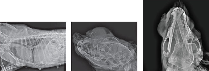
Fig. 2. Cytology reveals several mesenchymal cells without malignant features, including cells with a round morphology and cytoplasm vacuoles, coarse chromatin, or nucleoli. The radiology displayed an osteolytic expansile lesion, proliferative periosteal reaction, irregular margin, and stippled to speculated columnar periosteal reaction (Fig. 3). The tumor originated from the end of the nostrils to the ocular region. The transition zone is poorly demarcated from normal bone.
Fig. 3. X-ray showing osteolytic expansile lesion proliferative periosteal reaction irregular margin and stippled to speculated columnar periosteal reaction. The frontal bone, nasal bone, and maxilla depicted gross destruction. These are features of aggressive bone lesions. The differential diagnosis included multiple myeloma, Osteoma, Squamous cell carcinoma, malignant melanoma, acanthomatous ameloblastoma, and fibrosarcoma. Biopsy sample obtained from the rostral aspect of the mass shows cartilaginous tissue in the center of the tumor, and the chondrocytes are normal. There was no malignant tumor, and the pathological assessment showed a histiocytoma. Surgical approach was performed to remove the expansile tumor (Fig. 4 Post-op), general anesthesia was induced by ketamine-Diazepam (5 mg.kg-1, 0.5 mg.kg-1) and Isoflurane maintenance by 3% MAC (minimum alveolar concentration).
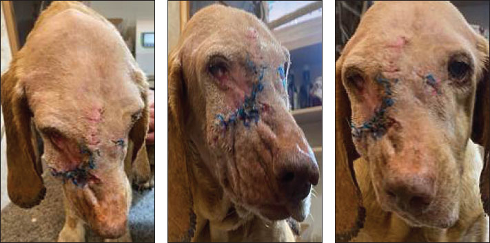
Fig. 4. Gross images after the surgical procedure showing good healing and at least tissue damage. Widespread antibiotics (Cefazolin 20 mg.kg-1 q24 hours) and analgesics (Tramadol 2 mg.kg-1) were administered to prevent infection and control pain management. Sections of the mass reveal a well-circumscribed region composed of sheets of round to polygonal histiocytic cells. The cells exhibit moderate amounts of pale eosinophilic cytoplasm and centrally to eccentrically located round to oval nuclei with finely stippled chromatin and indistinct nucleoli. A mild to moderate infiltrate of small lymphocytes is often observed at the periphery of the lesion (Fig. 5).
Fig. 5. The cells exhibit moderate amounts of pale eosinophilic cytoplasm and centrally to eccentrically located round to oval nuclei with finely stippled chromatin and indistinct nucleoli. DiscussionThis case report highlights the importance of a comprehensive diagnostic approach for an extended, cutaneous histiocytoma. The combination of cytologic, radiologic, and pathologic investigations allowed for an accurate diagnosis of the tumor and its invasion. Early recognition and treatment can improve outcomes for affected animals. Cutaneous histiocytoma is a rare neoplasm that accounts for only 0.3% of all canine skin tumors (Perez-Alenza, 2016; Hohenhaus et al., 2016). The clinical presentation of this case was consistent with that of previous studies, with a rapid growth pattern and painless development (Moore, 2015). The cytologic and pathologic features were also consistent with the diagnosis of histiocytoma. Surgical excision is usually recommended as the primary treatment modality due to the tumor's benign behavior and lack of evidence of metastasis (Vail DM Thamm and Liptak, 2019). In this case, cause of tumor invasion into the maxillary bone, nasal sinus destruction, orbital pressure, ocular discharge, and visual problems, surgical intervention was extremely recommended and radically performed. Several studies have reported cases of cutaneous histiocytoma in dogs, with a predilection in young animals and a benign clinical course. Treatment typically involves surgical excision, although spontaneous regression has been reported in some cases (Palus et al., 2014). The authors believe that any manipulation of the histiocytoma could accelerate tumor development and growth. This case had two time manipulations before surgery, and each manipulation changed most mass. Histopathologic examination of the excised tumor confirmed the presence of histiocytic cells with distinct cytoplasmic features, further supporting the diagnosis of cutaneous histiocytoma. ConclusionOverall, this case report of a massive cutaneous histiocytoma in a 12-year-old intact female dog highlights the importance of considering this benign tumor in the differential diagnosis of cutaneous masses in dogs. Further research is needed to better understand the underlying pathogenesis and behavior of histiocytoma in dogs. The authors believe that although the cutaneous histiocytoma is benign, the distance of the skin to underneath structures could be affected, thereby endangering the prognosis of treatment. AcknowledgmentsThe authors thank Dr. Hassanzadeh Specialized Veterinary Hospital for their kind support. Conflict of interestThe authors declare that they have no conflicts of interest. All data relevant to the study are included in the article. FundingThere was no specific grant for this research from any funding agency in the public, commercial, or not-for-profit sectors. Author’s contributionRN: draft editing, manuscript writing, surgical procedures, methodology, investigation, supervision, data curation. MNN: formal analysis, investigation, software, data curation. AAT: validation, review, editing, visualization. SS: writing, supervision, resources, and conceptualization. MAM: methodology, data curation, software. MR: visualization, validation, investigation, writing draft, resources. MT: validation, review and editing, visualization. AA: formal analysis, investigation, software, data curation. Data availabilityThe authors have no conflict with data availability. The authors have nothing to disclose. No AI-assisted technologies were used in the generation of this manuscript. ReferencesBenaduce Emanuelli Mello, C., Engelmann, A.M., Kommers, G.D., Martins Flores, M., Almeida Fighera, R., Rodrigues E Rodrigues, B., Ceolin Lamego, E., Bagolin Da Silva, C., Bueno, A. and Melazzo De Andrade, C. 2023. Fine needle aspiration cytology: high accuracy in diagnosing cutaneous and subcutaneous neoplasms in dogs. Comp. Clin. Pathol. 32(1), 155–164. Costa, D., Ferreira, R., Prada, J., Queiroga, F.L., Rodrigues, P., Silva, F. and Pires, I. 2020. A role for angiogenesis in canine cutaneous histiocytoma regression: insights into an old clinical enigma. Vivo 34, 3279–3284. García-Reynoso, I.C., Flores-Dueñas, C.A., Castro-del Campo, N., Jácome-Ibarra, M., Herrera-Ramírez, J.C., Gómez-Gómez, S.D., Rodríguez-Gaxiola, M.A. and Gaxiola-Camacho, S.M. 2025. Risk factors for the occurrence of cutaneous neoplasms in dogs: a retrospective study by cytology reports, 2019–2021. Animals 14(15), 2069. Goldschmidt, M.H. and Goldschmidt, K.H. 2018. Canine and feline skin tumors: a review. J. Vet. Dermatol. 29(2), 139–151. Goldstein, R.E., Stoyanov, M.A., Hargis, J.D., Temin, C.W. and DeBey, A.L. 2020. Clinical features and diagnostic imaging findings in dogs with cutaneous histiocytoma: a retrospective study of 54 cases. J. Vet. Inter. Med. 34(3), 847–854. Gualtieri, M., Marogna, G. and Vannozzi, I. 2018. Cutaneous histiocytoma in dogs: a retrospective study of 20 cases. J. Vet. Dermatol. 28(3), 246–253. Hohenhaus, A.E., Kelsey, J.L., Haddad, J., Barber, L., Palmisano, M., Farrelly, J. and Soucy, A. 2016. Canine cutaneous and subcutaneous soft tissue sarcoma: an evidence-based review of case management. J. Am. Anim. Hosp. Assoc. 52(2), 77 -89. Jeong Y.K., Lee, K.C. and Lee, H.B. 2020. CT findings in canine cutaneous histiocytoma: a study of 20 cases. J. Vet. Med. Sci. 82(6), 931–938. Kalosy, K.S., Keating, M.K., Rosenkrantz, W.S. and Moore, P.F. 2025. retrospective evaluation of clonality in canine erythema multiforme. Vet. Dermatol. 1–11. Mariani, C.L., Jennings, M.K., Olby, N.J., Borst, L.B., Brown Jr, J.C., Robertson, I.D., Seiler, G.S. and MacKillop, E. 2015. Histiocytic sarcoma with central nervous system involvement in dogs: 19 cases. J. Vet. Int. Med. 29(2), 607–613. McCue, D.A., Sibley, J.A., Schleis, S.E. and Kiupel, M. 2019. Canine cutaneous histiocytoma: a retrospective study of 114 cases. J. Vet. Inter. Med. 33(4), 1245–1252. Moore, P.F. 2015. A review of histiocytic diseases of dogs and cats. Vet. Pathol. 51(1), 167–184. Olaifa, O.S., Ohore, O.G., Ola, O.O., Usman, A.A., Antia, R.E., Jarikre, T.A., Tijani, M.O., Akinleye, A.O., Oyesiji, E.A., Ajumobi, F. and Oluwarore, K. 2025. Cytopathological comparison of well and poorly differentiated canine soft tissue sarcomas (Fibrosarcomas): diagnostic challenges in a resource-limited setting. Ukrainian J. Vet. Agricult. Sci. 2(82), 70–75. Palus, V., Guillermo, R.M. and Camus, A.C. 2014. Concurrent histiocytoma and mastocytoma in a young dog: case report. BMC. Vet. Res. 12(10), 102. Patterson, A.P. 2020. Pathological features of canine cutaneous histiocytoma a study of 25 cases. J. Comp. Pathol. 182, 19–27. Petal, T.S. 2020. Immunohistochemical characterization of canine cutaneous histiocytoma a study of 25 cases. J. Comp. Pathol. 182, 28–35. Pires, I., Rodrigues, P., Alves, A., Silva, F. and Lopes, C. 2024. Histopathological and ultrastructural study of a canine langerhans cell tumour (canine cutaneous histiocytoma). Cells 13(15), 1263. Ryan, S., Wouters, E.G.H., Nimwegen, S.V. and Kirpensteijn, J. 2022. Skin and subcutaneous tumors. Vet. Surgical Oncol. 29, 92–142. Vail, D.M.T. and Liptak, J.M. 2019. Withrow and MacEwen's small animal clinical oncology: a review of the literature. J. Am. Anim. Hosp. Assoc. 55(2), 93–102. Von Rueden, S.K. and Fan, T.M. 2021. Cancer-immunity cycle and therapeutic interventions-opportunities for including pet dogs with cancer. Front. Oncol. 11, 773420. Wang, Y., Huang, Y., Cai, W.X. and Tao, Q. 2022. Multiple benign fibrous histiocytomas of the mandible: a case report and review of the literature. Exp. Therap. Med. 24(3), 593. | ||


| How to Cite this Article |
| Pubmed Style Nikzad R, Nemati-nejad M, Tabarestani AA, Sahvieh S, Minaie MA, Ramezani M, Tavana M, Azizzadeh A. A case report of massive cutaneous histiocytoma in a 12-year-old intact bitch: Cytologic, radiologic, pathologic investigation. Open Vet. J.. 2025; 15(10): 5368-5372. doi:10.5455/OVJ.2025.v15.i10.54 Web Style Nikzad R, Nemati-nejad M, Tabarestani AA, Sahvieh S, Minaie MA, Ramezani M, Tavana M, Azizzadeh A. A case report of massive cutaneous histiocytoma in a 12-year-old intact bitch: Cytologic, radiologic, pathologic investigation. https://www.openveterinaryjournal.com/?mno=240918 [Access: January 25, 2026]. doi:10.5455/OVJ.2025.v15.i10.54 AMA (American Medical Association) Style Nikzad R, Nemati-nejad M, Tabarestani AA, Sahvieh S, Minaie MA, Ramezani M, Tavana M, Azizzadeh A. A case report of massive cutaneous histiocytoma in a 12-year-old intact bitch: Cytologic, radiologic, pathologic investigation. Open Vet. J.. 2025; 15(10): 5368-5372. doi:10.5455/OVJ.2025.v15.i10.54 Vancouver/ICMJE Style Nikzad R, Nemati-nejad M, Tabarestani AA, Sahvieh S, Minaie MA, Ramezani M, Tavana M, Azizzadeh A. A case report of massive cutaneous histiocytoma in a 12-year-old intact bitch: Cytologic, radiologic, pathologic investigation. Open Vet. J.. (2025), [cited January 25, 2026]; 15(10): 5368-5372. doi:10.5455/OVJ.2025.v15.i10.54 Harvard Style Nikzad, R., Nemati-nejad, . M., Tabarestani, . A. A., Sahvieh, . S., Minaie, . M. A., Ramezani, . M., Tavana, . M. & Azizzadeh, . A. (2025) A case report of massive cutaneous histiocytoma in a 12-year-old intact bitch: Cytologic, radiologic, pathologic investigation. Open Vet. J., 15 (10), 5368-5372. doi:10.5455/OVJ.2025.v15.i10.54 Turabian Style Nikzad, Reza, Masoud Nemati-nejad, Amirhossein Alizadeh Tabarestani, Sonia Sahvieh, Mohammad Amin Minaie, Mohammad Ramezani, Mehdi Tavana, and Azam Azizzadeh. 2025. A case report of massive cutaneous histiocytoma in a 12-year-old intact bitch: Cytologic, radiologic, pathologic investigation. Open Veterinary Journal, 15 (10), 5368-5372. doi:10.5455/OVJ.2025.v15.i10.54 Chicago Style Nikzad, Reza, Masoud Nemati-nejad, Amirhossein Alizadeh Tabarestani, Sonia Sahvieh, Mohammad Amin Minaie, Mohammad Ramezani, Mehdi Tavana, and Azam Azizzadeh. "A case report of massive cutaneous histiocytoma in a 12-year-old intact bitch: Cytologic, radiologic, pathologic investigation." Open Veterinary Journal 15 (2025), 5368-5372. doi:10.5455/OVJ.2025.v15.i10.54 MLA (The Modern Language Association) Style Nikzad, Reza, Masoud Nemati-nejad, Amirhossein Alizadeh Tabarestani, Sonia Sahvieh, Mohammad Amin Minaie, Mohammad Ramezani, Mehdi Tavana, and Azam Azizzadeh. "A case report of massive cutaneous histiocytoma in a 12-year-old intact bitch: Cytologic, radiologic, pathologic investigation." Open Veterinary Journal 15.10 (2025), 5368-5372. Print. doi:10.5455/OVJ.2025.v15.i10.54 APA (American Psychological Association) Style Nikzad, R., Nemati-nejad, . M., Tabarestani, . A. A., Sahvieh, . S., Minaie, . M. A., Ramezani, . M., Tavana, . M. & Azizzadeh, . A. (2025) A case report of massive cutaneous histiocytoma in a 12-year-old intact bitch: Cytologic, radiologic, pathologic investigation. Open Veterinary Journal, 15 (10), 5368-5372. doi:10.5455/OVJ.2025.v15.i10.54 |

