


| Research Article | ||
Open Vet. J.. 2025; 15(10): 5266-5272 Open Veterinary Journal, (2025), Vol. 15(10): 5266-5272 Research Article Frequency of erythrocyte antigen DEA 1 expression in dogs (Canis lupus familiaris) typed in veterinary clinical laboratories in the Valparaíso region, Chile: A retrospective studyMeyling Chang1, Fernando Pérez1, Diego Yañez1,2, Carlos Yañez2 and Cristian Larrondo1*1Facultad de Medicina Veterinaria y Agronomía, Universidad de Las Américas, Viña del Mar, Chile 2Laboratorio AnimaLab Diagnóstico, Quilpué, Chile *Corresponding Author: Cristian Larrondo. Facultad de Medicina Veterinaria y Agronomía, Universidad de Las Américas, Viña del Mar, Chile. Email: clarrondo [at] udla.cl Submitted: 01/06/2025 Revised: 27/08/2025 Accepted: 06/09/2025 Published: 31/10/2025 © 2025 Open Veterinary Journal
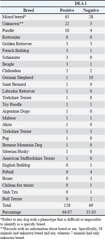
AbstractBackground: Several erythrocyte antigens have been identified in canines, specifically in dogs, such as dog erythrocyte antigen (DEA): 1, 3, 4, 5, 7; DAL, Kai 1, and Kai 2. Among these, only some are of significant clinical importance, while others require further research to determine their antigenic potential and the possible incompatibility reactions that could arise during transfusions. In Chile, only the DEA 1 is typed due to its high antigenic capacity and the risk of severe hemolytic reactions it can provoke; however, no frequency studies on this antigen have been conducted in the country. Additionally, there is limited information regarding possible associations between dog breed, health status, sex, and DEA1 expression. Aim: This study aimed to determine the frequency of DEA-1 expression in dogs from the Valparaíso region, Chile, and to assess its association with dog breed, health status, and sex. Methods: Data from Alvedia Quick Test BT DEA 1 immunochromatographic tests collected from February 2018 to September 2024 in three veterinary clinical laboratories in the Valparaíso region, Chile, were retrospectively analyzed. The frequency of DEA 1 expression was determined. Chi-square and Fisher’s exact tests were used to assess associations between breed, health status, sex, and DEA 1 expression. Results: The frequency of DEA-1 positivity was 64.97%. A statistically significant association was found between breed and DEA-1 expression (p=0.006). However, no significant association was observed between DEA-1 expression and sex (p=0.276) or health status (p=0.494). Conclusion: In the Valparaíso region of Chile, the observed frequency of DEA 1 is similar to the globally reported rate (60%). Certain breeds of dogs are highly likely to express DEA 1, whereas others rarely do. Keywords: Blood typing, DEA 1, Dogs, Frequency. IntroductionVeterinary medicine is subdivided into various specialties, many of which focus on ensuring patient survival (Hale, 1995). Transfusion medicine is often integrated into the field of internal medicine, where emergencies are a daily occurrence, and continuous updates are essential for the well-being of treated animals (Mangiattera et al., 2021). Transfusion medicine also encompasses subfields such as hematology and immunology, where statistical analysis is frequently required to identify and validate a wide range of observations or analytes that may later be utilized once they have been scientifically confirmed (Day and Kohn, 2012). Over the years, veterinary transfusion medicine has undergone constant evolution, continuously updating and adapting to new technologies. These advancements provide veterinarians with greater capabilities in emergency care and the stabilization of critically ill patients. Despite these improvements, ongoing research in this field remains essential. While transfusion medicine is characterized by its potential to improve patient prognosis, it also presents complications that may worsen the recipient’s condition or even be fatal in some cases (Mangiaterra et al., 2021). Certain fundamental principles must be followed in veterinary medicine to ensure a successful transfusion procedure. These include identification of the deficiency or pathology, determination of its etiology, and transfusion of the specific blood component needed (Madriz et al., 2014). Given these considerations, a wide variety of blood products are available on the market, each with different cellular compositions and incorporated substances. These products are designed to address various patient needs based on the associated clinical signs and pathologies (Madriz et al., 2014). By the 1970s, the International Canine Immunology Committee established a nomenclature system for canine blood groups. Initially, blood groups were labeled with letters from A to G; however, the classification was changed to the dog erythrocyte antigen (DEA) system, where each antigen is designated by the DEA prefix followed by a number (e.g., DEA 1.1, DEA 1.2, DEA 3, and so on) (Novais and Magron, 2018). Recent studies have described the discovery of additional erythrocyte antigens in patients who, despite being fully typed under the DEA system, continued to show incompatibility in cross-matching tests. Among these discoveries is the Dal blood group, named after its initial identification in a Dalmatian patient. However, this antigen is not exclusive to Dalmatians and has also been found in other breeds and mixed-breed dogs (Blais et al., 2007). Another recently described blood group system is the Kai system, which has been studied in South Korea. This system uses monoclonal antibodies—anti-Kai1 and anti-Kai2—to type canine patients. No severe transfusion reactions have been reported to date; however, crossmatching tests are recommended to prevent delayed adverse reactions (Mangiaterra et al., 2021). DEA 1 DEA 1 consists of three erythrocyte antigen expressions (DEA 1.1, DEA 1.2, and DEA 1.3), which are alleles with minor differences. DEA 1.1 exhibits Mendelian dominance over DEA 1.2 and DEA 1.3, meaning that a patient can express only one of these alleles or be negative for all of them. Due to its antigenic potential, DEA 1.1 is considered the most clinically significant, and there is a significantly larger body of research associated with it (Hale, 1995; Hohenhaus, 2012; Sink, 2017). Later studies determined that all three DEA 1 expressions share the same epitope but differ in membrane expression, making their clinically insignificant differentiation (Silverstein and Hopper, 2022). To date, no natural antibodies have been reported for these alleles, indicating that no first exposure would trigger a hemolytic reaction. However, this initial transfusion would induce the production of alloantibodies, which could lead to severe hemolytic reactions upon a second exposure (Kessler et al., 2010; Davidow, 2013; Novais and Magron, 2018). DEA 1 is the most antigenic blood group and the primary cause of severe transfusion reactions. For instance, a DEA 1-negative dog receiving DEA 1-positive erythrocytes would develop alloantibodies that could initiate a delayed hemolytic reaction within 1–2 weeks post-transfusion. Consequently, a second transfusion with the same blood type would result in an acute hemolytic reaction, with complete destruction of the transfused erythrocytes within approximately 12 hours post-exposure (Hale, 1995; Novais and Magron, 2018). The clinical importance of accurate DEA 1 blood typing lies in the absence of natural antibodies against this antigen. If a crossmatch test were performed on an untyped patient, no agglutination might be observed in the test, but a reaction could still occur upon a second transfusion exposure (Mesa, 2015). After a thorough review, we found no information on this topic originating from Chile. Understanding that a significant portion of the canine population is negative for the DEA 1 erythrocyte antigen may raise awareness of the severity of a second exposure, thereby encouraging the implementation of pre-transfusion blood typing as a prophylactic measure. Additionally, performing a crossmatching test in cases of repeated transfusions is recommended, given that at least seven other erythrocyte antigens cannot be identified but may still cause agglutination and, consequently, a hemolytic reaction (Davidow et al., 2021). The objective of this study was to determine the prevalence of DEA 1 expression in dogs from the Valparaíso region of Chile and to investigate its potential associations with breed, health status, and sex. Materials and MethodsData collectionAn initial search was conducted using Google Maps and Google to identify four clinical laboratories in the Valparaíso region that offer blood typing services for dogs. After completing the initial screening, each selected laboratory was contacted via email, outlining the main objectives of the study and providing a data collection template in Microsoft Excel format. Laboratories that accepted the proposal were asked about the blood typing method and the cities or provinces they served. For scientific purposes, the collected data remained anonymous and confidential. Data were collected between February 2018 and September 2024. Once the data were obtained, they were recorded in a Microsoft Excel sheet, including the dog’s breed, age, health status, sex, and DEA 1 positive or negative result, to later conduct an epidemiological frequency study and present the findings schematically. In this study, the identification of the breed was based on the phenotypic expression of each patient. This decision was made because of the difficulty in finding patients with certificates verifying their breed. Therefore, it is assumed that there may be instances where non-pure breeds were included. However, based on their phenotype, the predominant genes expressed correspond to those of the identified breed. All dogs were blood-typed using the Alvedia Quick Test BT immunochromatographic kit. This test has been reported to exhibit 100% specificity and approximately 98% sensitivity for detecting the DEA 1 antigen (Kessler et al., 2010; León and Mantilla, 2021). A list of the collaborating institutions that participated in the study and from which the data were collected is provided below. 1. Mevetlab: located in the city of Viña del Mar. The demographic coverage of the laboratory includes the cities of Viña del Mar, Quilpué, and Villa Alemana in the Valparaíso region of Chile. 2. Centro Diagnóstico Veterinario Quilpué: located in the city of Quilpué. The demographic coverage of the laboratory includes the cities of Quilpué, Villa Alemana, Valparaíso, Concón, and La Calera, in the Valparaíso region, Chile. 3. Laboratorio Veterinario Dr. Bunster: located in the city of Los Andes. The demographic coverage of the laboratory includes the provinces of the Aconcagua Valley, including the cities of Los Andes and San Felipe in the Valparaíso region of Chile. Data analysisThe inclusion criteria for blood sampling records were as follows: dogs from the Valparaíso region, blood-typed dogs, dogs that required a blood transfusion, and dogs identified as blood donors. Records that did not meet these criteria were excluded from the study. The frequency and percentage of DEA 1 erythrocyte antigen expression were calculated. Chi-square tests with their corrections and Fisher’s exact test were used to find associations between the categorical variables, such as the dog’s breed, sex, health status, and DEA 1 positivity. The confidence level used was p ≤0.05. The SPSS statistical software, version 26, was used for data analysis. Ethical approvalNot needed for this study. ResultsIn total, 197 records were collected from the three laboratories. The mean age of the dogs was 8.79 ± 4.29 years. The vast majority of records corresponded to dogs diagnosed with anemia (n=78), followed by animals with other pathologies (e.g., neoplasia, n=27), and healthy dogs identified as potential blood donors (n=12). A total of 64.97% of the samples tested positive for the DEA 1 erythrocyte antigen, while 35.03% of the individuals tested negative for DEA 1 (Table 1). Table 1. Frequency and percentage of DEA-1 by dog breed. Blood samples obtained from dogs in three veterinary laboratories in the Valparaíso region, Chile.
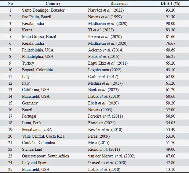
Regarding the sex of the dogs, of the 107 DEA 1 positive patients, 52.3% were females, while 47.7% were males. Of the 72 individuals with a negative result for DEA 1, 44.4% were females and 55.6% were males. Rottweiler (n=6) and Schnauzer (n=3) showed total positivity for the antigen (Table 1). In contrast, the Pitbull (n=4), Bull Terrier (n=2), and Boxer (n=3) were the breeds with 100% antigen absence. Finally, another breed worth mentioning is the German Shepherd, which showed a 91% absence of the antigen (10 out of 11 dogs), with only one dog testing positive for DEA 1. To compare the frequencies of the erythrocyte antigen DEA 1, all the international studies found through a bibliographic review were grouped in Table 2. Table 2. Percentage of dog erythrocyte antigen 1 (DEA 1) positivity reported in the literature.
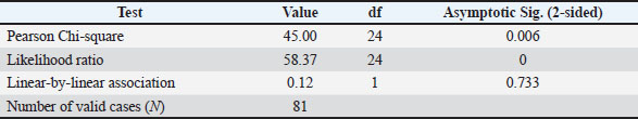
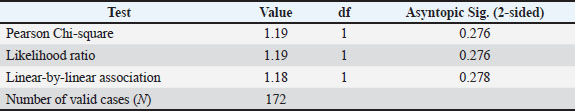
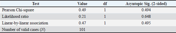
A significant association (X2=45, df=24, p=0.006) was observed between DEA 1 and breed (Table 3). Additionally, no statistically significant association was found between sex and the presence of DEA 1 (X2=1.19, df=1, p=0.276) (Table 4) or between health status and the presence of DEA 1 (X2=0.49, df=1, p=0.494) (Table 5). Table 3. Association between dog breed and DEA 1 expression. Results of the Pearson chi-square test.
Table 4. Association between dog sex and DEA 1 expression. Results of the Pearson chi-square test.
Table 5. Association between the health status of the dog and DEA 1 expression. Results of the Pearson chi-square test.
DiscussionOn the cell membrane of each canine's erythrocytes, there are glycoprotein receptors known as erythrocyte antigens. These are considered alloantigens because they do not exhibit antigenic properties within the same organism but can trigger an immune response in another individual of the same species (Enriquez, 2021). These antigens may have a glycoprotein or glycolipid origin, and some are known for their high immunogenicity (Hale, 1995). Their identification is crucial for the successful execution of blood transfusions in dogs. Additionally, considering the breed of the recipient could be beneficial in selecting an ideal donor, given the established association between certain breeds and the DEA 1 antigen. In recent years, a group of researchers analyzed the geographic distribution and prevalence of various erythrocyte antigens in dogs, compiling 41 epidemiological studies from around the world, including those from the Americas, Asia, Europe, and Africa. They reported that 60% of dogs worldwide carry the DEA 1 antigen. However, a more detailed analysis reveals that the study results vary between 39.89% and 91.3%, depending on the breed(s) studied (Mangiaterra et al., 2021). Comparable results were obtained in this study (64.97%). The obtained frequency contributes a new data point to the global statistics on DEA 1 epidemiology, addressing the lack of information regarding the prevalence of the antigen in Chile. Comparing the results obtained in Table 2, it can be concluded that the country with the highest similarity in the frequency of DEA 1 antigen positivity (64.97%) is Turkey, where Ergul Ekiz et al. (2011) obtained a positivity rate of 65.2%. A similar frequency of 66.2% was obtained in Philadelphia, United States (Polak et al., 2015). Next is Colombia, with 63.1% (Leguizamón, 2022), followed by Italy with 62% (Carli et al., 2017), and 61.2% (Medina et al., 2017). Additionally, Bank et al. (2023) obtained the same result as Medina et al. (2017) (61.2%) in California, United States. The two studies with the closest frequency to the one found in this study were conducted in North America. Izabik et al. (2010) found a frequency of 60.6% in Mansfield, United States, while a 69.6% positivity to the DEA 1 antigen was detected in Philadelphia, United States (Acierno et al., 2014). The study that differs the most from the one conducted presented a frequency of 93.2%, which occurs in Santo Domingo, Ecuador (Narváez-Rueda et al., 2022), possibly due to the breeds used for the study. The breed–DEA 1 relationship suggests a predisposition in certain breeds to carry the gene encoding DEA 1. This correlation has been previously identified in specific breeds, such as Rottweilers, Bernese Mountain Dogs, and Golden Retrievers (Riond et al., 2011; Carli et al., 2017; Silverstein and Hopper, 2022), whereas the absence of the antigen has been reported in Boxers (Riond et al., 2011). However, this relationship has not been established for the broader category of "breed" itself. Additionally, previous studies have supported the findings of the present study, which show no correlation between sex and DEA 1 expression (Carli et al., 2017; Enriquez, 2021). Future analyses should focus on purebred populations to either confirm or contrast these findings. This information could be valuable for internal medicine practitioners when selecting an optimal blood donor. However, these results must be interpreted with caution due to the limited sample sizes for many breeds, often fewer than five individuals. The high frequency observed for the most immunogenic antigen, DEA 1, must be considered due to the potential risks previously discussed regarding blood incompatibility, especially given that the ideal blood donor is classified as DEA 1-negative (Hale, 1995; Silverstein and Hopper, 2022). This highlights the importance of performing compatibility testing as a preventive measure. Additionally, the presence of other erythrocyte antigens (e.g., DEA 4, 5, and 7) should be considered, as they have been suggested to potentially cause delayed hemolytic reactions (Novais and Magron, 2018). Finally, considering these details will help veterinary clinicians make informed decisions when performing a successful blood transfusion. ConclusionThe study concludes that the observed frequency of DEA 1 in the Valparaíso region of Chile is similar to the global frequency. Therefore, analyzing this frequency at the national level, rather than just the regional level, would be valuable, as related results would be expected. A statistically significant association was found between a dog’s breed and the presence of DEA-1 antigen. This supports the scientific assertion that certain breeds have a high probability of carrying the antigen, whereas its presence is nearly nonexistent in others. This finding is crucial for characterizing the ideal blood donor. Additionally, the study confirms that genes on the sex chromosomes are not related to erythrocyte antigen expression. Conducting a genomic study on a breed that is 100% positive for DEA 1, such as Rottweilers, would be beneficial to identify the protein responsible for its expression. Furthermore, developing a rapid immunochromatography or agglutination test for DEA 7 would be particularly useful, given the high prevalence of alloantibodies against it, with reported incidence rates as high as 38%, which can lead to delayed hemolytic reactions (Spada et al., 2016). AcknowledgmentsThe first author of this article is immensely grateful to Dr. Fernando Pérez, Dr. Diego Yañez, Dr. Carlos Yañez, and Dr. Bunster for participating in the study. Conflict of interestAll authors declare no conflicts of interest with any financial organizations concerning the materials and data discussed in this manuscript. FundingThis study received no external funding. Authors’ contributionsAll authors have contributed to this study and have reviewed and approved the final version of the manuscript. Data availabilityAll data supporting this study’s findings are available within the manuscript. ReferencesAcierno, M.M., Raj, K. and Giger, U. 2014. DEA 1 expression on dog erythrocytes analyzed by immunochromatographic and flow cytometric techniques. J. Vet. Intern. Med. 28, 592–598. Bank, A.S., Farrell, K.S. and Epstein, S.E. 2023. Prevalence of dog erythrocyte antigen 1 in a population of dogs tested in California. J. Vet. Emerg. Crit. Care 33, 267–271. Blais, M.C., Berman, L., Oakley, D.A. and Giger, U. 2007. Canine Dal blood type: a red cell antigen lacking in some Dalmatians. J. Vet. Intern. Med. 21, 281–286. Carli, E., Carminato, A., Ravagnan, S., Capello, K., Antognoni, M.T., Miglio, A., Furlanello, T., Proverbio, D., Spada, E., Stefani, A., Mutinelli, F. and Vascellari, M. 2017. Frequency of DEA 1 antigen in 1037 mongrel and PUREBREED dogs in ITALY. BMC Vet. Res. 13, 364. Davidow, B. 2013. Transfusion medicine in small animals. Vet. Clin. North Am. Small. Anim. Pract. 43, 735–756. Davidow, E.B., Blois, S.L., Goy., Thollot, I., Harris, L., Humm, K., Musulin, S., Nash, K.J., Odunayo, A., Sharp, C.R., Spada, E., Thomason, J., Walton, J. and Wardrop, K.J. 2021. Association of Veterinary Hematology and Transfusion Medicine (AVHTM) Transfusion Reaction Small Animal Consensus Statement (TRACS). Part 1: definitions and clinical signs. J. Vet. Emerg. Crit. Care 31, 141–166. Day, M. and Kohn, B. 2012. BSAVA manual of canine and feline hematology and transfusion medicine. J. Small Anim. Pract. 53, 619. Ebelt, A.K., Fuchs, S., Weber, C., Müller, E. and Giger, U. 2020. Survey of blood groups DEA 1, DEA 4, DEA 5, Dal, and Kai 1/Kai 2 in different canine breeds from a diagnostic laboratory in Germany. Front. Vet. Sci. 7, 1–10.. Enriquez, A.V. 2021. Frecuencia del grupo sanguíneo DEA 1 en perros (Canis familiaris) con características clínico– hematológicas de potenciales donadores en Lima, Perú. B.S. thesis, Universidad Nacional Mayor de San Marcos, Perú. Ergul Ekiz, E., Arslan, M., Ozcan, M., Gultekin, G.I., Gulay, O.Y., Kirmizibayrak, T. and Giger, U. 2011. Frequency of dog erythrocyte antigen 1.1 in 4 breeds native to different areas in Turkey. Vet. Clin. Pathol. 40, 518–523. Ferreira, R.R.F., Gopegui, R.R. and Matos, A.J.F. 2011. Frequency of dog erythrocyte antigen 1.1 expression in dogs from Portugal. Vet. Clin. Pathol. 40, 198–201. Hale, A.S. 1995. Canine Blood Groups and their Importance in Veterinary Transfusion Medicine. Vet. Clin. North Am. Small Anim. Pract. 25, 1323–1332. Hohenhaus, A.E. 2012. Blood Transfusion and Blood Substitutes.In Fluid, Electrolyte, and Acid-Base Disorders in Small Animal Practice. Ed. Dibartola, S. MO: Elsevier Saunders Publishers, pp: 585–604. Iazbik, M.C., O'Donnell, M., Marin, L., Zaldivar, S., Hudson, D. and Couto, C.G. 2010. Prevalence of dog erythrocyte antigens in retired racing Greyhounds. Vet. Clin. Pathol. 39, 433–435. Kessler, R.J., Reese, J., Chang, D., Seth, M., Hale, A.S. and Giger, U. 2010. Dog erythrocyte antigens 1.1, 1.2, 3, 4, 7, and Dal blood typing and cross-matching by gel column technique. Vet. Clin. Pathol. 39, 306–316. Leguizamón, P. 2022. Caracterización de grupos sanguíneos en población de caninos y felinos de la sabana de Bogotá, por medio de la técnica de inmunocromatografía. B.S. thesis, Universidad de Ciencias Aplicadas y ambientales, Colombia. León, Y. and Mantilla, J. 2021. Revisión Bibliográfica de los Grupos Sanguíneos, Técnicas de Identificación y Reacciones Transfusionales Presentes en Animales Domésticos. Universidad de Santander, Bucaramanga. Madhavan Unny, N., Revathi, K., Prasanth, C.R., Pillai, U.N. and Ajithkumar, S. 2020. Studies on dog erythrocytic antigen 1 in two dog breeds in Kerala. Indian J. Canine Pract. 12((1–2), 1–2. Madriz, E.A., Mora Hernández, C.A. and Viñals, L.M. 2014. Manual de procedimientos para transfusiones sanguíneas en caninos. B.S. thesis, Universidad Nacional Agraria, Nicaragua. Medina, A.A., Gavazza, A. and Lubas, G. 2017. Prevalence of dog erythrocyte antigen 1 in 7,414 dogs in Italy. Vet. Med. Int. 2017, 5914629. Mesa, I. 2015. Estudio clinicopatológico del galgo español: hemograma, análisis de gases y equilibrio ácido-base, electrolitos, antígeno eritocitario canino dea 1.1, bioquímica sérica, electroforesis de proteínas séricas y haptoglobina. PhD thesis, Universidad de Córdoba, España. Mangiaterra, S., Rossi, G., Antognoni, M.T., Cerquetella, M., Marchegiani, A., Miglio, A. and Gavazza, A. 2021. Canine blood group prevalence and geographical distribution around the World: an updated systematic review. Animals 11, 342. Narváez-Rueda., Armas-Ariza, J.C. and Zapata-Saavedra, M.L. 2022. Tipificación sanguínea para la determinación de compatibilidad en perros (Canis lupus familiaris). Rev. Cient. FCV-LUZ. 32((1), 1–5. Novais, A. 2003. Prevalência dos antígenos eritrocitários caninos em cães domésticos (Canis familiaris) e investigação dos parâmetros hematológicos e da ocorrência de antígenos eritrocitários em lobos-guará (Chrysocyon brachyurus) e cachorros-do-mato (Cerdocyon thos) criados no Brasil. PhD thesis, Universidade Estadual Paulista, Brazil. Novais, A. and Magron, H. 2018. Canine Blood Groups: a review. Elektronische Zeitschrift Für Tierärztliche Labormedizin Und Pathobiochemie 11, 183–187.. Novais, A.A., Santana, A.E. and Vicentin, L.A. 1999. Prevalence of DEA 1 canine blood group system in dogs (Canis familiaris, Linnaeus, 1758) reared in Brazil. Braz. J. Vet. Res. Anim. Sci. 36, 23–27. Pereira, M.E., Rocha, M.N.A., Mendonça, A.J., Almeida, A.D.B.P.F.D., Canei, D.H. and Sousa, V.R.F. 2020. Occurrence of DEA1.1 in blood donor dogs in Cuiabá, Mato Grosso, Brazil. Semin. Cienc. Agr. 41, 1067. Pérez, N. 2009. Determinación de la prevalencia del grupo sanguíneo DEA 1.1 y valores hematológicos de caninos (Canis familiaris) en el Valle Central de Costa Rica. B.S. thesis, Universidad Nacional de Costa Rica, Costa Rica. Polak, K., Acierno, M.M., Raj, K., Mizukami, K., Siegel, D.L. and Giger, U. 2015. Dog erythrocyte antigen 1: mode of inheritance and initial characterization. Vet. Clin. Pathol. 44, 369–379. Proverbio, D., Lubas, G., Spada, E., Medina Valentin, A.A., Viñals Florez, L.M., Del Rosario Perlado Chamizo, M., Perego, R., Pennisi, M.G., Ferro, E., Baggiani, L., Gavazza, A. and Blais, M.C. 2020. Prevalence of Dal blood type and dog erythrocyte antigens (DEA) 1, 4, and 7 in canine blood donors in Italy and Spain. BMC Vet. Res. 16, 126. Riond, B., Schuler, E., Rogg, E., Hofmann-Lehmann, R. and Lutz, H. 2011. Prevalence of dog erythrocyte antigen 1.1 in dogs in Switzerland evaluated with the gel column technique. Schweiz. Arch. Tierheilkd. 153, 369–374. Silverstein, D. and Hopper, K. 2022. Small animal critical care medicine. Elsevier. Sink, C.A. 2017. Practical Transfusion Medicine for the Small Animal Practitioner. New York, NY: John Wiley & Sons. Spada, E., Proverbio, D., Viñals Flórez, L.M., Del Rosario Perlado Chamizo, M., Serra Y Gómez De La Serna, B., Perego, R. and Baggiani, L. 2016. Prevalence of naturally occurring antibodies against dog erythrocyte antigen 7 in a population of dog erythrocyte antigen 7-negative dogs from Spain and Italy. Am. J. Vet. Res. 77, 877–881. Van Der Merwe, L.L., Jacobson, L.S. and Pretorius, G.J. 2002. The breed prevalence of dog erythrocyte antigen 1.1 in the Onderstepoort area of South Africa and its significance in selection of canine blood donors. J. S. Afr. Vet. Assoc. 73, 53–56. Yi, S.W., Kim, E., Oh, S.I., Oh, S.I., Kim, J.S., Ha, J.H., Lee, B., Yoo, J.G. and Do, Y.J. 2022. Prevalence of dog erythrocyte antigens 1 and 7 in eleven canine breeds in the Republic of Korea. Korean J. Vet. 45, 269–275. | ||
| How to Cite this Article |
| Pubmed Style Chang M, Pérez F, Yañez D, Yañez C, Larrondo C. Frequency of erythrocyte antigen DEA 1 expression in dogs (Canis lupus familiaris) typed in veterinary clinical laboratories in the Valparaíso region, Chile: A retrospective study. Open Vet. J.. 2025; 15(10): 5266-5272. doi:10.5455/OVJ.2025.v15.i10.43 Web Style Chang M, Pérez F, Yañez D, Yañez C, Larrondo C. Frequency of erythrocyte antigen DEA 1 expression in dogs (Canis lupus familiaris) typed in veterinary clinical laboratories in the Valparaíso region, Chile: A retrospective study. https://www.openveterinaryjournal.com/?mno=241510 [Access: January 25, 2026]. doi:10.5455/OVJ.2025.v15.i10.43 AMA (American Medical Association) Style Chang M, Pérez F, Yañez D, Yañez C, Larrondo C. Frequency of erythrocyte antigen DEA 1 expression in dogs (Canis lupus familiaris) typed in veterinary clinical laboratories in the Valparaíso region, Chile: A retrospective study. Open Vet. J.. 2025; 15(10): 5266-5272. doi:10.5455/OVJ.2025.v15.i10.43 Vancouver/ICMJE Style Chang M, Pérez F, Yañez D, Yañez C, Larrondo C. Frequency of erythrocyte antigen DEA 1 expression in dogs (Canis lupus familiaris) typed in veterinary clinical laboratories in the Valparaíso region, Chile: A retrospective study. Open Vet. J.. (2025), [cited January 25, 2026]; 15(10): 5266-5272. doi:10.5455/OVJ.2025.v15.i10.43 Harvard Style Chang, M., Pérez, . F., Yañez, . D., Yañez, . C. & Larrondo, . C. (2025) Frequency of erythrocyte antigen DEA 1 expression in dogs (Canis lupus familiaris) typed in veterinary clinical laboratories in the Valparaíso region, Chile: A retrospective study. Open Vet. J., 15 (10), 5266-5272. doi:10.5455/OVJ.2025.v15.i10.43 Turabian Style Chang, Meyling, Fernando Pérez, Diego Yañez, Carlos Yañez, and Cristian Larrondo. 2025. Frequency of erythrocyte antigen DEA 1 expression in dogs (Canis lupus familiaris) typed in veterinary clinical laboratories in the Valparaíso region, Chile: A retrospective study. Open Veterinary Journal, 15 (10), 5266-5272. doi:10.5455/OVJ.2025.v15.i10.43 Chicago Style Chang, Meyling, Fernando Pérez, Diego Yañez, Carlos Yañez, and Cristian Larrondo. "Frequency of erythrocyte antigen DEA 1 expression in dogs (Canis lupus familiaris) typed in veterinary clinical laboratories in the Valparaíso region, Chile: A retrospective study." Open Veterinary Journal 15 (2025), 5266-5272. doi:10.5455/OVJ.2025.v15.i10.43 MLA (The Modern Language Association) Style Chang, Meyling, Fernando Pérez, Diego Yañez, Carlos Yañez, and Cristian Larrondo. "Frequency of erythrocyte antigen DEA 1 expression in dogs (Canis lupus familiaris) typed in veterinary clinical laboratories in the Valparaíso region, Chile: A retrospective study." Open Veterinary Journal 15.10 (2025), 5266-5272. Print. doi:10.5455/OVJ.2025.v15.i10.43 APA (American Psychological Association) Style Chang, M., Pérez, . F., Yañez, . D., Yañez, . C. & Larrondo, . C. (2025) Frequency of erythrocyte antigen DEA 1 expression in dogs (Canis lupus familiaris) typed in veterinary clinical laboratories in the Valparaíso region, Chile: A retrospective study. Open Veterinary Journal, 15 (10), 5266-5272. doi:10.5455/OVJ.2025.v15.i10.43 |

