


| Research Article | ||
Open Vet. J.. 2025; 15(8): 3871-3877 Open Veterinary Journal, (2025), Vol. 15(8): 3871-3877 Research Article Histopathological study of the neuroprotective effects of Gum Arabic and Fenchol on neuronal cells in an Alzheimer’s disease rat modelFadhil Abbas Khudhair Al Ghaliby1* and Layla Alhasan2*1Department of Physical, College of Education, Al-Shatrah University, Al-Dawaia, Iraq 2Biology Department, College of Education for Pure Sciences, Thiqar University, Iraq *Corresponding Author: Layla Alhasan. Biology Department, College of Education for Pure Sciences, Thiqar University, Iraq. Email: layla.alhasan [at] utq.edu.iq Submitted: 30/03/2025 Revised: 10/07/2025 Accepted: 24/07/2025 Published: 31/08/2025 © 2025 Open Veterinary Journal
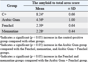
ABSTRACTBackground: Alzheimer’s disease (AD) is a progressive neurodegenerative disorder influenced by environmental and genetic factors. It is primarily characterized by beta-amyloid plaque deposition, neurofibrillary tangles, and impaired neuronal signaling. Given the lack of a definitive cure, research has increasingly focused on identifying natural compounds with neuroprotective and therapeutic potential. Aim: This study evaluates the effects of natural compounds (Fenchol and Gum Arabic) on immune modulation and neuroinflammatory markers, specifically interleukin-6 (IL-6), in a rat model of AD. By examining the effects of these natural products on the immune response and brain tissue pathology, this research aims to provide new insights into their potential therapeutic benefits for slowing AD progression. Methods: In this study, 36 adult male rats were randomly assigned to five groups: (1) a negative control group received standard feed and water, (2) a positive control group treated with aluminum chloride (17 mg/kg/day orally), (3) a Gum Arabic-treated group (2 ml, 10 g/100 ml orally) post-induction, (4) a Fenchol-treated group (2 ml, 5 mg/80 ml orally), and (5) a memantine-treated group (2 ml, 1.57 g/25 ml orally). After 1 month, histopathological assessments were performed to evaluate neuronal integrity, granule cell density, and beta-amyloid accumulation in the hippocampus. Additionally, serum IL-6 concentrations were measured via ELISA to assess systemic neuroinflammatory responses. Data were statistically analyzed using analysis of variance followed by least significant difference (LSD) post hoc tests. Results: Histopathological analysis revealed significant neurodegeneration in the positive control group, characterized by cytoplasmic vacuolation, reduced granule cell density, and elevated beta-amyloid levels. The Gum Arabic-treated group exhibited a partial neuroprotective effect, with a notable reduction in neurodegeneration, increased granule cell density, and a 50% decrease in amyloid plaques. The Fenchol-treated group demonstrated improved neuronal integrity and a marked reduction in beta-amyloid aggregates. The memantine-treated group exhibited the most substantial neuroprotective effect, significantly preserving granule cells and minimizing beta-amyloid deposition. Biochemical analysis revealed that IL-6 levels were markedly elevated in the positive control group (85.00 ± 3.00 ng/l) compared to the negative control (60.00 ± 2.00 ng/l). All treatment groups showed significant reductions in IL-6 levels. Memantine and combined treatments restored IL-6 levels close to normal. Fenchol and Gum Arabic alone reduced IL-6 levels, though to a lesser extent, indicating partial inflammatory effects. Conclusion: The findings suggested that Gum Arabic and Fenchol possess neuroprotective properties, suggesting their potential as therapeutic agents for AD, with efficacy comparable to that of memantine. Their ability to downregulate IL-6 further highlights their potential in mitigating neuroinflammation associated with Alzheimer’s pathology. Further investigations are warranted to elucidate their underlying mechanisms and evaluate their potential clinical applications. Keywords: Alzheimer’s disease, Neuroprotection, Natural compounds, Gum Arabic, Fenchol, IL-6, Hippocampus. IntroductionAlzheimer’s disease (AD) is an age-related neurodegenerative disorder that leads to memory loss and cognitive impairment (Shalan and Alhasan, 2024). The neurodegenerative process begins slowly and worsens over time due to multiple contributing factors, resulting in a gradual, irreversible destruction of specific neurons (Ayuob et al., 2018). As the disease progresses, affected individuals lose the ability to perform cognitive and intellectual functions, and as the disease advances, it significantly impairs their learning and memory capacities (Dhami, et al., 2023). Over time, AD leads to cumulative neurophysiological changes that affect mood and behavior (Zaher et al., 2020). Various factors contribute to the development of AD, with environmental and genetic factors playing a prominent role (Mahal et al., 2023). Oxidative stress, neuroinflammation, and genetic mutations are also significant contributors to the disease’s progression (Dhami, et al., 2023). However, age remains the primary risk factor, with prevalence increasing among individuals aged 65-85 years. This prevalence doubles every 5 years, reaching approximately 30%–40% by age 85 (Jeremic et al., 2021). Although numerous studies have attempted to elucidate the disease’s mechanisms, no cure has yet been developed. Therapeutic nutrition aims to utilize food as a therapeutic tool, harnessing its bioactive components to address and prevent various health issues. Nutritional therapy plays a fundamental role in managing many diseases. Plants and their natural derivatives are increasingly being studied as therapeutic agents. Recent years have witnessed a growing interest in medicinal plants as therapeutic products worldwide (Elshafie et al., 2023). Plants serve as a vital food source for both humans and animals and are also a source of numerous chemical substances. These compounds are known as natural products and are secondary metabolites produced through metabolic transformations within plant cells and tissues (Hilal et al., 2024). Medicinal plants serve as a natural reservoir of therapeutic bioactive compounds with therapeutic properties, playing a vital role in disease prevention and human health enhancement. In contrast, modern synthetic drugs, despite their widespread use, often have undesirable side effects (Tungmunnithum et al., 2018). Numerous natural products, containing bioactive compounds showing protective effects against neurodegenerative diseases, are utilized as neuroprotective strategies to counteract pathological physiological changes. The therapeutic potential of natural products and bioactive compounds in providing neuroprotection against neurological diseases is increasingly recognized. For instance, Gum Arabic and basil (Ocimum basilicum), an aromatic medicinal plant, play crucial roles in preventing neuropsychiatric conditions such as depression, anxiety, and nervous fatigue. Basil contains Fenchol, a plant-derived compound that offers neuroprotective effects, particularly against AD, by reducing the neurotoxic effects caused by the accumulation of amyloid-beta A in neurons. Additionally, Fenchol protects neurons from apoptosis by activating free fatty acid receptor 2 (FFAR2) signaling pathways, acting as a potent agonist for these signals. Fenchol also exhibits antimicrobial and antioxidant activities, which further enhance its neuroprotective properties (Razazan et al., 2020). Materials and MethodsAnimal modelIn this study, 30 mature male Sprague–Dawley rats, weighing between 200 and 250 g were used. The rats were obtained from the Animal House Colony at the College of Science, Thi-Qar University, Iraq. Experimental designThe animals were provided with a standard laboratory diet and clean water. After 1 week of acclimatization, the rats were placed in ordinary cages, where an appropriate ambient temperature was maintained with artificial lighting following a 12-hour light/dark cycle. The five groups, each consisting of six rats, were as follows: Group I: Rats (negative control)Rats received a normal diet and tap water throughout the experimental period. Group II: Rats (positive group control)Rats were administered with 17 mg/kg aluminum chloride (ALCl3) orally and once time per day along with a normal diet for 1 month. Group III (Gum Arabic treatment)Rats obtained 17 mg/kg of ALCl3 orally and treated with 2 ml (10 g/100 ml) Gum Arabic for 1 month. Group IV (Fenchol treatment)Rats were administered 17 mg/kg ALCl3 and treated with Fenchol orally and treated with 2 ml (5 mg/80 ml) Fenchol for 1 month. Both treatments were Fencol and Gum Arabic. Group V (memantine treatment)Rats were given 17 mg/kg ALCl3 orally and treated with 2 ml (1.5 g /25 ml) Memantine for 1 month. Sample collectionAt the end of the experiment, the rats were kept fasting for half a day. Blood samples were collected for further analysis using the orbital sinus technique of Sandford. The rats were then decapitated, and their entire brains were quickly dissected, thoroughly washed with isotonic buffer, dried, and immediately frozen at 80°C (Hoelbeek et al., 2021). Induction of ADRats were inducted with AD orally administration using AlCl3 dissolved in distilled water for 1 month at a dose of 17 mg/kg body weight every day (Shalan and Alhasan, 2023). HistopathologyBrain samples were collected and stored in a 10% formalin solution for 48 hours, and then transferred to fresh formalin solution after another 24 hours. The brain samples were sliced into sections with a thickness of 0.5 cm using the Italian automatic tissue processor (Histo-Line ATP700). Subsequently, the brain samples were sectioned into 4-m thick sections using a semi-automatic microtome (Histo-Line MRS3500, Italy). Brain sections were carefully placed in a water bath (FALC BI, Italy) and mounted on glass slides using a heating plate (K&K HYSH11, Korea). Subsequently, the brain sections were stained with hematoxylin and eosin. Ethical approvalAll experimental procedures were approved by the Ethical Committee of Medical Research at the National Research Center, College of Education for Pure Sciences at the University of Thi-Qar, Iraq (No. 63438/23/9). All animals received human care in compliance with institutional guidelines. Statistical analysisAll data were collected in triplicate and are presented as mean values ± SD. The normality of the data distribution was assessed using the Shapiro–Wilk test. As the data followed a normal distribution, a one-way analysis of variance was conducted using GraphPad Prism (version 9), followed by an LSD post hoc test for multiple comparisons to identify specific intergroup differences (Perfetto et al., 2006). ResultsHistological examination revealed significant differences between the experimental groups. In the negative control group, the hippocampal neurons appeared normal morphology with intact, with a well-preserved dentate gyrus and no detectable amyloid beta protein aggregates, indicating a healthy brain tissue (Fig. 1). In contrast, the positive control group, which only received AlCl3 treatment, exhibited pronounced histopathological changes. There was a significant reduction in the number of granule cells compared to the negative control group (Fig. 2A,B), along with clear signs of neuronal loss and degeneration in the dentate gyrus of the hippocampus. This was evidenced by cytoplasmic vacuolization and gaps, indicating neuronal damage. Furthermore, a statistically significant (p < 0.05) increase in amyloid beta protein aggregation was observed in this group, as shown in Table 1.
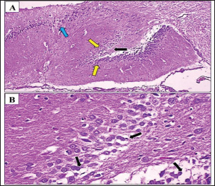
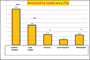
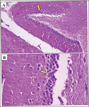
Fig. 1. Photomicrograph of the hippocampus of rat treated with Arabic Gum. (A, B) Neurodegeneration (black arrow) was observed in the dentate gyrus stratum granulosum cells within the affected hippocampus, characterized by the presence of cytoplasmic vacuoles in granule cells. Additionally, the density of granule cells (yellow arrow) was lower in the negative control group than in the positive control group, likely due to neuronal loss in the dentate gyrus. However, the number of granule cells was higher in the positive control group than in the positive control group. In addition, mild neurodegeneration (blue arrow) was observed in neurocyte of Cornu Ammonis region 3. H&E. A: 100x and B: 400x. The Gum Arabic-treated group demonstrated notable improvement relative to the positive control group. Histological analysis (Fig. 3A,B) revealed less severe neurodegeneration, with a higher number of granule cells, suggesting a partial neuroprotective effect. Amyloid beta accumulation was reduced by approximately 50% in the Cornu Ammonis region compared with that in the negative control group (Fig. 4). Statistical analysis confirmed a significant (p < 0.05) increase in cell preservation in the Gum Arabic group compared with the Fenchol and memantine groups. In the Fenchol-treated group, histological assessment showed reduced granule cell density compared with the negative control, but with minimal amyloid beta deposition, particularly in the hippocampus, where plaques were nearly absent (Fig. 5A,B). The histogram in Figure 6 demonstrated a statistically significant (p < 0.05) protective effect in this group compared with both the Gum Arabic and memantine groups. The memantine-treated group also exhibited a degree of neuroprotection. Although a decrease in granule cell density was observed relative to the negative control, it was higher than that observed in other treated groups (Fig. 7A). Table 1 highlights significant (p < 0.05) differences in amyloid beta levels in the memantine group compared with the other groups, suggesting moderate but incomplete protection.
Fig. 2. Photomicrograph of the cerebrum of Arabic Gum-treated rat. Gray matter. Many amyloid plaques (black arrow) were observed in the affected gray matter, with the amyloid plaques as focal areas in approximately 50% of the gray matter area. Table 1. Means of amyloid to total area score (%).
Fig. 3. Photomicrograph of the hippocampus of a Fenchol-treated rat. (A, B) Note the density of granule cells (yellow arrow) was lower in the negative control group than in the positive control group, likely due to neuronal loss in the dentate gyrus of the affected hippocampus. However, the number of granule cells was higher than that in the positive control and Arabic Gum groups. Also, mild neurodegeneration (black arrow) was observed in neurocyte of Cornu Ammonis region 2. H&E. A: 100x and B: 400x Analysis of interleukin-6 (IL-6) levels across the experimental groups revealed significant variations. In the negative control group, IL-6 levels remained within the normal physiological range, indicating the absence of inflammatory responses. In stark contrast, the positive control group exhibited a marked elevation in IL-6 levels, which was statistically significant (p < 0.05) compared to all other groups. This elevation correlates with the observed pathological changes, supporting the pro-inflammatory role of IL-6 in AlCl3-induced neuroinflammation.
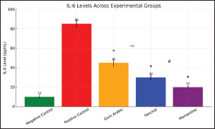
Fig. 4. Amyloid scoring level in the studied groups. *: indicates a significant (p < 0.05) increase in Fenchol and memantine groups compared with the Arabic Gum group. **: indicates a significant (p < 0.05) increase in Arabic Gum compared with Fenchol, memantine, and Arabic Gum Fenchol groups. ***: indicates a significant (p < 0.05) increase in control-positive group compared with other groups.
Fig. 5. Photomicrograph of the hippocampus of rats treated with memantine. (A, B) The density of granule cells (yellow arrow) was lower in the negative control group than in the positive control group due to neuronal loss in the dentate gyrus of the affected hippocampus. However, the number of granule cells was higher than that in the positive control and Arabic Gum groups H&E. A: 100x and B: 400x.
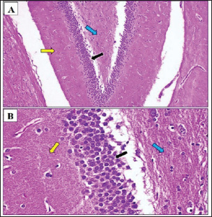
Fig. 6. Photomicrograph of the hippocampus of control negative rat. (A, B) Normal histological architecture of the dentate gyrus area of the hippocampus. Note the molecular stratum (yellow arrow), granulosum stratum (black arrow), and polymorphic layer (blue arrow). H&E. A: 100× and B: 400×.
Fig. 7. The standard curve generated from the ELISA technique for measuring IL-6 concentrations. The curve illustrates the relationship between known IL-6 concentrations and the optical density (OD) values, allowing for the quantification of IL-6 levels in experimental samples. The data points represent the average of triplicate measurements, and the curve was fitted using linear regression. In the Gum Arabic-treated group, IL-6 levels were notably reduced compared with those in the positive control group, although they remained slightly elevated relative to those in the negative control group. The Fenchol-treated group demonstrated a moderate regulatory effect, with a pronounced and statistically significant reduction in IL-6 levels compared with the positive control group (p < 0.05), suggesting a stronger anti-inflammatory action.
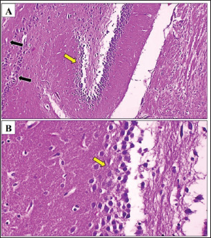
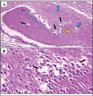
Fig. 8. Photomicrograph of the hippocampus of a control-positive rat. (A, B) Neurodegeneration (black arrow) was observed in stratum granulosum cells of dentate gyrus area of the affected hippocampus, where cytoplasmic vacuoles were observed within granule cells. However. The density of the granule cells (yellow arrow) was less compared with the control negative group due to neuron loss in the dentate gyrus area. Also, there was a significant loss in neurocyte number of the Cornu Ammonis (CA) regions 2 and 3 (blue arrow). H&E. A: 100x and B: 400x. Interestingly, these levels were significantly lower than those observed in both the Gum Arabic and Fenchol groups (p < 0.05), highlighting the superior anti-inflammatory efficacy of memantine under the experimental conditions. As shown in Figure 8. DiscussionThis study highlights the significant neuroprotective potential of the natural compounds Gum Arabic and Fenchol in an AlCl3-induced rat model of AD, with a particular focus on their dual histopathological and immunomodulatory effects. The findings reveal that both agents not only attenuated the extent of neurodegeneration in hippocampal tissue but also contributed to the downregulation of IL-6, a key pro-inflammatory cytokine implicated in AD. The histopathological results demonstrated that treatment with Gum Arabic and Fenchol reduced the amyloid plaque burden, preserved granule cell density, and decreased neuronal vacuolation compared to the positive control group. These results indicate their capacity to preserve hippocampal integrity. Concurrently, immunological findings showed significantly lower IL-6 levels in the treatment groups, especially in those receiving Gum Arabic treatment. These results indicated that the histological improvements were associated with a parallel decrease in inflammatory activity, indicating a potential mechanistic link between IL-6 suppression and structural brain preservation. The suppression of IL-6 expression observed in this study aligns with previous studies highlighting the anti-inflammatory and antioxidant properties of dietary fibers and plant-derived compounds. Hamid et al. (2023) and Salles et al. (2020) reported that the fermentable fibers in Gum Arabic modulate gut microbiota and inhibit oxidative stress, both of which are pathways linked to reduced systemic and neuroinflammation. Similarly, Fenchol’s role as an agonist of the FFAR2 receptor has been shown to activate anti-inflammatory cascades and modulate immune cell signaling, potentially contributing to its neuroprotective effects. The observed decline in IL-6 levels in the Fenchol group further supports previous findings by Shalan and Alhasan (2023), who demonstrated that O. basilicum extract, which contains Fenchol, downregulates tau aggregation, and improves memory in AD models. The membrane-stabilizing and anti-apoptotic actions of Fenchol—mediated by compounds such as caffeic acid and rosmarinic acid—may contribute to its ability to suppress IL-6 and protect neural architecture. Although memantine exhibited a reduction in IL-6 levels and provided degree of neuroprotection, it did not completely prevent neuronal loss in the dentate gyrus, consistent with previous reports of its limited efficacy in late-stage AD (Mohamed et al., 2020; Istifo et al., 2024). These comparative findings underscore the potential advantage of combining natural anti-inflammatory agents with standard therapies to enhance clinical outcomes. The immunohistochemical correlation observed in this study reinforces the importance of integrating molecular and histological endpoints when evaluating AD therapeutic candidates. The concurrent reduction in IL-6 levels and improvement in tissue pathology indicate that Gum Arabic and Fenchol exert neuroprotective effects by modulating key inflammatory pathways linked to disease progression. In conclusion, these findings support the therapeutic potential of Gum Arabic and Fenchol as multi-target agents with both anti-inflammatory and neuroprotective properties. Their effectiveness in reducing IL-6 expression while preserving hippocampal structure highlights their value as potential adjuncts or alternatives to conventional AD treatments. Future studies incorporating additional markers, such as caspase-3 and malondialdehyde, will be essential to further elucidate their mechanisms of action and confirm their efficacy in long-term disease models. AcknowledgmentThe authors would like to express their sincere gratitude to the Presidency of the University of Thi-Qar, the Deanery of the College of Education, and the Department of Biology in Thi-Qar, Iraq, for their continuous support and valuable facilities that contributed to the successful completion of this research. List of abbreviationsAD, Alzheimer’s disease; AlCl3, aluminum chloride; FFAR2, free fatty acid receptor2. Conflict of interestThe authors have no conflicts of interest to declare with the parties involved in this research. Author ContributionsAll authors contributed to design experiments and data analyses and writing the manuscript and approved the submitted article. FundingThe study was not funded by any sponsor or funder. ReferencesAyuob, N.N., El Wahab, M.G.A., Ali, S.S. and Abdel-Tawab, H.S. 2018. Ocimum basilicum improve chronic stress-induced neurodegenerative changes in mice hippocampus. Metab. Brain Dis. 33, 795–804; doi:10.1007/s11011-017-0173-3 Dhami, M., Raj, K. and Singh, S. 2023. Relevance of gut microbiota to Alzheimer’s disease (AD): Potential effects of probiotic in management of AD. Aging Health Res. 3(1), 100128; doi:10.1016/j.ahr.2023.100128 Elshafie, H.S., Camele, I. and Mohamed, A.A. 2023. A cComprehensive rReview on the bBiological, aAgricultural and pPharmaceutical pProperties of sSecondary mMetabolites bBased-pPlant oOrigin. Int. J. Mol. Sci. 24(4), 3266; doi:10.3390/ijms24043266 Hamid, N.S. 2023. The effect of Gum Arabic extract and arginine on certain hematological parameters, antioxidant activity, and the growth of lactic acid bacteria in female laboratory rats with type 2 diabetes. Master’s Thesis/Doctoral Dissertation, University of Thi-Qar, Faculty of Education for Pure Sciences, Department of Life Sciences. Hilal, B., Khan, M.M. and Fariduddin, Q. 2024. Recent advancements in deciphering the therapeutic properties of plant secondary metabolites: phenolics, terpenes, and alkaloids. Plant Physiol. Biochem. 211, 108674; doi:10.1016/j.plaphy.2024.108674 Hoelbeek, J.J., Kers, J., Steenbergen, E.J., Roelofs, J.J.T.H. and Florquin, S. 2021. Renal amyloidosis: validation of a proposed histological scoring system in an independent cohort. Clin. Kidney J. 14(3), 855–862; doi:10.1093/ckj/sfaa019 Istifo, N.N.H., Al-Zobaidy, M.A.J. and Abass, K.S. 2023. Long term effect of fluoxetine and memantine on biochemical markers of Alzheimer’s disease in scopolamine-induced mice. Regul. Mech. Biosyst. 15(2), 226–229; doi:10.15421/022433 Jeremic, D., Jiménez-Díaz, L. and Navarro-López, J.D. 2021. Past, present and future of therapeutic strategies against amyloid-β peptides in Alzheimer’s disease: a systematic review. Ageing Res. Rev. 72, 101496; doi:10.1016/j.arr.2021.101496 Mahal, M. H., Abed, N.A.N. and Al Jebur, L.A. 2023. Effect of oil extracted from rosemary and flaxseeds on oxidation factors and lipoproteins in male rabbits with Alzheimer’s disease. Samarra J. Pure Appl. Sci. 5(1), 151–159; doi:10.1016/j.ahr.2023.100128 Mohamed, A.B., Mohamed, A.Z. and Aly, M.S. 2020. Effect of thymoquinone against aluminum chloride-induced Alzheimer-like model in rats: a neurophysiological and behavioral study. Med. J. Cairo Univ. 88, 355–365; doi:10.21608/mjcu.2020.93997 Perfetto, S.P., Ambrozak, D., Nguyen, R., Chattopadhyay, P. and Roederer, M. 2006. Quality assurance for polychromatic flow cytometry. Nat. Protoc. 1(3), 1522–1530; doi:10.1038/nprot.2006.250 Razazan, A., Karunakar, P., Mishra, S.P., Sharma, S., Miller, B., Jain, S. and Yadav, H. 2021. Activation of microbiota sensing–free fatty acid receptor 2 signaling ameliorates amyloid-β induced neurotoxicity by modulating proteolysis-senescence axis. Front. Aging Neurosci. 13, 735933; doi:10.3389/fnagi.2021.735933 Salles, B.I.M., Cioffi, D. and Ferreira, S.R.G. 2020. Probiotics supplementation and insulin resistance: a systematic review. Diabetol. Metab. Syndr. 12, 98; doi:10.1186/s13098-020-00603-6 Shalan, M.A. and Alhasan, L. 2023. Evaluation of amyloid protein expression in a neurodegenerative rat model impairs memory abilities by Ocimum basilicum extract. J. Educ. Pure Sci. Univ. Thi-Qar 13(4); doi:10.32792/jeps.v13i4.376 Shalan, M.A. and Alhasan, L. 2024. Potential benefits of Ocimum basilicum extract on protein expression on ALCL3 induced rat models of Alzheimer’s disease. Univ. Dhi-Qar Coll. Educ. Pure Sci.; https://dspace.utq.edu.iq/items/a6c4f933-36cd-4b87-bc77-3e70b3db4b43/full Tungmunnithum, D., Thongboonyou, A., Pholboon, A. and Yangsabai, A. 2018. Flavonoids and oOther pPhenolic cCompounds from mMedicinal pPlants for pPharmaceutical and mMedical aAspects: an oOverview. Medicines 5(3), 93; doi:10.3390/medicines5030093 Zaher, M.A.F., Bendary, M.A. and Aly, A.S. 2020. Effect of thymoquinone against aluminum chloride-induced Alzheimer-like model in rats: a neurophysiological and behavioral study. Med. J. Cairo. 88(1), 165–176. Available via http://www.medicaljournalofcairouniversity.net University | ||

| How to Cite this Article |
| Pubmed Style Ghaliby FAKA, Alhasan L. Histopathological study of the neuroprotective effects of Gum Arabic and Fenchol on neuronal cells in an Alzheimer’s disease rat model. Open Vet. J.. 2025; 15(8): 3871-3877. doi:10.5455/OVJ.2025.v15.i8.53 Web Style Ghaliby FAKA, Alhasan L. Histopathological study of the neuroprotective effects of Gum Arabic and Fenchol on neuronal cells in an Alzheimer’s disease rat model. https://www.openveterinaryjournal.com/?mno=250057 [Access: June 26, 2026]. doi:10.5455/OVJ.2025.v15.i8.53 AMA (American Medical Association) Style Ghaliby FAKA, Alhasan L. Histopathological study of the neuroprotective effects of Gum Arabic and Fenchol on neuronal cells in an Alzheimer’s disease rat model. Open Vet. J.. 2025; 15(8): 3871-3877. doi:10.5455/OVJ.2025.v15.i8.53 Vancouver/ICMJE Style Ghaliby FAKA, Alhasan L. Histopathological study of the neuroprotective effects of Gum Arabic and Fenchol on neuronal cells in an Alzheimer’s disease rat model. Open Vet. J.. (2025), [cited June 26, 2026]; 15(8): 3871-3877. doi:10.5455/OVJ.2025.v15.i8.53 Harvard Style Ghaliby, F. A. K. A. & Alhasan, . L. (2025) Histopathological study of the neuroprotective effects of Gum Arabic and Fenchol on neuronal cells in an Alzheimer’s disease rat model. Open Vet. J., 15 (8), 3871-3877. doi:10.5455/OVJ.2025.v15.i8.53 Turabian Style Ghaliby, Fadhil Abbas Khudhair Al, and Layla Alhasan. 2025. Histopathological study of the neuroprotective effects of Gum Arabic and Fenchol on neuronal cells in an Alzheimer’s disease rat model. Open Veterinary Journal, 15 (8), 3871-3877. doi:10.5455/OVJ.2025.v15.i8.53 Chicago Style Ghaliby, Fadhil Abbas Khudhair Al, and Layla Alhasan. "Histopathological study of the neuroprotective effects of Gum Arabic and Fenchol on neuronal cells in an Alzheimer’s disease rat model." Open Veterinary Journal 15 (2025), 3871-3877. doi:10.5455/OVJ.2025.v15.i8.53 MLA (The Modern Language Association) Style Ghaliby, Fadhil Abbas Khudhair Al, and Layla Alhasan. "Histopathological study of the neuroprotective effects of Gum Arabic and Fenchol on neuronal cells in an Alzheimer’s disease rat model." Open Veterinary Journal 15.8 (2025), 3871-3877. Print. doi:10.5455/OVJ.2025.v15.i8.53 APA (American Psychological Association) Style Ghaliby, F. A. K. A. & Alhasan, . L. (2025) Histopathological study of the neuroprotective effects of Gum Arabic and Fenchol on neuronal cells in an Alzheimer’s disease rat model. Open Veterinary Journal, 15 (8), 3871-3877. doi:10.5455/OVJ.2025.v15.i8.53 |

