


| Case Report | ||
Open Vet. J.. 2025; 15(9): 4755-4758 Open Veterinary Journal, (2025), Vol. 15(9): 4755-4758 Case Report Cytological diagnosis of ganglion cyst in a dog: A case report from a resource-limited settingIssa Carolina García Reynoso1, Soila Maribel Gaxiola Camacho2*, César Augusto Flores Dueñas1, Nohemí Castro del Campo2, Miguel Ángel Rodríguez Gaxiola2, José Carlomán Herrera Ramírez1, Katya Montserrat Meza Silva1 and Sergio Daniel Gómez Gómez¹1Institute of Research in Veterinary Science, Autonomous University of Baja California, Mexicali, Mexico 2Faculty of Veterinary Medicine and Animal Science, Autonomous University of Sinaloa, Culiacan, Mexico *Corresponding Author: Soila Maribel Gaxiola Camacho. Faculty of Veterinary Medicine and Animal Science, Autonomous University of Sinaloa, Culiacan, Mexico. Email: soilagaxiola [at] uas.edu.mx Submitted: 08/05/2025 Revised: 08/08/2025 Accepted: 23/08/2025 Published: 30/09/2025 © 2025 Open Veterinary Journal
ABSTRACTBackground: Ganglion cysts (GCs) are rare conditions in both dogs and humans; thus, descriptions that aid in diagnosis are scarce; however, some differences exist that help us distinguish between them. Case Description: This case report presents a young female Dalmatian who developed a subcutaneous 6-cm nodule near the right ischial tuberosity, with irregular borders, slight mobility, and no pain. Fine needle aspiration cytology revealed a viscous, mucinous aspirate with sparse cellular content consistent with a GC. In this report, we discuss the most relevant findings observed under optical microscopy, present clinical data specific to this patient, and highlight the differences between ganglion and synovial cysts. Conclusion: This case highlights the diagnostic value of fine-needle aspiration cytology, especially in settings with limited access to advanced diagnostic techniques. Keywords: Ganglion cyst, Cytology, Synovial cyst, Nodule, Fine-needle aspiration. IntroductionGanglion cysts (GCs) are uncommon fluid-filled lesions that may occur in animals and are infrequently documented in the veterinary literature (Murata et al., 2014; Raskin and Conrado, 2023). These cysts contain viscous, gelatinous fluid and are typically associated with periarticular regions; however, they are not considered true synovial cysts (SCs) (Craig and Thompson 2017). Both cysts are soft tissue masses that could be asymptomatic or cause pain, weakness, swelling, or joint impairment (Giard and Pineda, 2015); however, in GCs, there are no synovial origin cells (O’Valle et al., 2013). Although the exact pathophysiology of GCs remains unclear, some authors speculate that they may originate from synovial fluid leakage due to joint instability or periarticular tissue degeneration (Murata et al., 2014; Bonelli and da Costa, 2019). GCs are cystic protrusions infiltrated with synovial-like fluid, potentially arising from adjacent joints or tendon sheaths (Crawford et al., 2011). According to Minotti and Taras (2002), repetitive stretching or microtrauma to joint structures may stimulate hyaluronic acid production, which accumulates within connective tissues and contributes to GC formation. Although cytological examination is commonly employed in veterinary diagnostics, detailed descriptions of GCs remain scarce (Krishnan et al., 2023). Nevertheless, cytology can aid in distinguishing inflammatory, degenerative, or neoplastic lesions and provides valuable diagnostic information (Dodd and Major, 2002). This case report presents relevant cytological findings that support a presumptive diagnosis of GC and emphasize the utility of fine-needle aspiration in environments with limited access to advanced diagnostic resources. Case DetailsA 1-year-old female Dalmatian presented with a 6-cm subcutaneous nodule near the right ischial tuberosity, with irregular borders, overlying erythema, and mild mobility. The nodule was not painful on palpation, was not discomforting, and did not interfere with walking, defecating, or lying down (Fig. 1). Fine needle aspiration cytology (FNAC) was performed to determine its inflammatory or neoplastic origin. The differential diagnoses included a perianal hernia, anal sac fistula or abscess, and hygroma. Microscopically, a clear, basophilic background with abundant acidophilic granules and a large number of erythrocytes was observed. Spindle cells with clear, irregular basophilic cytoplasm and poorly defined borders with an oval nucleus of coarse granular chromatin and 1–3 nucleoli were also found. These cells exhibited moderate anisocytosis and anisokaryosis. Other findings included round cells with basophilic foamy cytoplasm and a round nucleus of coarse granular chromatin with 1 nucleolus, which are consistent with histiocytes (Fig. 2). Cytological findings did not indicate neoplastic, inflammatory, or degenerative changes. A presumptive diagnosis of a GC was made based on the cytological and macroscopic features.
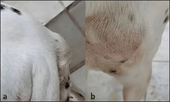
Fig. 1. (a) Nodule on the right ischial tuberosity. (b) Erythema and irregular borders. FNAC was performed using a 5-ml syringe and a 21-gauge needle. Serosanguineous, viscous fluid (5 ml) was aspirated and immediately smeared onto glass slides. Two smears were prepared using the blood smear technique, and two using the linear smear technique. All slides were air-dried and stained with a rapid hemocolorant (Hycell®) before being evaluated by a board-certified veterinary clinical pathologist.
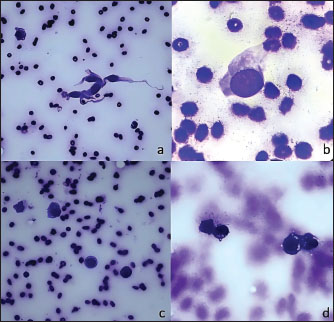
Fig. 2. (a) Fusiform cells in the center and a round foamy cell on left side 40×. (b) Granular background and fusiform cell 100×. (c) Round foamy cells 20×. (d) Round foamy cells with granular background 40×. Although histopathological confirmation was not possible, the lesion remained clinically stable during a follow-up period of 8 weeks, which was the last time we saw the patient. However, regarding the cytological study and evolution of the patient, we firmly believe in the benign nature of the lesion. DiscussionThe differential diagnosis between GC and SC remains a challenge in veterinary practice. This case report demonstrates the utility of cytology as a non-invasive and effective diagnostic tool, particularly in resource-limited settings. The cytological evaluation revealed fluid with distinctive characteristics of GC: moderate density, mucoid matrix, and amorphous material accompanied by macrophages, which differentiate these cysts from SC (Dodd and Major, 2002; Satturwar et al., 2022; Rana et al., 2023). GC tends to be less invasive than SC, which can infiltrate the synovial capsule (Steiner et al., 1996). The observed spindle-shaped cells with elongated basophilic cytoplasm and oval nuclei are consistent with myofibroblasts and mesenchymal cells in GC linings (Gude and Morelli, 2008; Giard and Pineda, 2015). A key differentiating feature in this case was the absence of synoviocytes, inflammatory cells, or joint effusion, as SCs are lined by the synovial membrane and communicate with the joint cavity, whereas GCs lack such a lining and connection. In veterinary practice throughout Latin America, economic constraints often limit access to advanced diagnostic methods. In these settings, cytology offers several advantages:
Cytology represents a particularly valuable diagnostic tool in Latin American contexts with limited resources. Sharkey et al. (2014) described cytology as a minimally invasive, rapid, and cost-effective diagnostic modality with broad usage in veterinary medicine. Marrinhas et al. (2022) demonstrated that this technique allows resource optimization by maximizing the diagnostic information obtained from a single cytological sample. The evidence-based application of cytopathology involves the management of preanalytical factors and comprehensive evaluation of diagnostic accuracy, which is particularly relevant when used as a basis for crucial medical decisions in critically ill patients or when financial considerations limit diagnostic and therapeutic options (Sharkey and Wellman, 2011). Christopher and Hotz (2004) emphasized the importance of properly expressing probability in cytological diagnosis when used as a presumptive diagnostic method. Hodges (2013) noted that cytology not only provides diagnostic value but also contributes to the economic sustainability of veterinary practice, a crucial factor in regions with limited resources. Although diagnostic confirmation typically requires a combination of methods (AlGhamdi et al., 2023), in this case, cytology provided a reliable presumptive diagnosis, reducing both the patient’s stress and the owner’s financial burden. Although surgical resection is the standard treatment with generally positive outcomes (Aikawa et al., 2014), the owner’s decision against surgery highlights the need to document alternative diagnostic and therapeutic approaches in veterinary literature, especially for practitioners in resource-limited regions, where the other solution is monitoring, palliative treatment, and sometimes only surgical resection of these lesions. ConclusionThis case highlights the diagnostic value of cytology in veterinary practice, establishing it as a minimally invasive and effective alternative to more invasive or costly diagnostic methods. The rarity of GCs in dogs, coupled with their morphological similarity to other cystic lesions, underscores the importance of accurate differentiation, especially when therapeutic decisions depend on it. This case contributes to the veterinary literature by exemplifying how cytological features can guide diagnosis and decision-making even in the absence of histopathology or imaging, particularly in resource-constrained settings, when interpreted in a clinical context. The combination of a comprehensive clinical evaluation with cytological analysis allowed a precise diagnostic approach in this case, emphasizing the relevance of cytology as a first-line technique in contexts where diagnostic resources are limited. AcknowledgmentsNone. Conflict of interestThe authors of this case report declare no conflicts of interest to declare with any person or institution. FundingThis study did not receive any specific grant. Authors’ contributionsICGR: Cytologic analysis, patient data recompilation, and writing of the original draft. SMGC: Project administration and supervision. CAFD: Conceptualization and writing. NCC: Methodology and supervision. MARG: Review and methodology. JCHR: Writing and supervision. KMMS: Cytologic analysis information. SDGG: Writing, review, and editing. All authors have read and approved the published version of the manuscript. Data availabilityAll data supporting this study’s findings are available in the manuscript. ReferencesAikawa T., Sadahiro S., Nishimura M., Miyazaki Y. and Shibata M. 2014 Ganglion cyst arising from the composite occipito-atlanto-axial joint cavity in a cat. Vet. Comp. Orthop. Traumatol. 27(4), 319–23. AlGhamdi, Y.S., Mahmoud, B.M., Alblaui, S.M., Altaraman, K.K., Alhumam, K.E., Alsinan, I.A. and Alhossan, A.M. 2023. A ganglion cyst in the anterior cruciate ligament of a 13 year-old boy. Cureus 15, e37692. Bonelli, M.D.A. and da Costa, R.C. 2019. Spontaneous regression of extradural intraspinal cysts in a dog: a case report. BMC Vet. Res. 15, 396. Christopher, M.M. and Hotz, C.S. 2004. Cytologic diagnosis: expression of probability by clinical pathologists. Vet. Clin. Pathol. 33, 84–95. Crawford, A., O’Donnell, M., Crowe, O., Eliashar, E. and Smith, R.K. 2011. Digital sheath synovial ganglion cysts in horses: digital sheath synovial ganglion cysts. Vet. Surg. 40, 66–72. Dodd, L.G. and Major, N.M. 2002. Fine-needle aspiration cytology of articular and periarticular lesions. Cancer 96, 157–166. Giard, M.C. and Pineda, C. 2015. Ganglion cyst versus synovial cyst? ultrasound characteristics through a review of the literature. Rheumatol. Int. 35, 597–605. Gude, W. and Morelli, V. 2008. Ganglion cysts of the wrist: pathophysiology, clinical picture, and management. Curr. Rev. Musculoskelet. Med. 1, 205–211. Hodges, J. 2013. Using cytology to increase small animal practice revenue. Vet. Clin. North Am. Small Anim. Pract. 43, 1385–1390. Krishnan, P., Dineshkumar, T., Divya, B., Krishnan, R. and Rameshkumar, A. 2023. Ganglion cyst of temporomandibular joint – A systematic review. Ann. Diagn. Pathol. 67, 152212. Marrinhas, C., Malhão, F., Lopes, C., Sampaio, F., Moreira, R., Caniatti, M., Santos, M. and Marcos, R. 2022. Doing more with less: multiple uses of a single slide in veterinary cytology. A practical approach. Vet. Res. Commun. 46, 641–654. Craig, L.E. and Thompson, K.G. 2017. Tumors of joints. 5th ed. In Tumors in domestic animals, Ed., Meuten D.J., IA: Wiley-Blackwell, pp. 352. Minotti, P. and Taras, J.S. 2002. Ganglion cysts of the Wrist. J. Am. Soc. Surg. Hand 2, 102–107. Murata, D., Sogawa, T., Tokunaga, S., Iwanaga, T., Kawaguchi, H., Miyoshi, N., Momoi, Y., Fujiki, M. and Miura, N. 2014. Ganglion cysts arising from a canine stifle joint. J. Vet. Med. Sci. 76, 457–459. O’Valle, F., Hernández Cortés, P., Aneiros-Fernández, J., Caba-Molina, M., Gómez-Morales, M., Cámara, M., Payá, J.A., Aguilar, D., Del Moral, R.G. and Aneiros, J. 2013. Morphological and immunohistochemical evaluation of ganglion cysts. Cross sectional study of 354 cases. Histol. Histopathol. 29, 601–607. Rana, S., Pradhan, A., Casaos, J., Mozaffari, K., Ghodrati, F., Sugimoto, B., Yang, I. and Nagasawa, D.T. 2023. Lumbar spinal ganglion cyst: a systematic review with case illustration. J. Neurol. Sci. 445, 120539. Raskin, R.E., and Conrado, F.O. 2023. Integumentary system: Non-neoplastic tumors. 4th ed. In Canine and feline cytopathology, Eds., Raskin, R.E., Meyer, D.J. and Boes, K.M. St. Loius, USA: Elsevier, pp. 63. Satturwar, S., Wakely, P.E. Jr. and Pantanowitz, L. 2022. Approach to FNA of myxoid soft tissue tumors. Adv. Anat. Pathol. 29, 380–388. Sharkey, L.C. and Wellman, M.L. 2011. Diagnostic cytology in veterinary medicine: a comparative and evidence-based approach. Clin. Lab. Med. 31, 1–19. Sharkey, L.C., Seelig, D.M. and Overmann, J. 2014. All lesions great and small, part 1: diagnostic cytology in veterinary medicine. Diagn. Cytopathol. 42, 535–543. Steiner, E., Steinbach, L.S., Schnarkowski, P., Tirman, P.F. and Genant, H.K. 1996. Ganglia and cysts around joints. Radiol. Clin. North Am. 34, 395–425. | ||
| How to Cite this Article |
| Pubmed Style Reynoso ICG, Camacho SMG, Dueñas CAF, Campo NCD, Gaxiola M�R, Ramírez JCH, Silva KMM, Gómez SDG. Cytological diagnosis of ganglion cyst in a dog: A case report from a resource-limited setting. Open Vet. J.. 2025; 15(9): 4755-4758. doi:10.5455/OVJ.2025.v15.i9.81 Web Style Reynoso ICG, Camacho SMG, Dueñas CAF, Campo NCD, Gaxiola M�R, Ramírez JCH, Silva KMM, Gómez SDG. Cytological diagnosis of ganglion cyst in a dog: A case report from a resource-limited setting. https://www.openveterinaryjournal.com/?mno=257097 [Access: June 22, 2026]. doi:10.5455/OVJ.2025.v15.i9.81 AMA (American Medical Association) Style Reynoso ICG, Camacho SMG, Dueñas CAF, Campo NCD, Gaxiola M�R, Ramírez JCH, Silva KMM, Gómez SDG. Cytological diagnosis of ganglion cyst in a dog: A case report from a resource-limited setting. Open Vet. J.. 2025; 15(9): 4755-4758. doi:10.5455/OVJ.2025.v15.i9.81 Vancouver/ICMJE Style Reynoso ICG, Camacho SMG, Dueñas CAF, Campo NCD, Gaxiola M�R, Ramírez JCH, Silva KMM, Gómez SDG. Cytological diagnosis of ganglion cyst in a dog: A case report from a resource-limited setting. Open Vet. J.. (2025), [cited June 22, 2026]; 15(9): 4755-4758. doi:10.5455/OVJ.2025.v15.i9.81 Harvard Style Reynoso, I. C. G., Camacho, . S. M. G., Dueñas, . C. A. F., Campo, . N. C. D., Gaxiola, . M. �. R., Ramírez, . J. C. H., Silva, . K. M. M. & Gómez, . S. D. G. (2025) Cytological diagnosis of ganglion cyst in a dog: A case report from a resource-limited setting. Open Vet. J., 15 (9), 4755-4758. doi:10.5455/OVJ.2025.v15.i9.81 Turabian Style Reynoso, Issa Carolina García, Soila Maribel Gaxiola Camacho, César Augusto Flores Dueñas, Nohemí Castro Del Campo, Miguel Ángel Rodríguez Gaxiola, José Carlomán Herrera Ramírez, Katya Montserrat Meza Silva, and Sergio Daniel Gómez Gómez. 2025. Cytological diagnosis of ganglion cyst in a dog: A case report from a resource-limited setting. Open Veterinary Journal, 15 (9), 4755-4758. doi:10.5455/OVJ.2025.v15.i9.81 Chicago Style Reynoso, Issa Carolina García, Soila Maribel Gaxiola Camacho, César Augusto Flores Dueñas, Nohemí Castro Del Campo, Miguel Ángel Rodríguez Gaxiola, José Carlomán Herrera Ramírez, Katya Montserrat Meza Silva, and Sergio Daniel Gómez Gómez. "Cytological diagnosis of ganglion cyst in a dog: A case report from a resource-limited setting." Open Veterinary Journal 15 (2025), 4755-4758. doi:10.5455/OVJ.2025.v15.i9.81 MLA (The Modern Language Association) Style Reynoso, Issa Carolina García, Soila Maribel Gaxiola Camacho, César Augusto Flores Dueñas, Nohemí Castro Del Campo, Miguel Ángel Rodríguez Gaxiola, José Carlomán Herrera Ramírez, Katya Montserrat Meza Silva, and Sergio Daniel Gómez Gómez. "Cytological diagnosis of ganglion cyst in a dog: A case report from a resource-limited setting." Open Veterinary Journal 15.9 (2025), 4755-4758. Print. doi:10.5455/OVJ.2025.v15.i9.81 APA (American Psychological Association) Style Reynoso, I. C. G., Camacho, . S. M. G., Dueñas, . C. A. F., Campo, . N. C. D., Gaxiola, . M. �. R., Ramírez, . J. C. H., Silva, . K. M. M. & Gómez, . S. D. G. (2025) Cytological diagnosis of ganglion cyst in a dog: A case report from a resource-limited setting. Open Veterinary Journal, 15 (9), 4755-4758. doi:10.5455/OVJ.2025.v15.i9.81 |

