


| Case Report | ||
Open Vet. J.. 2025; 15(10): 5420-5426 Open Veterinary Journal, (2025), Vol. 15(10): 5420-5426 Case Report Combined ophthalmic therapy and equine placental extract supplementation to treat corneal perforation in geriatric cats: A case report in the absence of surgical interventionTakae Natori1 and Eiichi Hirano2*1Ikeda Animal Hospital, Kawasaki, Japan 2Medical Affairs Department, Japan Bio Products Co., Ltd., Tokyo, Japan *Corresponding Author: Eiichi Hirano. Medical Affairs Department, Japan Bio Products Co., Ltd., Tokyo, Japan. Email: ehirano [at] placenta-jbp.co.jp Submitted: 26/05/2025 Revised: 22/08/2025 Accepted: 11/09/2025 Published: 31/10/2025 © 2025 Open Veterinary Journal
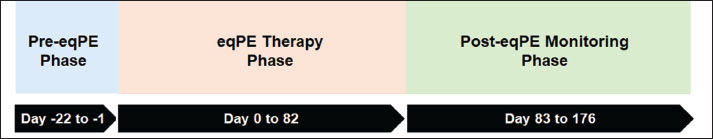
AbstractBackground: Environmental changes affecting pet animals, along with an increase in age-related conditions and diseases among pets, have become increasingly evident. Despite this trend, there remains a paucity of research on therapeutic approaches for geriatric companion animals. Case Description: The case report describes a 19-year-old spayed female cat that was diagnosed with chronic renal failure and recurrent corneal disease. Surgical intervention was not pursued at the owner’s request, leaving medical management as the only viable option. The patient’s corneal condition deteriorated, culminating in corneal perforation. Upon initiation of equine placental extract supplementation alongside medical treatment, substantial improvements were observed in hyperaemia, ocular discomfort, and additional corneal manifestations, including wound severity, ocular discharge, photophobia, lacrimation, and blepharospasm. Conclusion: The integration of medical therapy with placental extract supplementation may represent a novel therapeutic alternative for geriatric cats ineligible for surgical intervention. Keywords: Corneal perforation, Geriatric cat, Placental extract, Supplementation. IntroductionImproved nutrition, healthcare, and husbandry of domestic cats have increased their lifespan (Gunn-Moore, 2003). In 2017, 41.4% of UK cats at veterinary clinics were 7 years or older (Sánchez-Vizcaíno et al., 2017), while 20.4% of US cats were 10 or older in 2011 (Bellows et al., 2016a), demonstrating the considerably higher proportion of elderly cats in the pet population (Gunn-Moore, 2006; Bellows et al., 2016a). Routine ophthalmological and physical examinations, including blood pressure measurements, are recommended for older cats (Epstein et al., 2005, 2005; Kroll et al., 2001; Pittari et al., 2009). However, the classification of age-related changes and diseases is challenging in veterinary practice due to limited research (Paepe et al., 2013; Bellows et al., 2016a,b), particularly in feline geriatric ophthalmology (Karck et al., 2013; Paepe et al., 2013). Therefore, a more detailed understanding of the risks and progression of symptoms associated with eye diseases in older cats is needed. Animals are susceptible to corneal injuries that may cause significant vision impairment (Goulle, 2012), severe pain (Marfurt et al., 2001), secondary bacterial (Klatte et al., 2012; Bell et al., 2013) and fungal (Chew et al., 2010) infections, and traumatic lens capsule rupture. Animals are more susceptible to this condition for several reasons. These include anatomical factors, such as having large, exposed eyes that are inadequately protected; behavioural factors, such as walking through grassy areas and rubbing the eyes; environmental factors, such as frequent contact with sand, branches, and insects; and husbandry issues, such as owners being unaware of the early symptoms. These risks and other potential complications require early diagnosis and prompt treatment (Davidson and Nelms, 1999). Corneal perforation and laceration are among the leading causes of blindness related to corneal injuries. Corneal perforation may occur due to penetrating or blunt trauma or as a result of deep or lytic corneal ulcers (Brooks and Matthews, 2007). Older cats have an increased likelihood of developing conditions, such as kidney and dental diseases, hyperthyroidism, tumours, diabetes, and arthritis (Gunn-Moore, 2006). A retrospective study examining the medical records of cats with systemic hypertension revealed that chronic kidney disease (80%) was the most common cause of iris aneurysm in cases of hypertensive ophthalmopathy, followed by hyperthyroidism (20%) (Linek, 2020). Placental extract is derived from mammalian placentas through enzymatic digestion. Equine placental extract (eqPE) is used as a supplement for companion animals. Nakagaki et al. (2024) reported that two elderly dogs with refractory corneal disorders improved after treatment with eqPE for 6–9 weeks. Thus, eqPE supplementation may benefit elderly animals with ocular diseases and as a potential approach for feline geriatric ophthalmology (Karck et al., 2013; Paepe et al., 2013). However, existing research on the use of eqPE in cats is limited. This case report describes the effects of eqPE supplementation on corneal symptoms, chronic renal disease, and anaemia of an elderly cat with chronic renal disease, anaemia, and recurrent corneal damage progressing to corneal perforation. Case DetailsThe case included three phases: Pre-eqPE Phase (Day −22 to Day −1), eqPE therapy Phase (Day 0–Day 82), and Post-eqPE Monitoring Phase (Day 83 to Day 176; Fig. 1).
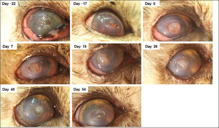
Fig. 1. The timeline diagram for this case. Pre-eqPE Phase: Day −22–Day −1. eqPE Therapy Phase: Day 0–Day 82. Post-eqPE Monitoring Phase: Day 83–Day 176. Patient background, initial presentation, and pharmacological treatment in the pre-eqPE phaseA 19-year-old spayed female Persian chinchilla cat (1.88 kg) presented with increased intraocular pressure, intraocular haemorrhage, and ocular pain for approximately 6 months, along with intermittent recurrent corneal disorders managed with topical ofloxacin (one drop, two to three times daily), 0.3% sodium hyaluronate (three to six drops, one to three times daily), enrofloxacin (6.25 mg/kg, once daily), and doxycycline hydrochloride hydrate (6.25 mg/kg, twice daily). Medical history included right mandibular gingival osteosarcoma, partially resected one year prior, and chronic kidney disease (CKD) for approximately 7 years treated with ferric chloride (Lenziaren packet: 0.125 g per head, twice daily), activated charcoal (capsule: 200 mg per head, twice daily), famotidine (tablet: 1.25 mg/head, twice daily), cyproheptadine hydrochloride hydrate (powder: 0.1 g/head, once daily), benazepril hydrochloride (tablet: 1.25 mg/head, once daily), and telmisartan (tablet: 2.5 mg/head, once daily). She also intermittently received amlodipine besilate (tablet: 0.21 mg, once daily) or sodium lactate, sodium chloride, potassium chloride, and calcium chloride hydrate (60 ml, twice daily) based on symptom severity. Anaemia was managed with subcutaneous polyethylene glycolylated recombinant feline erythropoietin (0.33 ml/kg, every 2 weeks). All events are reported relative to eqPE ingestion (Day 0). During a routine appointment for corneal disorder management, the right eye presented with severe conjunctivitis, elevation of the corneal area, and corneal perforation (Fig. 2, Day −22). The left eye showed no abnormalities, and the size of both eyes was comparable. Given the patient’s advanced age (19 years) and minimal to absent vision, the owner declined surgical intervention and referral to a secondary care facility. The patient was therefore discharged with an Elizabethan collar and continued on prescribed topical and oral treatments.
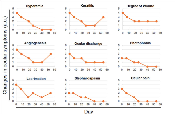
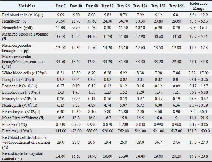
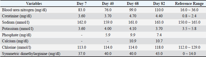
Fig. 2. Changes in the appearance of corneal damage before and during combined equine placental extract (eqPE) supplementation. From Days −22 to −17: before eqPE supplementation; Days 0–54: start of eqPE supplementation period. At the 5-day follow-up, conjunctivitis in the right eye had improved; however, corneal perforation, new edema oedema, and intraocular haemorrhage were noted, indicating disease progression (Fig. 2, Day −17). Intraocular pressure (IOP) was measured at 13 mmHg in the left eye and was unmeasurable in the right eye. A contact lens was fitted for protection, the Elizabethan collar was removed, and the patient was closely observed. The following day (Day −16), the patient returned after dislodging and losing the contact lens. Examination revealed bloody tears, reduced haemorrhage, increased anterior chamber turbidity, and persistent conjunctivitis in the right eye. IOPs were 13 mmHg (left) and 10 mmHg (right). Because of symptomatic improvement, a new contact lens was applied, and the Elizabethan collar was reapplied. The same medical therapies were maintained. After 10 days (Day 8), the patient returned due to another loss of the contact lens and elected to discontinue lens use. Subsequent examination of the right eye identified bloody tears and an elevated region at the site of corneal perforation; prescribed eye drops and oral medications were continued. Four days post-discontinuation of the contact lens (Day 4), bloody tears persisted in the right eye with ongoing treatment as before. After 4 days (Day 0), further signs included bloody tears, conjunctivitis, partial corneal clouding, angiogenesis, and expansion of Descemet’s membrane damage (Fig. 2). Although acetylcysteine was considered, its use was ultimately rejected because of its irritant properties. During-eqPE phase interventions and outcomesWe explored eqPE as a treatment option after finding a case report showing its potential to improve corneal disorders in two dogs (Nakagaki et al., 2024). With owner consent, the patient ingested 2 ml eqPE (Nakagaki et al., 2024) alongside previously described therapies. Ocular symptoms were subjectively elevated using a six-point scale: 0, good; 1, slightly better; 2, undecided; 3, slightly worse; 4, worse; 5, much worse (Fig. 3). On Day 0, hyperaemia, keratitis, degree of wound formation, angiogenesis and lacrimation were assigned a score of 4; ocular discharge and ocular pain a score of 3; and photophobia and blepharospasm a score of 2 (Fig. 3). Seven days after initiating eqPE intake, no significant changes were observed (Fig. 2, Day 7). Meanwhile, no changes were observed in blepharospasm, and a one-point decrease occurred in scores for all other items compared with Day 0 (Fig. 3). On Day 19, intraocular haemorrhage was not detected; however, the cornea was opaque and cloudy. Compared with the ocular symptoms on Day 7, ocular discharge remained unchanged, lacrimation scores decreased by two points, and other symptoms improved. On Day 26, fewer tears, increased conjunctival transparency, and reduced angiogenesis were observed (Fig. 2, Day 26). On Day 40, the transparency of the cornea and conjunctiva increased (Fig. 2, Day 40); compared with Day 26, the scores for hyperaemia, photophobia, lacrimation and blepharospasm decreased by one point, while the other parameters remained unchanged (Fig. 3). Blood tests further revealed a haematocrit (HCT) value of 28.9%, red blood cell distribution width coefficient of variation (RDW-CV) of 28.8% (Table 1), and blood urea nitrogen (BUN), creatinine, and symmetric dimethylarginine (SDMA) levels above reference levels (Table 2). Compared with Day 40, the scores for keratitis, angiogenesis, and lacrimation increased by one point on Day 54, while the scores for the other parameters remained unchanged (Fig. 3). Due to economic considerations, the eqPE dosing frequency was reduced to 2 ml/day after Day 68, with treatments continued except for doxycycline hydrochloride hydrate. Because the patient appeared to be in good health, she was monitored. On Day 82, increased corneal transparency and oedema were observed, without intraocular haemorrhage. Financial constraints faced by the owner caused eqPE treatment to be discontinued once the full supply was exhausted. At this point, HCT was 24.3% (Table 1), prompting subcutaneous administration of erythropoietin therapy (0.33 ml/kg biweekly). Haemoglobin (Hb) levels were marginally below the reference values, while the RDW-CV was slightly elevated (Table 1). Additionally, BUN, creatinine, and sSDMA levels were above the reference range (Table 2). As the patient displayed good general health, ongoing monitoring was conducted.
Fig. 3. Subjective assessment of corneal damage before and during combined equine placental extract (eqPE) supplementation. Subjective evaluation scores after eqPE supplementation. Higher values indicate worsening of symptoms, whereas lower values indicate improvement. Post-eqPE phase interventions and outcomesOn Day 96, doxycycline hydrochloride hydrate (6.25 mg/kg, twice daily) was resumed to treat conjunctivitis, with continued use of the original eye drops and oral medications. On Day 109, Descemet’s membrane protrusions increased and angiogenesis appeared, while corneal oedema persisted in the absence of hyperaemia; doxycycline hydrochloride hydrate was discontinued but other treatments continued. On Day 152, HCT reached 20.8% and recovery followed (Table 1); Hb and RDW-CV values were outside the reference range but monitored (Tables 1 and 2). Between the end of therapy (Day 68) and the final visit (Day 166), ocular symptoms remained stable, eosinophils were consistently low, plateletcrit and platelets were frequently high (Table 1), and Hb and RDW-CV values were outside the reference range (Tables 1 and 2). Nevertheless, the patient was monitored as she appeared healthy. No significant changes in ocular symptoms were observed between Days 124 and 166. The ophthalmic treatment was suspended; however, the same oral treatment prescribed previously was continued. Table 1. Hematological parameters between day 7 and day 166.
Table 2. Serum biochemical parameters between day 7 and day 82.
Follow-upOn Day 176, the owner observed the patient screaming and struggling, leading to emergency hospitalisation. Although clinicians suspected seizures, thromboembolisms, and blood clots, they detected no abnormalities through palpation and imaging tests. While the veterinarian in charge and the owner planned follow-up care, the patient died after returning home. Ethical approvalNo ethical approval was required for this case. DiscussionThis case describes a 19-year-old cat with chronic renal disease, anaemia, endemic corneal disorders, and severe progressive corneal damage leading to perforation. With domestic cats living longer, such cases are likely to increase (Gunn-Moore, 2003). Treatment was limited by the owner’s disagreement to pursue surgery or autologous serum eye drop therapy (Tsubota et al., 1999; Kim et al., 2018) and to visit a secondary care facility. Many owners may face similar challenges due to the physical and financial burden of invasive treatments for older cats. Therefore, the findings of this case report may assist other cat owners in managing comparable situations. Combined medical and surgical treatments for corneal perforation have shown favourable outcomes (Grahn and Cullen, 2000). However, there are no existing cases of combining medical and supplemental therapies for corneal perforation treatment in cats. We previously reported that two elderly dogs with refractory corneal disorders improved after 6–9 weeks of oral eqPE when surgery was declined (Nakagaki et al., 2024). Meanwhile, no such cases involving cats have been reported. In the current case report, a cat with similar symptoms was administered eqPE. Following eqPE administration, most corneal symptoms improved, with only minor keratosis and angiogenesis remaining, which were improved compared to baseline. These results indicate that eqPE administration is unlikely to exacerbate corneal damage, underlying CKD, or anaemia, or to cause adverse events. Therefore, it can be inferred that eqPE intake is beneficial and safe for cats with corneal disorders. Between Days 68 and 81, eqPE administration was modified from daily to every other day dosing. From day 82 onwards, the administration of eqPE was stopped. On Day 109, there was an observed increase in Descemet’s membrane protrusion, and worsened corneal symptoms. Thus, an uninterrupted supplementation with eqPE may be crucial, as symptom exacerbation has been documented upon discontinuation of therapy in dogs (Nakagaki et al., 2024). In previous cases, placental extract was administered orally, but it failed to achieve complete resolution of corneal damage (Nakagaki et al., 2024). Hence, topical administration such as eye drops might result in more effective outcomes. Notably, no ocular irritation was detected in a study where 0.1 ml of placental extract was administered to the conjunctival sac of a rabbit’s eye, per the Organisation for Economic Co-operation and Development Guideline 405 (2021) (data not shown). There is future potential for eqPE use as ophthalmic drops in companion animals with corneal disorders. ConclusionIn this case, the patient was an elderly cat with recurrent corneal damage and perforation, compounded by underlying systemic conditions. Standard treatment primarily comprised topical ophthalmic therapy. However, in this case, the addition of pharmacologic agents and eqPE led to considerable improvement in corneal symptoms. These results have the potential for therapeutic advancements, even in cases where conventional treatments are inaccessible or insufficient. Accordingly, this combined approach may represent a viable option for similarly challenging cases. AcknowledgmentsWe would also like to thank Editage (www.editage.jp) for English language editing. Conflict of interestTakae Natori has no competing interests to declare. Eiichi Hirano is an employee of Japan Bio Products Co., Ltd. FundingThis study received no financial support. Authors' contributionTN conceived and designed the clinical study, analysed and interpreted the data, and reviewed the manuscript. EH interpreted the data and wrote the manuscript. All authors have approved the final draft of the manuscript. Data availabilityData supporting this study’s findings are available upon request from the corresponding author. ReferencesBell, C.M., Pot, S.A. and Dubielzig, R.R. 2013. Septic implantation syndrome in dogs and cats: a distinct pattern of endophthalmitis with lenticular abscess. Vet. Ophthalmol. 16, 180–185. Bellows, J., Center, S., Daristotle, L., Estrada, A.H., Flickinger, E.A., Horwitz, D.F., Lascelles, B.D.X., Lepine, A., Perea, S., Scherk, M. and Shoveller, A.K. 2016a. Aging in cats: common physical and functional changes. J. Feline Med. Surg. 18, 533–550. Bellows, J., Center, S., Daristotle, L., Estrada, A.H., Flickinger, E.A., Horwitz, D.F., Lascelles, B.D.X., Lepine, A., Perea, S., Scherk, M. and Shoveller, A.K. 2016b. Evaluating aging in cats: how to determine what is healthy and what is disease. J. Feline Med. Surg. 18, 551–570. Brooks, D.E. and Matthews, A.G. 2007. Equine ophthalmology.In Veterinary ophthalmology. Gelatt, K.N 4th, Oxford, UK: Blackwell Publishing, pp: 1193–212. Chew, H.F., Jungkind, D.L., Mah, D.Y., Raber, I.M., Toll, A.D., Tokarczyk, M.J. and Cohen, E.J. 2010. Post-traumatic fungal keratitis caused by Carpoligna species. Cornea 29, 449–452. Davidson, M.G. and Nelms, S.R. 1999. Diseases of the lens and cataract formation.In Veterinary ophthalmology. Gelatt, K.N 3rd, Philadelphia, PA: Lippincott Williams & Wilkins, pp: 797–825. Goulle, F. 2012. Use of porcine small intestinal submucosa for corneal reconstruction in dogs and cats: 106 cases. J. Small Anim. Pract. 53, 34–43. Grahn, B.H. and Cullen, C.L. 2000. Equine phacoclastic uveitis: the clinical manifestations, light microscopic findings, and therapy of 7 cases. Can. Vet. J. 41, 376–382. Gunn-Moore, D.A. 2003. Considering older cats. Compend. Contin. Educ. Pract. Vet. 26A (Suppl), 1–4. Gunn-Moore, D. 2006. Considering older cats. J. Small Anim. Pract. 47, 430–431. Karck, J., Von Spiessen, L., Rohn, K. and Meyer-Lindenberg, A.M. 2013. Interrelation between the degree of a chronic renal insufficiency and/or systemic hypertension and ocular changes in cats article in German. Tierarztl. Prax. Ausg. K. Kleintiere Heimtiere 41, 37–45. Kim, S.E., Lee, M.K. and Seo, K. 2018. Clinical application of serum eye drops for herpetic keratitis in cats: a pilot study. Int. J. Appl. Res. Vet. Med. 16, 221–225. Klatte, J.M., Dastjerdi, M.H., Clark, K., Harrison, C.J., Grigorian, F. and Stahl, E.D. 2012. Hyperacute infectious keratitis with Plesiomonas shigelloides following traumatic lamellar corneal laceration. Pediatr. Infect. Dis. J. 31, 1200–1201. Kroll, M.M., Miller, P.E. and Rodan, I. 2001. Intraocular pressure measurements obtained as part of a comprehensive geriatric health examination from cats seven years of age or older. J. Am. Vet. Med. Assoc. 219, 1406–1410. Linek, J. 2020. Iris aneurysm in feline hypertensive oculopathy. Vet. Ophthalmol. 23, 436–441. Marfurt, C.F., Murphy, C.J. and Florczak, J.L. 2001. Morphology and neurochemistry of canine corneal innervation. Investig. Ophthalmol. Vis. Sci. 42, 2242–2251. Nakagaki, T., Nakari, M., Tahara, K. and Hirano, E. 2024. Supplementation of equine placenta extract on corneal wound in two dogs: case report. Open Vet. J. 14, 1503–1508. Paepe, D., Verjans, G., Duchateau, L., Piron, K., Ghys, L. and Daminet, S. 2013. Routine health screening: findings in apparently healthy middle-aged and old cats. J. Feline Med. Surg. 15(8), 8–19. Pittari, J., Rodan, I., Beekman, G., Gunn-Moore, D., Polzin, D., Taboada, J., Tuzio, H. and Zoran, D. 2009. American Association of Feline Practitioners: senior care guidelines. J. Feline Med. Surg. 11, 763–778. Sánchez-Vizcaíno, F., Noble, P.J.M., Jones, P.H., Menacere, T., Buchan, I., Reynolds, S., Dawson, S., Gaskell, R.M., Everitt, S. and Radford, A.D. 2017. Demographics of dogs, cats, and rabbits attending veterinary practices in Great Britain as recorded in their electronic health records. BMC Vet. Res. 13, 218. Senior Care Guidelines Task Force, Epstein., Kuehn, N.F., Landsberg, G., Lascelles, B.D.X., Marks, S.L., Schaedler, J.M. and Tuzio, H. 2005. AAHA senior care guidelines for dogs and cats. J. Am. Anim. Hosp. Assoc. 41, 81–91. Tsubota, K., Goto, E., Shimmura, S. and Shimazaki, J. 1999. Treatment of persistent corneal epithelial defect by autologous serum application. Ophthalmology 106, 1984–1989. | ||
| How to Cite this Article |
| Pubmed Style Natori T, Hirano E. Combined ophthalmic therapy and equine placental extract supplementation to treat corneal perforation in geriatric cats: A case report in the absence of surgical intervention. Open Vet. J.. 2025; 15(10): 5420-5426. doi:10.5455/OVJ.2025.v15.i10.61 Web Style Natori T, Hirano E. Combined ophthalmic therapy and equine placental extract supplementation to treat corneal perforation in geriatric cats: A case report in the absence of surgical intervention. https://www.openveterinaryjournal.com/?mno=260630 [Access: January 25, 2026]. doi:10.5455/OVJ.2025.v15.i10.61 AMA (American Medical Association) Style Natori T, Hirano E. Combined ophthalmic therapy and equine placental extract supplementation to treat corneal perforation in geriatric cats: A case report in the absence of surgical intervention. Open Vet. J.. 2025; 15(10): 5420-5426. doi:10.5455/OVJ.2025.v15.i10.61 Vancouver/ICMJE Style Natori T, Hirano E. Combined ophthalmic therapy and equine placental extract supplementation to treat corneal perforation in geriatric cats: A case report in the absence of surgical intervention. Open Vet. J.. (2025), [cited January 25, 2026]; 15(10): 5420-5426. doi:10.5455/OVJ.2025.v15.i10.61 Harvard Style Natori, T. & Hirano, . E. (2025) Combined ophthalmic therapy and equine placental extract supplementation to treat corneal perforation in geriatric cats: A case report in the absence of surgical intervention. Open Vet. J., 15 (10), 5420-5426. doi:10.5455/OVJ.2025.v15.i10.61 Turabian Style Natori, Takae, and Eiichi Hirano. 2025. Combined ophthalmic therapy and equine placental extract supplementation to treat corneal perforation in geriatric cats: A case report in the absence of surgical intervention. Open Veterinary Journal, 15 (10), 5420-5426. doi:10.5455/OVJ.2025.v15.i10.61 Chicago Style Natori, Takae, and Eiichi Hirano. "Combined ophthalmic therapy and equine placental extract supplementation to treat corneal perforation in geriatric cats: A case report in the absence of surgical intervention." Open Veterinary Journal 15 (2025), 5420-5426. doi:10.5455/OVJ.2025.v15.i10.61 MLA (The Modern Language Association) Style Natori, Takae, and Eiichi Hirano. "Combined ophthalmic therapy and equine placental extract supplementation to treat corneal perforation in geriatric cats: A case report in the absence of surgical intervention." Open Veterinary Journal 15.10 (2025), 5420-5426. Print. doi:10.5455/OVJ.2025.v15.i10.61 APA (American Psychological Association) Style Natori, T. & Hirano, . E. (2025) Combined ophthalmic therapy and equine placental extract supplementation to treat corneal perforation in geriatric cats: A case report in the absence of surgical intervention. Open Veterinary Journal, 15 (10), 5420-5426. doi:10.5455/OVJ.2025.v15.i10.61 |

