


| Case Report | ||
Open Vet. J.. 2026; 16(1): 763-767
Open Veterinary Journal, (2026), Vol. 16(1): 763-767 Case Report Uterine leiomyoma and cystic endometrial hyperplasia in a captive older adult Asian elephant (Elephas maximus): A case reportGustavo Willian Pandolfo1, Aline Ruediger Baron1, Maria Augusta Fornara1, Anna Laura de Oliveira Cunha1, Jéssica Aline Withoeft1, José Daniel Luzes Fedullo2, Renata Assis Casagrandre1 and Claudia Salete Wisser1*1Laboratório de Patologia Animal, Centro de Ciências Agroveterinárias, Universidade do Estado de Santa Catarina (CAV/UDESC), Lages, SC, Brazil 2Médico Veterinário Autônomo, Penha, SC, Brazil *Corresponding Author: Claudia Salete Wisser. Laboratório de Patologia Animal, Centro de Ciências Agroveterinárias, Universidade do Estado de Santa Catarina, Florianópolis, Brazil. Email: claudia.wisser [at] udesc.br Submitted: 05/08/2025 Revised: 30/11/2025 Accepted: 15/12/2025 Published: 31/01/2026 © 2026 Open Veterinary Journal
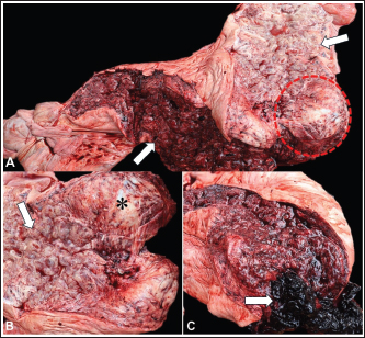
AbstractBackground: This report describes a case of uterine leiomyoma and cystic endometrial hyperplasia in an Asian elephant (Elephas maximus). Case Description: A 74-year-old female elephant in captivity was necropsied. At necropsy, the uterus was markedly enlarged, and a firm, white nodule measuring 10 × 10 × 8 cm protruded from the uterine wall into the lumen of the right uterine segment. Additionally, multiple cystic dilations (0.5–4 cm in diameter) were observed throughout the uterine segment. The mucosa was diffusely red with a moderate amount of crumbly clots in the lumen. Histologically, a benign mesenchymal neoplastic proliferation composed of multidirectional bundles of muscle tissue expanded the uterine muscular layer. The mucosa exhibited cystic dilations lined by simple squamous epithelium. In Masson’s trichrome staining, the neoplastic cells showed intense red staining, consistent with muscle tissue, whereas blue staining was restricted to the supporting fibrous connective tissue. Immunohistochemistry revealed mild cytoplasmic vimentin staining, moderate desmin immunoreactivity, and strong smooth muscle actin expression in neoplastic cells. Based on the anatomopathological, histochemical, and immunohistochemical findings, uterine leiomyoma and cystic endometrial hyperplasia were diagnosed. Conclusion: This case highlights the importance of diagnosing neoplasms and other conditions in captive elephants to better understand the diseases affecting these animals. Keywords: Megavertebrates, Neoplasm, Uterus, Zoo. IntroductionNeoplasms affecting the reproductive tract are most frequently observed in female elephants, although there is limited literature on the development of neoplasms in this species. Knowledge about these neoplasms is important because they can significantly impact reproduction, compromising the conservation of this species (Montali et al., 1997; Hermes et al., 2004; Pringproa et al., 2015; Landolfi et al., 2021). In a study in the United States, reproductive neoplasms in Asian elephants were observed in 80% (64/80) of the cases, with the majority being in the uterus (63/64; 98%) and only one case of ovarian neoplasm. Myometrial leiomyomas were present in 90% (57/63) of the cases with uterine neoplasm. The remaining cases included uterine adenocarcinoma, endometrial adenoma, in situ focal carcinoma in endometrial polyps, anaplastic carcinoma, endometrial hemangioma, primitive neuroectodermal tumor, and angiosarcoma (Landolfi et al., 2021). In most cases, cystic endometrial hyperplasia is frequently described in conjunction with uterine leiomyoma (Landolfi et al., 2021). In Brazil, there are no reports of neoplasms involving the reproductive tract in elephants, representing the first documented case in Brazil and one of the very few reported cases worldwide. This study aims to describe the anatomopathological findings of a case of uterine leiomyoma and cystic endometrial hyperplasia in an older adult Asian elephant (Elephas maximus) in southern Brazil. Case DetailsA necropsy was performed on a 74-year-old female Asian elephant (Elephas maximus) at a zoo in the state of Santa Catarina, southern Brazil. The elephant presented with hemorrhagic discharge from the vulvar region on the day preceding death. At necropsy, the elephant was in good body condition, with moderately pale mucous membranes. The uterus was markedly enlarged, and projecting from the uterine wall into the lumen of the right uterine segment, there was a poorly defined, white, firm nodule measuring 10 × 10 × 8 cm in diameter. In addition, multiple cystic dilations ranging from 0.5 to 4 cm in diameter were observed throughout the uterine segment (Fig. 1A–C). The mucosa was diffusely red with a moderate amount of crumbly clots in the lumen (Fig. 1C).
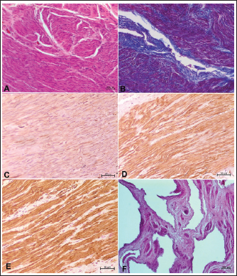
Fig. 1. Macroscopic findings of uterine leiomyoma and cystic endometrial hyperplasia in an Asian elephant (Elephas maximus). A) Uterus: markedly enlarged; projecting from the uterine wall into the lumen of the right uterine segment, white nodule measuring 10 × 10 × 8 cm (dashed); multiple cystic dilations with a thin wall, ranging from 0.5 to 4 cm (arrow), and diffusely red mucosa. B) Right uterine segment: showing the nodule (*) and cystic dilations in the mucosa (arrow). C) Left uterine segment: showing cystic dilatations and red mucosa; moderate amount of crumbly clot in the lumen. Uterine fragments were collected and fixed by immersion in 10% buffered formalin for routine histopathological processing and staining with hematoxylin and eosin (H&E). Histological sections of the uterus were subjected to Masson’s trichrome histochemical staining (Êxodo Científica, SP, Brazil). Immunohistochemistry was performed for vimentin, desmin, and smooth muscle actin. Tissue sections of 3 μm thickness were placed on charged glass slides, and endogenous peroxidase activity was blocked with Peroxidase Block (Novolink™ Kit, Cat. #RE7150-CE) for 10 minutes at room temperature (approximately 20°C). Antigen retrieval was performed in a water bath for 20 minutes at 100°C using Tris-EDTA buffer (pH 9.5) for desmin and smooth muscle actin, and in a microwave oven for 13 minutes with citrate buffer (pH 6) for vimentin. Nonspecific binding was blocked with Protein Block (Novolink™ Kit, Cat. #RE7150-CE) for 10 minutes at room temperature in a humid chamber (approximately 20°C). The following primary antibodies were used: vimentin (1:200, clone V9, Cat. #EP-12-54271, Biocare Medical, Concord, California, USA), desmin (1:200, clone D33, Cat. #EP-12-54441, Biocare Medical, Concord, California, USA), and smooth muscle actin (1:200, clone 1A4, Cat. #EP-12-52831, Biocare Medical, Concord, California, USA), all diluted in phosphate-buffered saline. Sections were incubated in a humid chamber at 4°C overnight. Subsequently, the secondary antibody (Novolink™ Post Primary) and the polymer (Novolink™ Polymer, Cat. #RE7150-CE) were applied and incubated for 30 minutes each at room temperature in a humid chamber. Visualization was achieved using 3,3’-diaminobenzidine (Novolink™ Kit, Cat. #RE7150-CE), followed by hematoxylin counterstaining. Positive controls included canine fibrosarcoma tissue (for vimentin), intestinal smooth muscle (for smooth muscle actin), and myocardium (for desmin). A universal negative control reagent (Cat. #EP-11-20567, EasyPath, São Paulo, Brazil) was simultaneously used with the tested samples. Histopathological examination revealed a poorly defined and nonencapsulated benign mesenchymal neoplastic proliferation expanding the uterine muscular layer, organized in multidirectional bundles and supported by moderate fibrovascular stroma. The cells were fusiform, with round to oval nuclei, fine chromatin, and inconspicuous nucleoli, sometimes single and prominent. The cytoplasm was moderately eosinophilic and poorly defined. Mild anisocytosis and anisokaryosis were observed, with zero mitotic figures in 2.37 mm² (Fig. 2A). In Masson’s trichrome staining, the neoplastic cells showed intense red staining, consistent with muscle tissue, whereas blue staining was restricted to the supporting fibrous connective tissue (Fig. 2B).
Fig. 2. Histopathological, histochemical, and immunohistochemical findings of uterine leiomyoma and cystic endometrial hyperplasia in an Asian elephant (Elephas maximus). A) Uterus: benign mesenchymal neoplastic proliferation, organized in multidirectional bundles and supported by moderate fibrovascular stroma (hematoxylin and eosin [HE], 10). B) Intense red staining, consistent with muscle tissue, while blue staining was restricted to the supporting fibrous connective tissue (Masson’s trichrome, 10x). C) Mild cytoplasmic staining of the neoplastic mesenchymal cells for vimentin (IHC, 20x). D) Moderate cytoplasmic immunolabeling of the neoplastic cells for desmin (IHC, 20x). E) Strong cytoplasmic immunolabeling of the neoplastic cells for smooth muscle actin (IHQ, 20x). F) Uterus: cystic dilatations lined by simple squamous epithelium (HE, 5x). Immunohistochemical evaluation revealed mild vimentin staining in the cytoplasm of the neoplastic cells, moderate desmin immunoreactivity, and strong smooth muscle actin expression in the neoplastic cells (Fig. 2C–E). Additionally, cystic dilations lined by simple squamous epithelium were observed in the mucosa (Fig. 2F), sometimes containing eosinophilic amorphous material, as well as moderate multifocal hemorrhage and congestion. Based on the anatomopathological, histochemical, and immunohistochemical findings, uterine leiomyoma and cystic endometrial hyperplasia were diagnosed. DiscussionElephants affected by uterine neoplasms or cystic endometrial hyperplasia range in age from 12 to 65 years (Agnew et al., 2004; Sapundzhiev et al., 2007; Pringproa et al., 2015; Landolfi et al., 2021). In our case, the 74-year-old elephant was the oldest elephant to have a neoplasm and the second oldest elephant ever reported in Brazil. Clinical signs are often absent or unreported, with cases in previous studies being diagnosed at necropsy, found incidentally, and not directly related to the cause of death (Agnew et al., 2004; Sapundzhiev et al., 2007; Pringproa et al., 2015). Although the leiomyoma and cystic endometrial hyperplasia were not the cause of death in the present case, they likely contributed to the clinical debilitation, particularly due to the animal’s advanced age and moderate intrauterine hemorrhage, which probably resulted in anemia, as evidenced by the pale mucous membranes. The cause of death was determined to be chronic renal disease and congestive heart failure. In a recent study of 57 cases of myometrial leiomyoma, 51 had multiple masses, and six had solitary masses. In more severe cases, the nodules may coalesce, distorting the uterine architecture and invading the uterine lumen (Landolfi et al., 2021). In the reported case, the neoplasm was solitary and extensive and invaded the lumen. The high involvement of endometrial hyperplasia associated with leiomyomas (24/28; 86%) is also significant, with no cases of metrorrhagia recorded (Landolfi et al., 2021). Cystic endometrial hyperplasia and intrauterine hemorrhage were observed in this case. Leiomyomas exhibit morphological characteristics that can resemble those of other smooth muscle tumors, making differentiation challenging when based solely on macroscopic and histological evaluation. In elephants, these neoplasms may present as single or multiple, white, firm nodules of varying sizes that expand from the myometrium (Agnew et al., 2004; Sapundzhiev et al., 2007; Pringproa et al., 2015; Landolfi et al., 2021). In contrast, the nodule in our case was solitary. Furthermore, microscopically, the cellular morphology of leiomyoma is characteristic of mesenchymal cells, and our findings are similar to those described. The number of mitoses is generally low, ranging from 0 to 4, and metastasis development is not observed (Mikaelian et al., 2000; Agnew et al., 2004; Souza et al., 2012). No mitoses were observed in the present case. Possible differential diagnoses were considered based on the anatomopathological findings, with the main being, fibroleiomyoma and fibrosarcoma. Masson’s trichrome histochemical technique was used to differentiate them, as it is difficult to distinguish collagen and smooth muscle in H&E staining. In a study on genital mesenchymal neoplasms in female dogs, fibroleiomyomas accounted for 41.9% (18/43) of the cases, being differentiated from other neoplasms by Masson’s trichrome staining, where leiomyomas and leiomyosarcomas have less than 50% collagen (connective tissue) among the neoplastic smooth muscle cells, and fibroleiomyomas have more than 50% (Souza et al., 2012). In this case, Masson’s trichrome staining confirmed a leiomyoma, with a predominance of smooth muscle cells and restricted to the supporting connective tissue. Uterine neoplasms can affect fecundity and pose challenges for managed elephant conservation. Age and nulliparity are key risk factors likely due to prolonged exposure to endogenous estrogens (Agnew et al., 2004; Landolfi et al., 2021). Elevated glucocorticoid levels, disrupted estrous cycles, and extended non-reproductive periods may dysregulate hormone receptors or local tissue responses in captivity, promoting proliferative uterine lesions (Hermes et al., 2004; Kumar et al., 2014). Immunohistochemistry is another frequently used method for diagnostic confirmation. The most commonly used markers for muscular tumors are vimentin, desmin, and smooth muscle actin (Pringproa et al., 2015; Veiga-Parga et al., 2016). Immunohistochemistry was performed for all three markers in the present case. Regarding vimentin immunostaining, a study on uterine tumors in dogs reported staining in >50% of neoplastic cells (Souza et al., 2012). Similar findings were also described in two Asian elephants with uterine leiomyoma, showing diffuse vimentin staining; however, the staining intensity was not specified (Pringproa et al., 2015). The mild immunostaining observed in our case may be associated with the low affinity of the primary antibody for the tissue or the loss of cellular receptors, as highlighted in the literature (Souza et al., 2012). Desmin, a specific muscle marker, is used to differentiate neoplasms originating from connective tissue, such as fibromas, from mesenchymal-origin tumors, such as leiomyomas and fibroleiomyoma (Meuten, 2017). Because desmin is a specific muscle marker, fibromas do not exhibit immunoreactivity for desmin, whereas leiomyomas typically show moderate immunolabeling in neoplastic cells (Souza et al., 2012). In this case, the observed moderate desmin positivity, with the results of Masson’s trichrome histochemical staining, supported the diagnosis of leiomyoma. Smooth muscle actin immunostaining showed strong cytoplasmic labeling, highlighting the muscular component of the tissue and corroborating findings previously described in elephants (Pringproa et al., 2015). Macroscopically, cystic endometrial hyperplasia lesions were similar to those described in the literature, both in elephants and other species, including disseminated endometrial cysts of varying sizes (Agnew et al., 2004; Radi, 2005; Ilha et al., 2010). Furthermore, none of the reported cases had associated metrorrhagia, which was only linked to uterine leiomyoma (Sapundzhiev et al., 2007; Landolfi et al., 2021). In our case, metrorrhagia may have resulted from impairment of endometrial integrity, cyst rupture, or local compression due to tumor size. Histologically, in addition to the observations made in this case, larger cysts may exhibit endometrial polypoid projections into the lumen. The epithelium of these larger cystic glands can vary from cuboidal to low and attenuated columnar, and adenomatous hyperplasia may be observed in some areas (Agnew et al., 2004; Radi, 2005; Ilha et al., 2010); however, such changes were not detected in this case. ConclusionThis report describes the first documented case of uterine leiomyoma and cystic endometrial hyperplasia in an Asian elephant (Elephas maximus) in Brazil. This case underscores the importance of diagnosing neoplasms and other reproductive conditions in captive elephants to improve the knowledge of diseases affecting these animals and inform their management and conservation. AcknowledgmentsNone. Conflict of interestThe authors declare no conflicts of interest about the research, authorship, or publication of this article. FundingThis work was financially supported by the Fundação de Amparo à Pesquisa e Inovação do Estado de Santa Catarina (FAPESC No. 35/2025; Process: 910/2025), the Programa de Apoio à Pós-Graduação maintained by the Coordenação de Aperfeiçoamento de Pessoal de Nível Superior (PROAP/CAPES, Finance Code 001), and the Conselho Nacional de Desenvolvimento Científico e Tecnológico (CNPq Call No. 4/2021; Process: 313408/2021-1). Authors’ contributionsAll authors contributed to the conception of the study, data analysis, and preparation of the final version of the manuscript. All authors performed material preparation and necropsy. Gustavo W. Pandolfo, Renata Assis Casagrande, and Claudia Salete Wisser performed histopathological and immunohistochemical evaluations. Gustavo W. Pandolfo, Renata Assis Casagrande, and Claudia Salete Wisser wrote the original draft. All authors have critically revised and approved the final manuscript. Data availability statementThe authors declare that all data used in this article are available. ReferencesAgnew, D.W., Munson, L. and Ramsay, E.C. 2004. Cystic endometrial hyperplasia in elephants. Pathol 41, 179–183. Hermes, R., Hildebrandt, T.B. and Göritz, F. 2004. Reproductive problems directly attributable to long-term captivity—asymmetric reproductive aging. Anim. Reprod. Sci. 82–83(83), 49–60; doi:10.1016/j.animrepsci.2018.09.010 Ilha, M.R.S., Newman, S.J., Van Amstel, S., Fecteau, K.A. and Rohrbach, B.W. 2010. Uterine lesions in 32 female miniature pet pigs. Pathol 47, 1071–1075. Kumar, V., Palugulla Reddy, V., Kokkiligadda, A., Shivaji, S. and Umapathy, G. 2014. Non-invasive assessment of reproductive status and stress in captive Asian elephants in three south Indian zoos. Gen. Comp. Endocrinol. 201, 37–44; doi:10.1016/j.gence.2019.03.010 Landolfi, J.A., Gaffney, P.M., McManamon, R., Gottdenker, N.L., Ellis, A.E., Rech, R.R., Han, S., Lowenstine, L.J., Agnew, D., Garner, M.M., McAloose, D., Hollinger, C., Leger, S., Terrell, S.P., Duncan, M. and Pessier, A.P. 2021. Reproductive tract neoplasia in adult female Asian elephants (Elephas maximus). Pathol 58, 1131–1141. Meuten, D.J. Tumors in Domestic Animals, 5th edition, Ames, IA: Wiley-Blackwell, 2017. Mikaelian, I., Labelle, P., Doré, M. and Martineau, D. 2000. Fibroleiomyomas of the tubular genitalia in female beluga whales. Diagn. Invest. 12, 371–374. Montali, R.J., Hildebrandt, T., Göritz, F., Hermès, R., Ippen, R. and Ramsay, E. 1997. Ultrasonography and pathology of genital tract leiomyomas in captive Asian elephants: implications for reproductive soundness. Zoothera 38, 253–258. Pringproa, K., Madarame, H., Sritun, J., Bumpenpol, P., Pedsri, P., Somgird, C. and Thitaram, C. 2015. Histopathological and immunohistochemical characterization of spontaneous uterine leiomyomas in two captive Asian elephants in a rat model. Thai J. Vet. Med. 45, 289–294. Radi, Z.A. 2005. Endometritis and cystic endometrial hyperplasia in a goat. Diagn. Invest. 17, 393–395. Sapundzhiev, E., Pupaki, D., Zahariev, P., Georgiev, G. and Ivanov, I. 2007. Fibroleiomyoma in elephant uterus. J. Vet. Med. A. 54, 499–500. Souza, S.O., Watanabe, T.T.N., Casagrande, R.A., Wouters, A.T.B., Wouters, F. and Driemeier, D. 2012. Caracterização histopatológica e imuno-histoquímica de neoplasmas mesenquimais da genitalia de 43 cameras. Pesq. Vet. Bras. 32, 1313–1318. Veiga-Parga, T., La Perle, K.M.D. and Newman, S.J. 2016. Spontaneous reproductive pathology in female guinea pigs. Diagn. Invest. 28, 656–661. | ||
| How to Cite this Article |
| Pubmed Style Pandolfo GW, Baron AR, Fornara MA, Cunha ALDO, Withoeft JA, Fedullo JDL, Casagrandre RA, Wisser CS. Uterine leiomyoma and cystic endometrial hyperplasia in a captive older adult Asian elephant (Elephas maximus): A case report. doi:10.5455/OVJ.2026.v16.i1.74 Web Style Pandolfo GW, Baron AR, Fornara MA, Cunha ALDO, Withoeft JA, Fedullo JDL, Casagrandre RA, Wisser CS. Uterine leiomyoma and cystic endometrial hyperplasia in a captive older adult Asian elephant (Elephas maximus): A case report. https://www.openveterinaryjournal.com/?mno=275515 [Access: February 04, 2026]. doi:10.5455/OVJ.2026.v16.i1.74 AMA (American Medical Association) Style Pandolfo GW, Baron AR, Fornara MA, Cunha ALDO, Withoeft JA, Fedullo JDL, Casagrandre RA, Wisser CS. Uterine leiomyoma and cystic endometrial hyperplasia in a captive older adult Asian elephant (Elephas maximus): A case report. doi:10.5455/OVJ.2026.v16.i1.74 Vancouver/ICMJE Style Pandolfo GW, Baron AR, Fornara MA, Cunha ALDO, Withoeft JA, Fedullo JDL, Casagrandre RA, Wisser CS. Uterine leiomyoma and cystic endometrial hyperplasia in a captive older adult Asian elephant (Elephas maximus): A case report. doi:10.5455/OVJ.2026.v16.i1.74 Harvard Style Pandolfo, G. W., Baron, . A. R., Fornara, . M. A., Cunha, . A. L. D. O., Withoeft, . J. A., Fedullo, . J. D. L., Casagrandre, . R. A. & Wisser, . C. S. (2026) Uterine leiomyoma and cystic endometrial hyperplasia in a captive older adult Asian elephant (Elephas maximus): A case report. doi:10.5455/OVJ.2026.v16.i1.74 Turabian Style Pandolfo, Gustavo Willian, Aline Ruediger Baron, Maria Augusta Fornara, Anna Laura De Oliveira Cunha, Jéssica Aline Withoeft, José Daniel Luzes Fedullo, Renata Assis Casagrandre, and Claudia Salete Wisser. 2026. Uterine leiomyoma and cystic endometrial hyperplasia in a captive older adult Asian elephant (Elephas maximus): A case report. doi:10.5455/OVJ.2026.v16.i1.74 Chicago Style Pandolfo, Gustavo Willian, Aline Ruediger Baron, Maria Augusta Fornara, Anna Laura De Oliveira Cunha, Jéssica Aline Withoeft, José Daniel Luzes Fedullo, Renata Assis Casagrandre, and Claudia Salete Wisser. "Uterine leiomyoma and cystic endometrial hyperplasia in a captive older adult Asian elephant (Elephas maximus): A case report." doi:10.5455/OVJ.2026.v16.i1.74 MLA (The Modern Language Association) Style Pandolfo, Gustavo Willian, Aline Ruediger Baron, Maria Augusta Fornara, Anna Laura De Oliveira Cunha, Jéssica Aline Withoeft, José Daniel Luzes Fedullo, Renata Assis Casagrandre, and Claudia Salete Wisser. "Uterine leiomyoma and cystic endometrial hyperplasia in a captive older adult Asian elephant (Elephas maximus): A case report." doi:10.5455/OVJ.2026.v16.i1.74 APA (American Psychological Association) Style Pandolfo, G. W., Baron, . A. R., Fornara, . M. A., Cunha, . A. L. D. O., Withoeft, . J. A., Fedullo, . J. D. L., Casagrandre, . R. A. & Wisser, . C. S. (2026) Uterine leiomyoma and cystic endometrial hyperplasia in a captive older adult Asian elephant (Elephas maximus): A case report. doi:10.5455/OVJ.2026.v16.i1.74 |

