


| Research Article | ||
Open Vet J. 2023; 13(9): 1195-1204 Open Veterinary Journal, (2023), Vol. 13(9): 1195–1204 Original Research Etiologies of nontraumatic feline uveitis in the UK: A retrospective observational study of 72 catsAmna Salih1*, Stamatina Giannikaki1,2, Natalia Escanilla1, Christopher S. F. K. Ioannides-Hoey1 and Matthew Best31Optivet Referrals, Hampshire, UK 2MeyeVet, Athens, Greece 3Eastcott Referrals, Swindon, UK *Corresponding Author: Amna Salih. Optivet Referrals, Hampshire, UK. Email: amna.salih [at] optivet.com Submitted: 27/03/2023 Accepted: 30/08/2023 Published: 30/09/2023 © 2023 Open Veterinary Journal
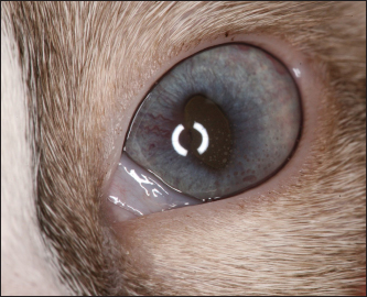
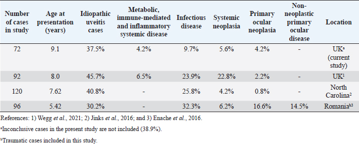
AbstractBackground: Uveitis is a common ophthalmic diagnosis in cats, that can lead to discomfort and loss of vision. Identification of nonidiopathic cases facilitates treatment and could reduce morbidity associated with this condition. Aim: To evaluate etiologies of nontraumatic uveitis in the UK, to compare diagnostic features between idiopathic cases and those with an established underlying etiology, and to investigate the association of clinical signs and abnormal diagnostic findings with a confirmed etiology. Methods: Records of cats diagnosed with uveitis at a UK referral center between August 2009 and April 2018 were retrospectively reviewed, excluding traumatic (and reflex) cases. Cases were categorized based on whether an underlying etiology had been established in cases with confirmed etiology, idiopathic, and inconclusive cases. All cases had a minimum of 12-month follow-up unless an underlying etiology had been established. Population characteristics, clinical signs, diagnostic investigation features, and results were reported. Results: 72 cases of uveitis were included, of which male cats and domestic breeds were overrepresented. An underlying etiology was determined in 23.6% of cases: 9.7% had infectious diseases, 5.6% had systemic neoplasia, 4.2% had primary ocular neoplasia, and 4.2% had metabolic disease. Idiopathic uveitis comprised 37.5% of cases, and the remaining 38.9% were inconclusive, of which 35.7% died or were euthanased within the follow-up period. Among the study population, no significant age difference was found between cats with idiopathic disease or confirmed etiology. The unilateral disease was reported in 56.9% of cases and was not different across the idiopathic cases and confirmed etiology groups. The most common ophthalmic clinical sign was an aqueous flare, followed by keratic precipitates and hypotony. Iris color change (p =0.015) and the presence of an intraocular mass (p =0.025) were associated with an underlying etiology. Conclusion: Idiopathic uveitis was found to be the most common diagnosis in this study population. However, a similar proportion of cases had possible underlying etiologies as a high proportion manifested systemic disease within the follow-up time. An underlying etiology could be established only in a quarter of cases. Further studies are required to standardize the investigations required when assessing cats with uveitis to minimize patient morbidity. Keywords: Cat, Eyes, Hypertension, Aqueous flare, Inflammation. IntroductionUveitis is defined as inflammation of the vascular tunic of the eye. A common ophthalmic diagnosis in cats, uveitis may cause both loss of vision and significant discomfort which may necessitate enucleation. Reported etiologies of uveitis in cats include infectious agents (bacterial, viral, protozoal, fungal, and parasitic), immune-mediated (lens-induced uveitis and periarteritis nodosa), neoplastic (lymphosarcoma, melanoma, other primary intraocular tumors, and other metastatic tumors), metabolic (systemic hypertension and hyperlipidemia), traumatic, toxic, reflex, or idiopathic (Miller, 2018). A significant proportion of feline uveitis cases do not have a known underlying cause detected despite an exhaustive internal medicine investigation; these cases are termed idiopathic. However, immune-mediated or idiopathic uveitis may be caused by organisms not yet recognized as pathogenic (Hendrix, 2021). Studies vary concerning the proportion of idiopathic cases, with 40.8% (Jinks et al., 2016), 45.7% (Wegg et al., 2021), and 30.2% (Enache et al., 2016) previously reported (Supplementary Table 1). The ocular signs are similar despite differing underlying etiologies, although unilateral uveitis has been suggested to be more likely with exogenous or idiopathic causes, and bilateral with underlying systemic disease (Gemensky et al., 1996). Clinical signs of anterior uveitis include aqueous flare, hypotony, conjunctival and episcleral hyperemia, ciliary flush, corneal edema, keratic precipitates, corneal neovascularization, iritis, iridal nodules, rubeosis iridis, miosis, synechiae, lacrimation, blepharospasm, and nictitating membrane protrusion (Glaze et al., 2021). Keratic precipitates, cataracts, synechiae, lens luxation, glaucoma, retinal detachment, and phthisis bulbi indicate chronicity (Stiles, 2013). Clinical signs are often subtler than in dogs (Fig. 1), resulting in cats presenting in the chronic stages (Glaze et al., 2021). Chronic uveitis is also a common cause of glaucoma in cats (McLellan and Teixiera, 2015). Identification of nonidiopathic cases facilitates early, specific treatment and could reduce associated morbidity. While an exhaustive diagnostic work-up is required to exclude nonidiopathic causes this is not always possible in clinical practice due to financial and other constraints which may contribute to the undertreatment of potential reversible disease. This study aimed to describe cases of feline uveitis focusing on nontraumatic and nonreflex causes. The objectives were to: 1) establish etiology based on recorded clinical findings, diagnostic investigation, and follow-up information and 2) compare cases of idiopathic uveitis to cases with a confirmed etiology (signalment and ophthalmic examination findings). We hypothesized that performing diagnostic testing would increase the likelihood of finding an underlying disease and used a 12-month follow-up period to allow underlying diseases to become apparent.
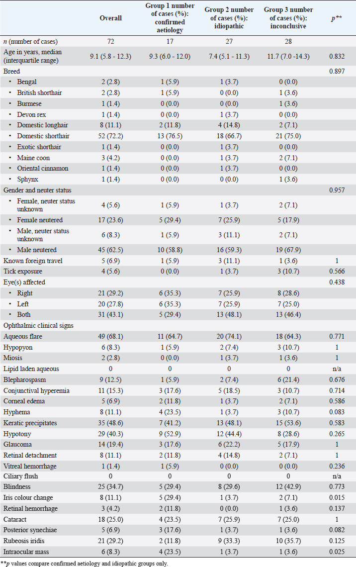
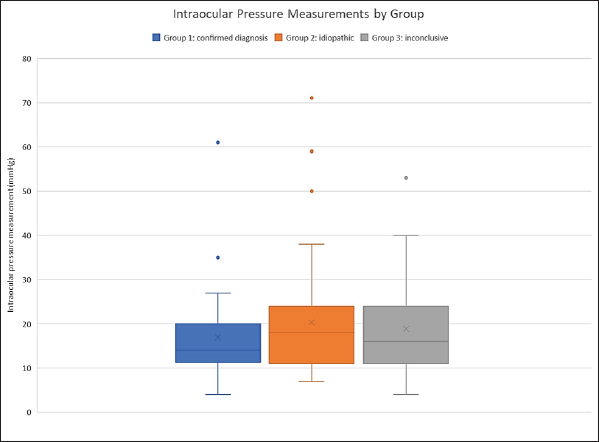
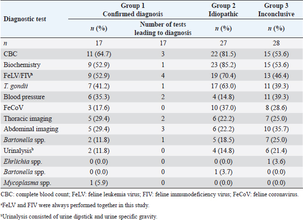
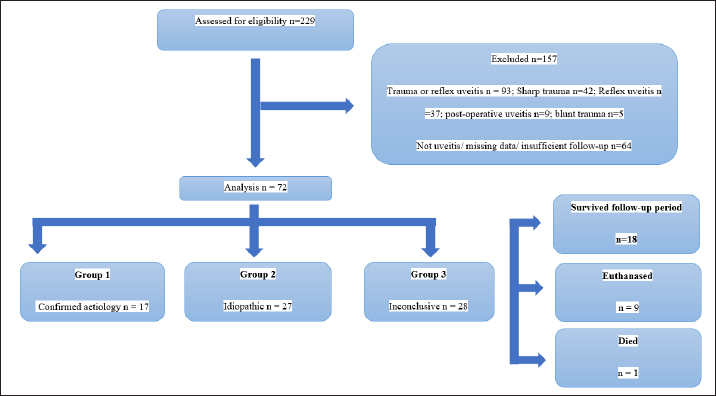
Fig. 1. A 4-month-old female entire Snowshoe kitten with bilateral uveitis. Clinical signs included corneal edema, keratic precipitates, aqueous flare, rubeosis iridis and miosis. The aetiology was FIP Materials and MethodsMedical records of feline cases presenting to a referral center in the south of the UK for nontraumatic uveitis between August 26, 2009 and April 21, 2018, were retrospectively reviewed, using the keyword “uveitis.” The inclusion criteria was a confirmed diagnosis of uveitis with a 12-month minimum follow-up period unless an underlying etiology had been established. Cases lost to follow-up or with inadequate follow-up were excluded. If an underlying etiology could not be determined, collaboration and further review of the patients’ clinicopathological data were performed by an internal medicine specialist. Cases with a confirmed traumatic cause or reflex uveitis were excluded. Data collected for each case included signalment, affected eye/s, known travel history from outside of the UK, ophthalmic examination findings, and clinicopathologic abnormalities [laboratory investigations such as hematology, biochemistry, infectious disease titers (Encephalitozoon cuniculi serology: IgM and IgG, Ehrlichia serology, Toxoplasma gondii serology: IgM and IgG, feline leukemia virus (FeLV) antigen, feline immunodeficiency virus (FIV) antibody, feline coronavirus serology, Bartonella serology, Mycoplasma PCR) urinalysis, diagnostic imaging, and cytology and/or histopathologic results]. In all cases, a full ophthalmic examination was performed by, or under the supervision of an ophthalmology diplomate (European College of Veterinary Ophthalmologists (ECVO) or Royal College of Veterinary Surgeons (RCVS)). This included evaluation of palpebral reflex, menace response, dazzle, and pupillary light reflexes, Schirmer tear testing (Schirmer Tear test strips, Intervet Inc. Merck Animal Health), slit lamp examination (SL 15, SL 17; Kowa, Tokyo, Japan), applanation or rebound tonometry (TonoPen Avia Vet; Reichert Inc.; TonoVet, Icare tonometer; and Revenio Group Corporation), direct and indirect ophthalmoscopy (Keeler Professional direct ophthalmoscope; Keeler Ltd., Keeler Vantage Plus), following pupil dilation (Tropicamide eye drops, Minims, Bausch & Lomb) and fluorescein staining (Fluorescein sodium; Minims, Bausch & Lomb). Pupil dilation was not performed in cases of secondary glaucoma or lens luxation. An intraocular pressure measurement of <15 mmHg in the presence of clinical signs (Miller et al., 1991), or ≥5 mmHg difference from the contralateral eye was recorded as hypotony. The internal medicine investigation consisted of a full clinical examination performed by a specialist in internal medicine (RCVS), and further testing including some or all of the following: hematology, biochemistry, infectious disease titers (as detailed above), urinalysis, diagnostic imaging and sampling of any abnormal lesions. Clinicopathologic data was reviewed from the primary care facility and the referral center (including external results from diagnostic laboratories). Imaging was performed primarily at the referral center where modalities included computed tomography (Aquilion 128 Slice CT, Toshiba Medical Systems), ultrasound (Vivid I, GE Healthcare Solutions; CX-50, Philips Healthcare), and thoracic radiography (HF200A+, Veterinary X-rays). Abdominal ultrasonography was performed by RCVS-recognized specialists in small animal internal medicine with extensive ultrasonography experience. These investigations varied depending on patient history and clinical examination findings, response to therapy, clinician and client preference, and financial constraints. Additional follow-up information was extracted from the referring vets, or by directly contacting the owners to determine how the animal had responded to treatment. This is standard practice for ophthalmology cases at the hospital and does not reflect intervention beyond clinical need. After recording data, cases were categorized into groups as follows: 1) Established underlying etiology based on clinical and diagnostic investigation findings, and/or case follow-up data (nonidiopathic disease process). 2) Idiopathic uveitis (where diagnostic tests performed and a minimum 12-month follow-up period failed to identify an underlying cause). 3) Inconclusive (a clear cause of uveitis was not identified from available data, but also could not be reasonably excluded due to co-morbidities, or the patient died or was euthanized within 12 months, without an established underlying etiology). Cases were not excluded from the study if a complete work-up was not available unless follow-up information was also not available. Diagnostic tests were classified as normal or abnormal based on reference ranges of the in-house machine or laboratory methodology used in each case. Signalment, ocular findings, and diagnostic workup were reported in groups using descriptive statistics. Groups were compared using Fisher’s exact test for categorical variables (due to small numbers in some categories) and analysis of variance for continuous variables. Univariate logistic regression models were constructed to explore the association between abnormal diagnostic test results and underlying etiology. Analysis was performed in Microsoft Excel and R 4.1.1. Significance was assigned where p < 0.05. Ethical approvalNot needed as this is a retrospective study. ResultsDemographic data and clinical characteristicsA database search using the keyword “uveitis” retrieved 229 cases. Ninety-three cases were excluded due to a traumatic or reflex etiology and 64 cases due to nonconfirmed uveitis or missing data. The study population, therefore, comprised 72 cases (Supplementary Fig. 1). Baseline data are displayed in Table 1. No significant difference was found when comparing the average age of cats in the idiopathic group to those with confirmed etiology (7.4 years and 9.3 years, respectively; p =0.83). Ten breeds were represented, with domestic shorthair being the most common across all three diagnostic groups. No significant differences in breeds were seen between the idiopathic and confirmed etiology groups. A high proportion of cats were male neutered (62.5%), with no significant differences in gender between idiopathic and confirmed etiology groups. Forty-one cases were unilaterally affected (56.9%) with the right eye in 21 cases (29.2%), and left in 20 cases (27.8%), whereas 31 cases (43.1%) had a bilateral presentation, with no significant difference between idiopathic and confirmed etiology groups. Of ophthalmic signs, significant differences were found only for iris color change, and for the presence of an intraocular mass, which were more common (p=0.015 and p=0.025, respectively) in the confirmed etiology group. The most common ophthalmic clinical signs were aqueous flare (68.1%), followed by keratic precipitates (48.6%) and hypotony (40.3%). Median intraocular pressure was 13 mmHg, with a range of 3–71 mmHg, with no significant differences between groups (p =0.57; Fig. 2). Diagnostic tests performed are presented in Table 2. A detailed diagnostic workup consisting of complete blood count (CBC) and biochemistry, urinalysis including culture, and imaging of the thorax and abdomen was performed in 7/72 cats (9.7%). A less complete diagnostic work up with either one or two of the above, but not all three, was performed in 52 cats (72.2%). From these, 3/52 lacked only complete urinalysis with culture, while 1/52 lacked only thoracic imaging. Three cats (3/72) had minimal work up consisting of infectious disease testing, while 10/72 had no additional diagnostic tests performed. Some infectious disease testing was performed in 47/72 cases (65.2%). A detailed diagnostic work up was performed in 3/28 inconclusive cases, a less complete work up in 16/28, and no work up in 9/28 cases. Of hematological abnormalities, anemia (defined as PCV/HCT of less than 35%) was detected in 5/72 cases (6.9%). Leukocytosis was present in 3/72 cases (4.2%), and leukopenia in 2/72 (2.7%). Azotemia was detected in 2/72 cases (2.7%). Hypoalbuminemia was detected in 2/72 cases (2.7%), and hyperglobulinemia in 3/72 cases (4.2%). Imaging of the abdomen was performed in 21 cases, of which 10 cases had abnormal findings. Four cases of those with abnormal findings were assigned an underlying etiology. One case without abnormal imaging findings was also found to have an underlying etiology. Six (6/21 cases) were idiopathic (with no significant imaging findings) and ten cases were classified as inconclusive (6 of these had significant findings, but an underlying cause for the could not be established). Thirteen cases underwent both abdominal and thoracic imaging, and a further five cases underwent thoracic imaging alone. Only three cases had significant thoracic findings, with two assigned an underlying etiology, and one was categorized as inconclusive. Of all cases undergoing thoracic imaging (18/72), an underlying etiology was confirmed in one case, five were categorized as idiopathic, and six were categorized as inconclusive. Of all the cases that underwent abdominal or thoracic imaging, 8/26 were found to have an underlying etiology, compared to 2 of the 13 which had both abdominal and thoracic imaging. Table 1. Patient characteristics and ocular signs grouped by cause of uveitis.
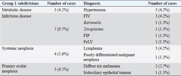
Fig. 2. Box and whisker chart showing intraocular pressure measurements by group DiagnosesAn underlying etiology was achieved in 17 of the 72 cats (23.6%). A complete list of etiological diagnoses is shown in Table 3. Systemic neoplasia was diagnosed in four cases (three lymphomas, and one poorly differentiated malignant neoplasm), primary ocular neoplasia in three (diffuse iris melanoma in two, iridociliary epithelial tumor in one), systemic infectious disease in seven [FIV in three; bartonellosis, toxoplasmosis, feline infectious peritonitis (FIP), and FeLV in one case each], and three cases with metabolic disease resulting in systemic hypertension. Two cases in this study were assigned an underlying nonidiopathic etiology based on follow-up data (both of these cases had lymphoma). A total of 27 cats (37.5%) were diagnosed with idiopathic uveitis. Table 2. Diagnostic tests performed by group.
Table 3. Individual diagnoses in group 1 where aetiology was established.
An inconclusive etiology was assigned to the remaining 28 (38.9%; Table 1), with insufficient evidence to establish causation or exclude underlying disease. Of these, nine were euthanased and a further one cat died without an established etiology for their uveitis (36%) during the study period. Eight of the 10 cats with inconclusive uveitis did not have a cause of death identified or recorded. Contributing factors included neurological signs, abdominal effusion, severe panuveitis, lymphadenopathy, inflammatory cerebrospinal fluid (CSF) changes, and gastrointestinal disease. A further eight cases had metabolic, immune-mediated, or inflammatory systemic disease (e.g., pancreatitis and biliary system disease), which were suspected to be related to the uveitis; however, it was not possible to establish causation. However, in one case of unilateral uveitis, a road traffic accident was the cause of death (censored from comparative analysis). Ocular histopathology was available for 14 cases in this study. A histopathologic diagnosis of lymphoplasmacytic uveitis was found in nine cases; diffuse iris melanoma in two, and one each of iridociliary epithelial tumor, suppurative endophthalmitis, and a poorly differentiated malignant neoplasm. Univariate logistic regression was used to test the association of a diagnostic test abnormality to a confirmed etiology for uveitis. This largely failed to identify tests (hematology, serum biochemistry, thoracic imaging, abdominal imaging, and infectious disease testing) as statistically significant, other than systemic hypertension (p =0.042), with an odds ratio of 5.33, but a confidence interval of 1.06–30.02. DiscussionThis study described cases of nontraumatic and nonreflex feline uveitis presented to a referral center in the UK. Etiology was confirmed in 23.6% of cases whereas 37.5% were considered idiopathic and 38.9% inconclusive (censored from comparative analysis). A minimum 12-month follow-up period was chosen to reduce the bias of an incomplete diagnostic work-up (unless prior aetiologic diagnosis had been achieved), as we would expect the majority of systemic diseases presenting with uveitis to progress. Examples include the 8–9 day survival time of untreated FIP (Ritz et al., 2007; Fischer et al., 2011), 69 days survival for systemic lymphoma with intraocular involvement (Musciano et al., 2020) but over 6 months for ocular coccidiodomycosis (Tofflemire and Betbeze, 2010). The follow-up period can also detect systemic disease, such as lymphoma, that may be occult at the time of presentation despite full clinical staging (Musciano et al., 2020). Some cases may have been misclassified as idiopathic due to incomplete diagnostic work-up (despite follow-up) with no additional clinical signs to indicate underlying disease. The immunodeficiency stage of FIV infection may present with subtle clinical signs, without significant progression for years (Walker et al., 1994; Nelson and Couto, 2020), with one study finding no significant effect on lifespan (Spada et al., 2018). In addition to the immunodeficiency stage, uveitis can also occur in the acute stage (Orandle and Baldwin, 1995). All three FIV-positive cats in the current study survived for at least 18 months after their diagnosis. On the other hand, some cases may have been excluded from the idiopathic group due to the development of unrelated systemic diseases during the follow-up period. The majority of cats in this study were male; in agreement with other recent studies (Enache et al., 2016; Jinks et al., 2016; Wegg et al., 2021). The median age was similar to two other retrospective studies (Jinks et al., 2016; Wegg et al., 2021), but older than a Romanian study (Enache et al., 2016), with higher infectious disease prevalence in the latter study. The proportion of domestic shorthair/longhair compared to purebred cats was similar in our study and other retrospective studies (85.8%; Jinks et al., 2016; 76%; Enache et al., 2016; and 79.6%; Wegg et al., 2021). This may relate to relative proportions within the feline population, and increased outdoor access resulting in increased risk of infectious diseases. There was a similar proportion of unilaterally and bilaterally affected cats with idiopathic uveitis (48.1% affected bilaterally), similar to a previous study by Gemensky et al. (1996; 48%). A wide range of intraocular pressures was observed in cases (Fig. 2). An explanation for this would be that a proportion of cats presented later in the course of the disease with acute or chronic secondary glaucoma present or developing. The presence of an intraocular mass was significantly associated with an underlying etiology; commonly associated in the UK with primary or secondary neoplasia. Iris color change was also associated with the underlying disease with 25% (2/8) of these cats diagnosed with diffuse iris melanoma; however, iris color change may occur due to different disease processes leading to secondary iris inflammation, and therefore, it is considered nonspecific (Maggs, 2009). An underlying etiology was detected in approximately a quarter of cases which is lower compared than in other retrospective studies (Enache et al., 2016; Jinks et al., 2016; Wegg et al., 2021; Supplementary Table 1). This was likely due to some of the inconclusive cases having an unidentified underlying cause; reflected in those that did not survive within the follow-up period. More standardized thorough investigations may have resulted in a higher number of cases being assigned an underlying etiology. In some cases, a diagnosis of a concurrent systemic disease had been made (e.g., pancreatitis, cholangitis); however, causation could neither be established nor excluded. Compared to other retrospective studies (Enache et al., 2016; Jinks et al., 2016; Wegg et al., 2021), infectious disease cases were considerably lower. This could be due to variations in diagnostic tests and interpretation of their results, different patient populations, and geographical variation [e.g., leishmaniosis (Pimenta et al., 2015), not endemic in the UK (Pimenta et al., 2015)]. FIP cases were notably lower than in other similar retrospective studies which reported 15.8% (Jinks et al., 2016), 16.3% (Wegg et al., 2021), and 2.0% (Enache et al., 2016). This is likely due to the multisystemic involvement of the disease meaning the cases may have presented to other services compared to an ophthalmology referral centre. Bartonellosis was diagnosed in one cat. Although of zoonotic importance, cats rarely exhibit clinical signs, although chronic relapsing bacteremia is possible (Álvarez-Fernández et al., 2018). Systemic lymphoma was diagnosed in two cases in this study during the follow-up period, comprising 11.8% of cases with a confirmed underlying etiology. Both of these cases received chemotherapy; however, remission was not achieved, and the owners elected euthanasia due to quality of life concerns. Lymphoma is the most common metastatic neoplastic cause of uveitis in cats, although only a small proportion have ocular involvement and true solitary ocular lymphoma is rare (Musciano et al., 2020). Many primary intraocular tumors are benign; however, secondary glaucoma, uveitis, and hemorrhage may develop causing can cause both pain and blindness (Grahn and Peiffer, 2021). Cases are commonly defined as idiopathic as a diagnosis of exclusion if an underlying cause cannot be established (Bergstrom et al., 2017). To avoid over-classifying cases as idiopathic we had 1) an inconclusive category where causation could not be established or excluded and 2) a minimum 12-month follow-up if an underlying etiology had not been previously established. In this study, 37.5% of cases were classified as idiopathic; comparable with previous studies (30.2%; Enache et al., 2016; 40.8%; Jinks et al., 2016; and 45.7%; Wegg et al., 2021). Lymphoplasmacytic uveitis is a nonspecific histologic diagnosis that may be uni or bilateral (Grahn and Peiffer, 2013). It is associated with FIV, feline herpesvirus 1 (FHV-1), FeLV, toxoplasmosis, and bartonellosis, but many cases remain idiopathic, presumed to be immune-mediated uveitis. With an ever-expanding understanding, a higher proportion of uveitis cases are likely to be assigned an underlying etiology. Other examples include eosinophilic uveitis identified in an 8-year-old cat (Newbold and Premanandan, 2022), and a case series describing intraocular Cytauxzoon felis as a causative agent (Meekins and Cino-Ozuna, 2018). Univariate logistic regression modeling largely failed to identify an association between abnormal diagnostic test results and confirmed etiology of uveitis. This approach was chosen to try to identify the most useful diagnostic tests in cases of feline uveitis. Significance was identified for systemic hypertension; however, the relatively wide confidence interval was likely to be a reflection of an imprecise odds ratio. These findings may have resulted from a small proportion of patients having all diagnostic tests and selection bias. The main limitation of this study was the lack of standardization of diagnostic testing, with a complete diagnostic work-up performed in relatively few cases (7/72), meaning that underlying disease will likely not have been identified in a significant proportion. Thorough diagnostic investigation was offered to all owners as per the gold standard assessment; however, this was often declined, e.g., due to financial limitations. To mitigate this bias, if an underlying etiology could not be confirmed or excluded from available data, cases were classified as inconclusive (rather than idiopathic) and censored from the comparative statistical analysis. Some cases may have been more likely to have further investigations due to additional clinical signs. An inconsistency in ophthalmic examination was found with the tonometry equipment over a relatively long period, leading to additional bias. In conclusion, excluding traumatic and reflex uveitis, idiopathic uveitis was the most common diagnosis in 37.5% (27/72) cats. An underlying disease process to account for uveitis was identified in 23.6% (17/72); the most commonly infectious disease, followed by systemic neoplasia, metabolic disease, and primary ocular neoplasia. The remainder of cases [38.9% (28/72)] were categorized as inconclusive where an underlying etiology could not be excluded or confirmed; some of these cases would have benefited from diagnostic investigations; however, this was not possible due to owner considerations. Although an underlying etiology was suspected in many of these cases due to additional clinical signs, this study design helped to avoid cases being misclassified. A minimum 12-month follow-up was incorporated to reduce the bias of an often incomplete work-up. Further studies with larger datasets are required to standardize the investigations performed when assessing cats with uveitis, which would allow tailored treatment and prognostic information to reduce patient morbidity. AcknowledgmentThe authors thank Tom Salih for the critical review of this manuscript. Conflict of interestThe authors declare that there is no conflict of interest. FundingThe authors received no financial support for the research, authorship, and publication of this article. Author contributionsAmna Salih: Data collection and writing of the manuscript. Stamatina Giannikaki: Study design and manuscript review. Natalia Escanilla: Assistance with manuscript writing. Christopher Ioannides-Hoey: Assistance with manuscript writing. Matthew Best: Study design and statistical design. Data availabilityData supporting the findings in this study are available within the manuscript. ReferencesÁlvarez-Fernández, A., Breitschwerdt, E.B. and Solano-Gallego, L. 2018. Bartonella infections in cats and dogs including zoonotic aspects. Parasites Vectors 11, 1–21. Bergstrom, B.E., Stiles, J. and Townsend, W.M. 2017. Canine panuveitis: a retrospective evaluation of 55 cases (2000–2015). Vet. Ophthalmol. 20, 390–397. Enache, A., Ionaşcu, I., Şonea, A. and Cucoş, A. 2016. Causes of feline uveitis: a retrospective study of 96 cases at the Faculty of Veterinary Medicine Bucharest, 2012-2015. Agric. Agric. Sci. Proc. 10, 396–402. Fischer, Y., Ritz, S., Weber, K., Sauter-Louis, C. and Hartmann, K. 2011. Randomized, placebo controlled study of the effect of propen-tofylline on survival time and quality of life of cats with feline infectious peritonitis. J. Vet. Intern. Med. 25, 1270–1276. Gemensky, A., Lorimer, D. and Blanchard, G. Feline uveitis: a retrospective study of 45 cases. In Annual Meeting of the American College of Veterinary Ophthalmologists, Maui, Hawaii, 1996, p: 19. Glaze, M.B., Maggs, D.L. and Plummer, C.E. 2021. Feline ophthalmology. In Veterinary ophthalmology, 6th edn. Eds., Gelatt, K., Ben-Shlomo, G., Gilger, B.C., Hendrix, D.V.H., Kern, T.J. and Plummer, C.E. Ames, IA: John Wiley & Sons, pp: 1738–1739. Grahn, B.H. and Peiffer, R.L. 2021. Veterinary ophthalmic pathology. In Veterinary ophthalmology, 6th edn. Eds., Gelatt, K., Ben-Shlomo, G., Gilger, B.C., Hendrix, D.V.H., Kern, T.J. and Plummer, C.E. Ames, IA: John Wiley & Sons, pp: 542–547. Grahn, B.H. and Peiffer, R.L. 2013. Veterinary ophthalmic pathology. In Veterinary ophthalmology, 5th edn. Eds., Gelatt, K., Gilger, B. and Kern, T.J. Ames, IA: John Wiley & Sons, p: 477. Hendrix, D.V.H. 2021, Diseases and surgery of the canine anterior uvea. In Veterinary ophthalmology, 6th edn. Eds., Gelatt, K., Ben-Shlomo, G., Gilger, B.C., Hendrix, D.V.H., Kern, T.J. and Plummer, C.E. Ames, IA: John Wiley & Sons, p: 1266. Jinks, M.R., English, R.V. and Gilger, B.C. 2016. Causes of endogenous uveitis in cats presented to referral clinics in North Carolina. Vet. Ophthalmol. 19, 30–37. Maggs, D. 2009. Feline uveitis: an intraocular lymphadenopathy. J. Feline Med. Surg. 11, 167–182. McLellan, G.J. and Teixeira, L.B.C. 2015. Feline glaucoma. Vet. Clin. North Am. Small Anim. Pract. 45, 1307–1333. Meekins, J. and Cino-Ozuna, A.G. 2018. Histologic identification of intraocular Cytauxzoon felis in three cats. JFMS Open Rep. 4, 1–5. Miller, P.E. 2018. Diseases of the uvea. In: Slatter’s fundamentals of veterinary ophthalmology, 6th edn. Eds., Maggs, D.J., Miller, P.E. and Ofri, R. St. Louis, MO: Elsevier, p: 266. Miller, P.E., Pickett, J.P., Majors, L.J. and Kurzman, I.D. 1991. Evaluation of two applanation tonometers in cats. Am. J. Vet. Res. 52, 1917–1921. Musciano, A.R., Lanza, MR, Dubielzig, R.R., Teixeira, L.B.C. and Durham, A.C. 2020. Clinical histopathological classification of feline intraocular lymphoma. Vet. Ophthalmol. 23, 77–89. Newbold, G.M. and Premanandan, C. 2022. An unusual case of eosinophilic uveitis in a cat. Vet. Ophthalmol. 25, 73–77. Nelson, R.W. and Couto, C.G. 2020. Polysystemic viral diseases. In: Small animal internal medicine, 6th edn. St. Louis, MO: Elsevier, p: 1492. Orandle, M.S. and Baldwin, C.J. 1995. Clinical and pathological features associated with feline immunodeficiency virus infection in cats. Ames, IA: Iowa State University Digital Repository, vol. 57, pp: 61–65. Pimenta, P., Alves-Pimenta, S., Barros, J., Barbosa, P., Rodrigues, A., Pereira, M.J., Maltez, L., Gama, A., Cristovao, J.M., Campino, L., Maia, C. and Cardoso, L. 2015. Feline leishmaniosis in Portugal: 3 cases (year 2014). Vet. Parasitol. Reg. Stud. Rep. 1–2, 65–69. Ritz, S., Egberink, H. and Hartmann, K. 2007. Effect of feline interferon-omega on the survival time and quality of life of cats with feline infectious peritonitis. J. Vet. Intern. Med. 21, 1193–1197. Spada, E., Perego, R., Sgamma, E.A. and Proverbio, D. 2018. Survival time and effect of selected predictor variables on survival in owned pet cats seropositive for feline immunodeficiency and leukemia virus attending a referral clinic in northern Italy. Prev. Vet. Med. 1, 38–46. Stiles, J. 2013. Feline ophthalmology. In: Veterinary ophthalmology, 5th edn. Eds., Gelatt, K., Gilger, B. and Kern, T.J. Ames, IA: John Wiley &Sons, p: 1504. Tofflemire, K. and Betbeze, C. 2010. Three cases of feline ocular coccidioidomycosis: presentation, clinical features, diagnosis, and treatment. Vet. Ophthalmol. 13, 166–172. Walker, C., Canfield, P.J. and Love, D.N. 1994. Analysis of leucocytes and lymphocyte subsets for different clinical stages of naturally acquired feline immunodeficiency virus infection. Vet. Immunol. Immunopathol. 44, 1–12. Wegg, M.L., Jeanes, E.C., Pollard, D., Fleming, L. and Dawson, C. 2021. A multicenter retrospective study into endogenous causes of uveitis in cats in the United Kingdom: ninety two cases. Vet. Ophthalmol. 24, 591–598. Supplementary MaterialsSupplementary Table 1. Comparison of results with other retrospective feline uveitis studies.
Supplementary Fig. 1. CONSORT chart | ||
| How to Cite this Article |
| Pubmed Style Salih A, Giannikaki S, Escanilla N, Ioannides-Hoey CS, Best M. Etiologies of nontraumatic feline uveitis in the UK: A retrospective observational study of 72 cats. Open Vet J. 2023; 13(9): 1195-1204. doi:10.5455/OVJ.2023.v13.i9.15 Web Style Salih A, Giannikaki S, Escanilla N, Ioannides-Hoey CS, Best M. Etiologies of nontraumatic feline uveitis in the UK: A retrospective observational study of 72 cats. https://www.openveterinaryjournal.com/?mno=147654 [Access: May 19, 2024]. doi:10.5455/OVJ.2023.v13.i9.15 AMA (American Medical Association) Style Salih A, Giannikaki S, Escanilla N, Ioannides-Hoey CS, Best M. Etiologies of nontraumatic feline uveitis in the UK: A retrospective observational study of 72 cats. Open Vet J. 2023; 13(9): 1195-1204. doi:10.5455/OVJ.2023.v13.i9.15 Vancouver/ICMJE Style Salih A, Giannikaki S, Escanilla N, Ioannides-Hoey CS, Best M. Etiologies of nontraumatic feline uveitis in the UK: A retrospective observational study of 72 cats. Open Vet J. (2023), [cited May 19, 2024]; 13(9): 1195-1204. doi:10.5455/OVJ.2023.v13.i9.15 Harvard Style Salih, A., Giannikaki, . S., Escanilla, . N., Ioannides-Hoey, . C. S. & Best, . M. (2023) Etiologies of nontraumatic feline uveitis in the UK: A retrospective observational study of 72 cats. Open Vet J, 13 (9), 1195-1204. doi:10.5455/OVJ.2023.v13.i9.15 Turabian Style Salih, Amna, Stamatina Giannikaki, Natalia Escanilla, Christopher S.F.K. Ioannides-Hoey, and Matthew Best. 2023. Etiologies of nontraumatic feline uveitis in the UK: A retrospective observational study of 72 cats. Open Veterinary Journal, 13 (9), 1195-1204. doi:10.5455/OVJ.2023.v13.i9.15 Chicago Style Salih, Amna, Stamatina Giannikaki, Natalia Escanilla, Christopher S.F.K. Ioannides-Hoey, and Matthew Best. "Etiologies of nontraumatic feline uveitis in the UK: A retrospective observational study of 72 cats." Open Veterinary Journal 13 (2023), 1195-1204. doi:10.5455/OVJ.2023.v13.i9.15 MLA (The Modern Language Association) Style Salih, Amna, Stamatina Giannikaki, Natalia Escanilla, Christopher S.F.K. Ioannides-Hoey, and Matthew Best. "Etiologies of nontraumatic feline uveitis in the UK: A retrospective observational study of 72 cats." Open Veterinary Journal 13.9 (2023), 1195-1204. Print. doi:10.5455/OVJ.2023.v13.i9.15 APA (American Psychological Association) Style Salih, A., Giannikaki, . S., Escanilla, . N., Ioannides-Hoey, . C. S. & Best, . M. (2023) Etiologies of nontraumatic feline uveitis in the UK: A retrospective observational study of 72 cats. Open Veterinary Journal, 13 (9), 1195-1204. doi:10.5455/OVJ.2023.v13.i9.15 |

