


| Research Article | ||
Open Vet. J.. 2024; 14(5): 1130-1134 Open Veterinary Journal, (2024), Vol. 14(5): 1130–1134 Research Article Saline volume required to achieve peristaltic intraluminal pressure during leak testing of canine colotomies, using two methods of luminal occlusionEleni Prastiti1, Ioannis Savvas2, Vasileia Angelou2 and Lysimachos G. Papazoglou2*1Limassol Veterinary Clinic, Limassol, Cyprus 2Companion Animal Clinic, School of Veterinary Medicine, Aristotle University of Thessaloniki, Thessaloniki, Greece *Corresponding Author: Lysimachos Papazoglou. Companion Animal Clinic, School of Veterinary Medicine, Aristotle University of Thessaloniki, Thessaloniki, Greece. Email: makdvm [at] vet.auth.gr Submitted: 22/01/2024 Accepted: 22/04/2024 Published: 31/05/2024 © 2024 Open Veterinary Journal
AbstractBackground: No studies have appeared in the literature evaluating the intraluminal volume of injected saline in the canine colon for performing leak tests in colotomy incisions. Aim: To determine the volume of the injected intraluminal saline necessary to achieve an intraluminal pressure of 17.3 cm H2O in 10 cm colonic segments containing a closed colotomy occluded with intestinal forceps or by digital pressure. Methods: Fresh colon was obtained from 8 canine cadavers and divided into 10 cm segments. A 3 cm antimesenteric colonic incision was performed at each intestinal segment which was closed using a 3–0 polydioxanone suture in a simple continuous pattern. Each colonic construct was occluded with Doyen intestinal forceps or by digital pressure and a leak test was performed by saline infusion. The saline volume needed to achieve a predetermined intraluminal pressure of 17.3 cm H2O, following occlusion was recorded. Results: The mean volume of injected saline with the Doyen intestinal forceps occlusion (20.4 ± 8.2 ml) was significantly larger than that of the digital occlusion technique (17.5 ± 6.8 ml) [p=0.021]. Conclusion: For 10 cm canine colonic constructs containing a closed colotomy, saline volumes of 20.4 ml with Doyen occlusion and 17.5 ml with digital occlusion can be utilized to achieve intraluminal pressures of 17.3 cm H2O. Keywords: Leak test, Colon, Colotomy, Dog. IntroductionColotomy is used in dogs for the collection of full-thickness samples, removal of impacted feces, foreign bodies, or small masses, and for addressing colonic perforations (Fossum and Hedlund, 2003; Williams, 2018; Smeak, 2020). Intestinal dehiscence is the most serious complication of colorectal surgery in small animals (Wylie and Hosgood, 1994; Latimer et al., 2019). Postoperative intestinal dehiscence rates are reported to be between 7% and 14% in dogs and morbidity rates associated with dehiscence are reported to be between 55% and 85% in dogs and cats (Allen et al., 1992; Wylie and Hosgood, 1994; Ralphs et al., 2003; Grimes et al., 2011; Duell et al., 2016; Latimer et al., 2019). Intraoperative performance of leak test is recommended to assess the security of the enteric suture line and decrease the risk of postoperative leakage by detection and management of errors in closure technique and suture line leaks (Saile et al., 2010; Huss, 2014; Matz et al., 2014; Giuffrida and Cimino Brown, 2018). Studies reviewing the importance of leak testing in colorectal surgeries in humans emphasized that intraoperative leak testing is a good method for the prevention of anastomotic leakage and reduces the risk of postoperative clinical and radiological leaks (Gilbert and Trapnell, 1988; Beard et al., 1990; Griffith and Hardcastle, 1990; Dixon and Holmes, 1991; Wheeler and Gilbert, 1999; Smith et al., 2007; Ivanov et al., 2011). Ideally, the leak test would achieve but not exceed normal peristaltic intraluminal pressures, but in fact, intraluminal pressure is not routinely measured in practice (Giuffrida and Cimino Brown, 2018). The normal intraluminal peristaltic pressure of the canine colon has been documented (Hofmann et al., 1993). The volume of intraluminal injected saline needed to achieve normal peristaltic pressure in the small intestine has been reported in two studies in dogs (Saile et al., 2010; Matz et al., 2014). To the best of authors’ knowledge, no studies have appeared in the literature evaluating the intraluminal volume of injected saline in the canine colon for performing leak tests in colotomy incisions. The study presented here aimed to determine the volume of the injected intraluminal saline necessary to achieve an intraluminal pressure of 17.3 cm H2O in 10 cm colonic segments containing a closed colotomy occluded with intestinal forceps or by digital pressure. We hypothesized that intraluminal pressure of 17.3 cm H2O could be achieved with both methods of occlusion in the 10 cm segments of the canine colon containing a closed colotomy site. Materials and MethodsHealthy colon was harvested from eight client-owned dogs, that were euthanized for reasons other than digestive tract pathology using an IV infusion of propofol and 10% potassium chloride. Immediately after euthanasia, a midline celiotomy was performed and the colon was harvested. Intestinal contents were gently removed by milking them away and the remaining intestinal contents were flushed with saline. The colon was divided into 10 cm segments and 26 colonic segments were obtained from the 8 cadavers in total. Following colonic preparation, colonic segments were stored in a container filled with saline at room temperature (18°C–23°C) until their use within 2 hours following harvesting. In each segment, an antimesenteric 3 cm incision was performed and the incision was closed using a 3–0 polydioxanone suture armed in a taper needle (PDSTM, ETHICON) in a full-thickness simple continuous pattern. After closure, the leak test was performed. Each construct was digitally occluded with the index and middle finger placed across the edges of the construct (DO) or cross-clamped with straight Doyen intestinal forceps placed on the edges of the construct (DIF) for the leak test. Digital or Doyen placement was standardized to include the same length of each construct inside digital or Doyen occlusion. Each construct was first occluded with the DO technique the leak test was performed and after 5 minutes the test was repeated using the DIF technique. A 23 G needle was inserted into the lumen of the occluded construct 2 cm away from the incision line at the one end of the incision and was attached to a pressure transducer (DTXPlusTM, Argon Medical Devices). The transducer was connected to a monitor (iPM12 Vet, Mindray) through an invasive blood pressure cable (12 Pin Becton Dickinson IBP Cable, Mindray). With a 21 G butterfly needle attached to a 60 ml syringe and placed 2 cm away from the incision line at the other end of the incision and at the same site of the construct, normal saline was infused into the lumen of the occluded construct (Figs. 1 and 2). When intraluminal pressure reached 17.3 cm H2O (Hofmann et al., 1993), the leak testing was completed and the total volume of the injected saline was measured and recorded for both occlusion techniques. Fifty-two measurements were recorded in total. Statistical analysisData were presented as mean ± SD. The body weight of the dogs included in the study was assessed for normality using the hypothesis and the Shapiro-Wilk test. For comparing the intraluminal infused volume needed to achieve the intraluminal pressure of 17.3 cm H2O with the two occlusion methods, a paired t-test was used. For all data analyses, SPSS software was used and significance was set at values of p < 0.05. The power was performed with G*power v.3.1 software. For the calculation of the sample size, data from Saile et al. (2010) were used. To detect a 20% reduction of the volume of saline required (from 12.2 ± 4.2 to 9.76 ± 3.2 ml), which we assumed to be clinically significant, at α=0.05 and β=0.8 with a two-tailed t-test, a total number of eight subjects were required.
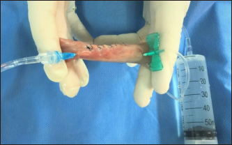
Fig. 1. A 10 cm colonic construct containing the incision line was digitally occluded. A blue needle on the left of the picture attached to a manometer was used for intraluminal pressure monitoring and a green butterfly needle on the right of the picture attached to a 60 ml syringe was used for saline infusion and volume measurements.
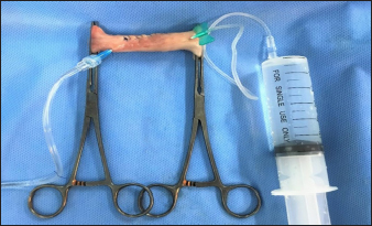
Fig. 2. A 10 cm colonic construct containing the incision line was occluded with Doyen intestinal forceps. A blue needle on the left of the picture attached to a manometer was used for intraluminal pressure monitoring and a green butterfly needle on the right of the picture attached to a 60 ml syringe was used for saline infusion and volume measurements. Ethical approvalThe study was approved by the ethical committee of the Companion Animal Clinic (1/2020) and a written consent of the owners was obtained.
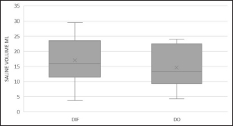
Fig. 3. Box and whisker plots for saline volume are required to achieve a predetermined intraluminal pressure for canine colotomies with two different occlusion techniques (DIF: Doyen intestinal forceps occlusion and DO: Digital occlusion). The median is represented as the central line inside each box (marked with an x). The upper and lower whiskers represent the maximum and minimum values of each technique. ResultsAccording to the Shapiro-Wilk test, the weight of the dogs followed a normal distribution. The mean weight was 26.9 ± 8.5 kg (95% CI: 19.8–34 kg, range: 17–40 kg, median: 25 kg). The mean volume of injected saline needed for achieving the intraluminal pressure of 17.3 cm H2O with the DIF technique (20.4 ± 8.2 ml; 95% CI: 13.6–27.2 ml, range: 7–48 ml, median: 21.4 ml) was significantly greater than that of DO technique (17.5 ± 6.8 ml; 95% CI: 11.8–23.2 ml, range: 5–39 ml, median: 17.6 ml) [p=0.021] (Fig. 3). DiscussionIn the study presented here, we found that the mean volume of saline required to achieve the intraluminal pressure of 17.3 cm H2O following colotomy in dogs using the DIF technique was larger than that of the DO technique. Thus, our hypothesis was rejected. Colotomy was chosen in the present study as it is an easily reproducible technique that has been reported by others (Wylie and Hosgood, 1994; Williams, 2018; Latimer et al., 2019) and is performed in our hospital for biopsy purposes and removal of impacted fecaliths. Colotomy can be closed in a simple interrupted or continuous appositional pattern. In our study, the 3 cm colotomy was closed with a 3–0 polydioxanone suture in a full-thickness continuous pattern. Dehiscence related to large intestinal surgery in small animals has been reported to range from 9% to 10 % (Wylie and Hosgood, 1994; Latimer et al., 2019). Septic peritonitis caused by dehiscence of full-thickness large intestinal incisions is correlated with a high morbidity rate, reaching 55% in dogs (Wylie and Hosgood, 1994; Latimer et al., 2019). Performance of the leak test perioperatively plays a crucial protective role against dehiscence, as the security of the intestinal suture line is assessed by the surgeon with minimal equipment. In our study during leak testing, the occlusion of the intestinal segments was achieved by two techniques. First, the constructs were digitally occluded, followed by occlusion with Doyen intestinal forceps. Both of these occlusion techniques are commonly used in practice. Digital occlusion is the choice of preference because is the less traumatic occlusion method for the intestine. Doyen intestinal forceps should be used for the shortest period and to the lowest compression possible, to minimize the risk of inducing an intestinal wall or large mesenteric vessel trauma (Radlinsky, 2013). In our study, a leak test was performed in 10 cm colonic constructs obtained from canine cadavers that weighed>15 kg. In each construct, a leak test was performed by the intraluminal injection of saline until intraluminal pressure reached 17.3 cm H2O, which corresponds to the normal mean peristaltic colonic pressure in dogs (Hofmann et al., 1993). Canine small or large intestines are considered to be low-pressure systems. The normal peristaltic pressure of the canine colon (17.3 cm H2O) was less than that reported for the canine jejunum (20–34 cm H2O) (Hofmann et al., 1993; Ellison, 2010). Saile et al. (2010) reported that the volume of saline needed to achieve 20 and 34 cm H2O intraluminal pressure in canine jejunum containing a closed intestinal biopsy was 11–14 and 16–19 ml, respectively, with digital occlusion and 9–11 and 12–15 ml, respectively, with Doyen occlusion (Saile et al., 2010). Matz et al. (2014) found that in 10 cm healthy jejunum segments occluded with Doyen forceps and containing a closed biopsy, the volume of saline needed to achieve the predetermined pressures of 20 and 34 cm H2O during leak testing, was similar for each closure orientation, even though luminal circumference was narrower in longitudinally closed biopsy sites (Matz et al., 2014). The saline volumes reported by Saile et al. (2010) and Matz et al. (2014) were smaller than those reported in our study. However, no reliable comparison can be made between their volumes and our volumes of saline used. These differences may be related to the different parts of the intestine examined as the large intestine has a greater diameter lumen than the small intestine, to the different peristaltic pressures that occurred, the different methodology employed, and the different state of pathology of the intestines examined. In the study reported here, we found that the volume of saline injected to achieve the intraluminal pressure of 17.3 cm H2O was larger for DIF compared to DO. In a recent study, however, evaluating leak testing between handsewn and stapled intestinal anastomosis in dogs no association between intraoperative anastomotic leak testing and a decrease in postoperative dehiscence was detected (Mullen et al., 2021). This difference in our study might be attributed to the completeness of the occlusion between Doyen forceps and the assistant’s fingers. Assistant’s fingers may result in less complete occlusion of intestinal lumen than that of Doyen forceps of the 10 cm colonic constructs, allowing for a smaller saline-filled volume. Our study had several limitations. Dogs that took part in the study had no digestive tract pathology and their gastrointestinal tract was macroscopically healthy in all of its length. Infiltration of the bowel with neoplastic or inflammatory cells in gastrointestinal diseases such as lymphoma or inflammatory bowel disease, could alter intestinal distensibility and affect the leak test. Another limitation is the fact that colonic segments were maintained in a container filled with saline at room temperature, instead of being maintained at the canine’s body temperature. Intestinal smooth muscle contracts in a low-temperature environment and as a result colonic segments’ original length, diameter, and distensibility decrease (Somlyo et al., 1971; Magaribuchi et al., 1973; Nasu et al., 1984). In contrast, no difference in initial leak pressure among four anastomosis techniques evaluated in cooled canine cadaveric jejunum has been identified (Fealey et al., 2020). Additionally, cadaveric canine jejunum evaluated immediately after harvesting or after freezing and subsequent thawing does not affect maximal intraluminal pressure (Duffy et al., 2020). In the present study, the saline volume needed to achieve the predetermined intraluminal pressure might be affected when tested at room temperatures. Another limitation of the present study was the lack of randomization of occlusion techniques. In all colonic constructs leaking test was performed first by DO and then with occlusion with DIF. It is possible that distention of the lumen with saline during the first leak test performed by DO increased the distensibility of the enteric wall and the saline volume needed to achieve the predetermined intraluminal pressure, during the second leak test performed by DIF. Moreover, the performance of colotomy rather than the performance of colectomy and anastomosis was another limiting factor. Colotomy is a less invasive surgical procedure compared to resection and anastomosis affecting the saline volume required. Finally, measurements in our study were performed in cadaveric colon derived from dogs weighing >15 kg. Testing in cadaveric intestines harvested from dogs of different weights might affect peristaltic intraluminal pressures and the volume of saline necessary for the leak test. Published data regarding peristaltic pressure in the canine colon are scarce. The need for such studies is important, as in surgical procedures performed in daily clinical practice, intraoperative measurement of intraluminal pressure is usually not performed when conducting the leak test. Further prospective studies on pressures performed in vivo in healthy and diseased canine colons derived from dogs of different weights following full-thickness incisions should be performed to determine the saline volume required for leak tests. ConclusionWe still believe that leak testing is a helpful method that trainee surgeons can use to help eliminate surgical errors. Both DIF and DO techniques can be used for performing leak testing in the canine cadaveric colon. The mean volume of saline required to achieve a predetermined intraluminal pressure following colotomy in dogs using the DIF technique was larger than that of the DO technique. AcknowledgmentsNone. Conflict of interestThe authors declare that there is no conflict of interest. FundingThis research received no specific grant. Data availabilityAll data supporting the findings of this study are available within the manuscript. Authors’ contributionsEleni Prastiti: Conceptualization; Data curation; Formal analysis; Investigation; Methodology; Resources; Software; Writing—original draft; Ioannis Savvas: Investigation; Methodology; Resources; Software; Writing—review and editing; Vasileia Angelou: Conceptualization; Methodology; Writing—review and editing; Lysimachos G Papazoglou: Conceptualization; Data curation; Formal analysis; Investigation. Methodology; Project administration; Supervision; Validation; Visualization; Writing - original draft; Writing—review and editing. ReferencesAllen, D.A., Smeak, D.D. and Schertel, E.R. 1992. Prevalence of small intestinal dehiscence and associated clinical factors: a retrospective study of 121 dogs. J. Am. Anim. Hosp. Assoc. 28, 70–76. Beard, J.D., Nicholson, M.L., Sayers, R.D., Lloyd, D. and Everson, N.W. 1990. Intraoperative air testing of colorectal anastomoses: a prospective, randomized trial. Br. J. Surg. 77, 1095–1097. Dixon, A.R. and Holmes, J.T. 1991. Colorectal anastomotic integrity after anterior resection: Is there a role for intraoperative testing? J. R. Coll. Surg. Edinb. 36, 35–36. Duell, J.R., Thieman Mankin, K.M., Rochat, M.C., Regier, P.J., Singh, A., Luther, J.K., Mison, M.B., Leeman, J.L. and Budke, C.M. 2016. Frequency of dehiscence in hand-sutured and stapled intestinal anastomoses in dogs. Vet. Surg. 45, 100–103. Duffy, D.J., Chang, Y.J., Balko, J.A. and Moore, G.E. 2020. Ex vivo comparison pf the effect of storage temperature on canine intestinal leakage pressures. Vet. Surg. 49, 496–501. Ellison, G.W. 2010. Intestinal obstruction. In Mechanisms of diseases in small animal surgery, 3rd ed. Ed., Bojrab, M.J. Jackson, WY: Teton NewMedia, pp: 183–187. Fealey, M.J., Regier, P.J., Steadman, C., Case, J.B. and Garcia-Pereira, F. 2020. Initial leak pressures of four anastomosis techniques in cooled cadaveric canine jejunum. Vet. Surg. 49, 480–486. Fossum, T.W. and Hedlund, C.S. 2003. Gastric and intestinal surgery. Vet. Clin. Small Anim. 33, 1117–1145. Gilbert, J.M. and Trapnell, J.E. 1988. Intraoperative testing of the integrity of left-sided colorectal anastomoses: a technique of value to the surgeon in training. Ann. R. Coll. Surg. Engl. 70, 158–160. Giuffrida, M.A. and Cimino Brown, D. 2018. Small intestine. In Veterinary surgery small animal, 2nd ed. Eds., Johnston, S.A. and K.M. Tobias. St Louis, MO: Elsevier, pp: 1730–1176. Griffith, C.D. and Hardcastle, J.D. 1990. Intraoperative testing of anastomotic integrity after stapled anterior resection for cancer. J. R. Coll. Surg. Edinb. 35, 106–108. Grimes, J.A., Schmiedt, C.W., Cornell, K.K. and Radlinksy, M.G. 2011. Identification of risk factors for septic peritonitis and failure to survive following gastrointestinal surgery in dogs. J. Am. Vet. Med. Assoc. 238, 486–494. Hofmann, R., Gomez, R., Tanagho, E.A. and Mcaninch, J.W. 1993. Motility and intraluminal pressure of the ileocolonic junctional zone and adjacent bowel in a canine model. Urol. Res. 21, 329–332. Huss, B.T. 2014. Surgery of the colon and rectum. In Current techniques in small animal surgery, 5th ed. Eds, Bojrab, M.J., Waldron D.R. and J.P. Toombs. Jackson, WY: Teton NewMedia, pp: 289–303. Ivanov, D., Cvijanović, R. and Gvozdenović, L. 2011. Intraoperative air testing of colorectal anastomoses. Srp. Ark. Celok. Lek. 139, 333–338. Latimer, C.R., Lux, C.N., Grimes, J.A., Benitez, M.E., Culp, W.T., Ben-Aderet, D. and Brown, D.C. 2019. Evaluation of short-term outcomes and potential risk factors for death and intestinal dehiscence following full-thickness large intestinal incisions in dogs. J. Am. Vet. Med. Assoc. 255, 915–925. Magaribuchi, T., Ito, Y. and Kuriyama, H. 1973. Effects of rapid cooling on the mechanical and electrical activities of smooth muscles of guinea pig stomach and taenia coli. J. Gen. Physiol. 61, 323–341. Matz, B.M., Boothe, H.W., Wright, J.C. and Boothe, D.M. 2014. Effect of enteric biopsy closure orientation on enteric circumference and volume of saline needed for leak testing. Can. Vet. J. 55,1255–1257. Mullen, K.M., Regier, P.J., Fox-Alvarez, W.A., Case, J.B., Ellison, G.W. and Colee, J. 2021. Evaluation of intraoperative leak testing of small intestinal anastomoses performed by hand-sewn and stapled techniques in dogs: 131 cases (2008-2019). J. Am. Vet. Med. Assoc. 258, 991–998. Nasu, T., Sakai, N., Washibe, T. and Ishida, Y. 1984. Cooling-induced contraction in ileal longitudinal smooth muscle of guinea-pig. J. Pharm. Pharmacol. 36, 322–325. Radlinsky, M.G. 2013. Surgery of the digestive system. In Small animal surgery, 4th ed. Ed., Fossum, T.W. St Louis, MO: Elsevier, pp: 386–583. Ralphs, C.C., Jessen, C.R. and Lipowitz, A.J. 2003. Risk factors for leakage following intestinal anastomosis in dogs and cats: 115 cases (1991-2000). J. Am. Vet. Med. Assoc. 223, 73–77. Saile, K., Boothe, H.W. and Boothe, D.M. 2010. Saline volume necessary to achieve predetermined intraluminal pressures during leak testing of small intestinal biopsy sites in the dog. Vet. Surg. 39, 900–903. Smeak, D.D. 2020. Colotomy. In Gastrointestinal surgical techniques in small animals. Eds., Monnet, E. and Smeak, D.D. Hoboken, NJ: Wiley and Sons, pp: 219–220. Smith, S., McGeehin, W., Kozol, R.A. and Giles, D. 2007. The efficacy of intraoperative methylene blue enemas to assess the integrity of a colonic anastomosis. BMC Surg. 7, 5–9. Somlyo, A.P., Devine, C.E., Somlyo, A.V. and North, S.R. 1971. Sarcoplasmic reticulum and the temperature-dependent contraction of smooth muscle in calcium-free solutions. J. Cell Biol. 51, 722–741. Wheeler, J.M. and Gilbert, J.M. 1999. Controlled intraoperative water testing of left-sided colorectal anastomoses: are ileostomies avoidable? Ann. R. Coll. Surg. Engl. 81, 105–108. Williams, J.M. 2018. Colon. In Veterinary surgery small animal, 2nd ed. Eds., Johnston, S.A. and Tobias, K.M. St Louis, MO: Elsevier, pp: 1761–1783. Wylie, K.B. and Hosgood, G. 1994. Mortality and morbidity of small and large intestinal surgery in dogs and cats: 74 cases (1980-1992). J. Am. Anim. Hosp. Assoc. 30, 469–474. | ||
| How to Cite this Article |
| Pubmed Style Prastiti E, Savvas I, Angelou V, Papazoglou LG. Saline volume required to achieve peristaltic intraluminal pressure during leak testing of canine colotomies, using two methods of luminal occlusion. Open Vet. J.. 2024; 14(5): 1130-1134. doi:10.5455/OVJ.2024.v14.i5.6 Web Style Prastiti E, Savvas I, Angelou V, Papazoglou LG. Saline volume required to achieve peristaltic intraluminal pressure during leak testing of canine colotomies, using two methods of luminal occlusion. https://www.openveterinaryjournal.com/?mno=186977 [Access: January 24, 2026]. doi:10.5455/OVJ.2024.v14.i5.6 AMA (American Medical Association) Style Prastiti E, Savvas I, Angelou V, Papazoglou LG. Saline volume required to achieve peristaltic intraluminal pressure during leak testing of canine colotomies, using two methods of luminal occlusion. Open Vet. J.. 2024; 14(5): 1130-1134. doi:10.5455/OVJ.2024.v14.i5.6 Vancouver/ICMJE Style Prastiti E, Savvas I, Angelou V, Papazoglou LG. Saline volume required to achieve peristaltic intraluminal pressure during leak testing of canine colotomies, using two methods of luminal occlusion. Open Vet. J.. (2024), [cited January 24, 2026]; 14(5): 1130-1134. doi:10.5455/OVJ.2024.v14.i5.6 Harvard Style Prastiti, E., Savvas, . I., Angelou, . V. & Papazoglou, . L. G. (2024) Saline volume required to achieve peristaltic intraluminal pressure during leak testing of canine colotomies, using two methods of luminal occlusion. Open Vet. J., 14 (5), 1130-1134. doi:10.5455/OVJ.2024.v14.i5.6 Turabian Style Prastiti, Eleni, Ioannis Savvas, Vasileia Angelou, and Lysimachos G. Papazoglou. 2024. Saline volume required to achieve peristaltic intraluminal pressure during leak testing of canine colotomies, using two methods of luminal occlusion. Open Veterinary Journal, 14 (5), 1130-1134. doi:10.5455/OVJ.2024.v14.i5.6 Chicago Style Prastiti, Eleni, Ioannis Savvas, Vasileia Angelou, and Lysimachos G. Papazoglou. "Saline volume required to achieve peristaltic intraluminal pressure during leak testing of canine colotomies, using two methods of luminal occlusion." Open Veterinary Journal 14 (2024), 1130-1134. doi:10.5455/OVJ.2024.v14.i5.6 MLA (The Modern Language Association) Style Prastiti, Eleni, Ioannis Savvas, Vasileia Angelou, and Lysimachos G. Papazoglou. "Saline volume required to achieve peristaltic intraluminal pressure during leak testing of canine colotomies, using two methods of luminal occlusion." Open Veterinary Journal 14.5 (2024), 1130-1134. Print. doi:10.5455/OVJ.2024.v14.i5.6 APA (American Psychological Association) Style Prastiti, E., Savvas, . I., Angelou, . V. & Papazoglou, . L. G. (2024) Saline volume required to achieve peristaltic intraluminal pressure during leak testing of canine colotomies, using two methods of luminal occlusion. Open Veterinary Journal, 14 (5), 1130-1134. doi:10.5455/OVJ.2024.v14.i5.6 |

