| Original Article | ||
Open Vet. J.. 2022; 12(2): 281-289 Open Veterinary Journal, (2022), Vol. 12(2): 281–289 Original Research Horizontal and vertical beam radiographs in vomiting dogs to diagnose mechanical gastrointestinal ileus: A diagnostic imaging comparative studyMaria Frau Tascon1*, Hock Gan Heng2, Rosa Novellas Torroja1, Yvonne Espada Gerlach1 and Carlo Anselmi1,**1Fundació Hospital Clínic Veterinari, Universitat Autònoma de Barcelona, 08193, Bellaterra, Barcelona, Spain 2Veterinary Clinical Sciences, Purdue University College of Veterinary Medicine, West Lafayette, IN, USA **Current address: Pride Veterinary Centre-Scarsdalevets, Derby, UK *Corresponding Author: Maria Frau Tascon. Fundació Hospital Clínic Veterinari, Universitat Autònoma de Barcelona, Carrer de l’Hospital s/n, 08193, Bellaterra, Barcelona, Spain. Email: frutascon [at] gmail.com Submitted: 22/12/2021 Accepted: 30/03/2022 Published: 20/04/2022 © 2022 Open Veterinary Journal
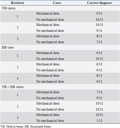
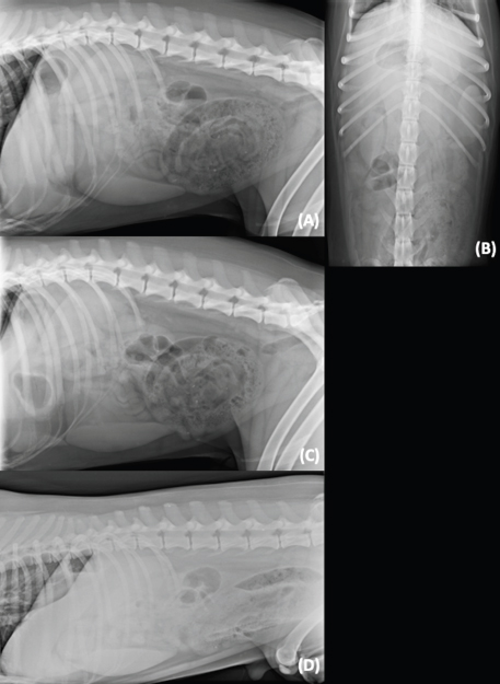
AbstractBackground: The horizontal beam (HB) view has been used in the identification of pneumothorax, pleural effusion, and pneumoperitoneum in small animals. Based on the literature, there were no published data evaluating the utility of HB radiography in vomiting dogs to differentiate between patients with or without mechanical gastrointestinal ileus. Aim: The purpose of this prospective pilot study was to determine the utility of HB radiograph as an additional view in vomiting dogs to differentiate patients with or without mechanical gastrointestinal ileus; and describe if there are any radiographic image characteristics associated with the HB view for patients with mechanical gastrointestinal ileus. Methods: A prospective study was carried out on dogs presented with acute vomiting. For all dogs, four radiographic views [ventrodorsal (VD), right lateral, left lateral, and left-to-right lateral HB in sternal recumbency] of the abdomen and abdominal ultrasound were obtained. If a mechanical ileus was detected ultrasonographically, an exploratory laparotomy or endoscopy was performed, otherwise medical treatment was elected. Results: A total of 22 patients were recruited, 11 diagnosed with mechanical ileus and 11 without mechanical ileus. Three blinded reviewers independently assessed the radiographs in three sets: vertical beam (VB) views, HB view alone, and a combination of both views. No statistical difference was found in the differentiation between patients with or without mechanical gastrointestinal ileus between HB views alone or added to VB views. Conclusion: This study suggests that the HB view in sternal recumbency may be an alternative for patients who are not stable enough to be positioned in lateral or VD recumbency. Keywords: Canine, Radiographic projection, Gastrointestinal obstruction, Foreign body. IntroductionHorizontal beam (HB) radiography was first used in the diagnosis of human pneumothorax in 1966 (Kurlander and Helmen, 1966). Horizontal beam radiography has been recommended as an useful additional view in the identification of pneumothorax, pleural effusion, and pneumoperitoneum in small animals, and it has been reported that HB was approximately twice as likely to identify concurrent free fluid and air as vertical beam (VB) radiography (Lynch et al., 2012; Ng et al., 2019). Survey radiography and abdominal ultrasonography are the current preferred imaging modalities for evaluation of acute vomiting in dogs, especially when foreign body ingestion is suspected and other systemic causes (e.g., renal disease) have been ruled out. Multiple veterinary studies have shown that abdominal ultrasonography improves detection of gastrointestinal foreign bodies and has improved accuracy for diagnosing mechanical ileus when survey radiographic studies are equivocal (Sharma et al., 2011; Ciasca et al., 2013). Abdominal ultrasonography, however, has its limitations such as being operator-dependent, possible interference/lack of visibility of organs of interest due to presence of intraluminal gas in the overlying bowel or peritoneal free gas, patient weight, poor restraint, possible patient discomfort, and body conformation (Shanaman et al., 2013; Garcia and Froes, 2014). Abdominal radiography in horses is always performed in the standing position with HB because it is not feasible to perform VB studies due to the large size of these patients (Butler et al., 2017). Vertical beam radiography is feasible in small size foals. Gas-capped fluid levels in hollow organs can only be evaluated on radiographs obtained in the standing position with HB views (Gibbs and Pearson, 1973; Butler et al., 2017). Horizontal beam views allow clinicians to take advantage of the physical nature of gas and fluid (Lynch et al., 2012). In the HB view, different levels of the gas-capped fluid lines in the same intestinal loop are commonly seen in the intestinal mechanical ileus, instead in paralytic ileus, gas-capped fluid lines tend to be at the same level in a given U-shaped section of the intestine (Gibbs and Pearson, 1973; Dennis et al., 2010; Geng et al., 2018). Although HB view is commonly practiced in equine medicine, this technique has not been systematically studied for small animal abdominal pathologies (Lynch et al., 2012). The objectives of this study were to (1) determine the diagnostic accuracy of HB radiograph as an additional view in vomiting dogs to differentiate patients with or without mechanical gastrointestinal ileus; and (2) describe if there are any radiographic image characteristics associated with the HB view for patients with mechanical gastrointestinal ileus. The authors hypothesized that the HB view, alone or added to VB views in patients presented with a history of acute vomiting, would increase the diagnostic accuracy to differentiate patients with or without mechanical gastrointestinal ileus. Material and MethodsThis pilot study was a prospective, diagnostic case-control study. Privately owned dogs were prospectively recruited for an observational diagnostic accuracy study at the veterinary hospital (Fundació Hospital Clínic Veterinari Universistat Autònoma de Barcelona) between January 2015 and June 2016. Written consent from each patient’s owner was obtained on admission. Subject inclusion for the study was determined by one of the authors (C.A. at that time, third-year diagnostic imaging resident). Patients selected were those who presented with signs of acute vomiting (less than 2 weeks), without other gastrointestinal signs such as diarrhea and without any chronic disease identified at serum biochemistry and hematology (e.g., chronic renal disease). Patients were excluded if there were obvious radiographic signs of mechanical gastrointestinal ileus caused by a large mineral or metallic opaque foreign object(s) or an unequivocal obstruction pattern due to linear-textile foreign body in the standard VB radiographic views. For all included dogs, four radiographic views (standard VB radiographic views [ventrodorsal (VD), right lateral, and left lateral (LL)] and an additional left-to-right lateral HB in the sternal recumbency of the abdomen were obtained (System: Premium Vet-variable focal distance, Sedecal, Madrid, Spain; X-ray tube: Rotanode E7239X, Toshiba, Tokyo, Japan), using variables mAs and kVp determined by the thickness of each individual patient. For the HB view, a mobile antiscatter grid provided by the manufacture of the X-ray machine was used. A radiolucent soft pad was placed between the table top and the patient during the HB view: this was found to be important to increase the distance between the patient and the table, allowing all the ventral portion of the patient’s abdomen to be included in the view. The image quality of each study was evaluated and approved by C.A. After the radiographic study, all the patients included underwent abdominal ultrasound (Mylab 70; Esaote, Genova, Italy) performed by C.A. The studies were performed in dorsal recumbency using microconvex (CA 123; 3-9 MHz; Esaote) and linear (LA523, Esaote 4-13) transducers. If a mechanical ileus was detected ultrasonographically, an exploratory laparotomy or endoscopy was performed, otherwise medical treatment was elected. For patients treated surgically, the location and type of foreign body were documented. For patients in the medically treated group, 2 weeks’ follow-up by phone call or at the hospital was performed. According to the outcome, the patients were classified into two groups: dogs with mechanical ileus (group A) and dogs without mechanical ileus (group B). Images were stored in the Digital Imaging and Communications in Medicine (DICOM) format to be reviewed later, in individual review sessions using different workstation, by two board certified radiologists (H.G.H and R.N.T) and a senior professor veterinary radiology specialist (Y.E.G). All reviewers had more than 15 years of experience in film reading. The cases were reviewed in three sittings, the first sitting consisted of only VB views, the second sitting consisted of only lateral HB view and the third sitting consisted of a combination of VB and lateral HB views. The cases were randomly assigned a number using an open-source web-based random integer generator (http://www.random.org) for each sitting. The reviewers evaluated the three sets of images independently with at least 1 week apart from each sitting. The reviewers were only provided with the history of acute vomiting and were blinded to the outcome of the cases. The following criteria were examined by the reviewers: small intestinal distension and disposition, presence of gas-capped fluid lines, presence of sign of intussusception, presence of foreign bodies (number and location), serosal margins visualization, and presence or absence of peritoneal effusion and pneumoperitoneum. The reviewers were required to document the presence of small-sized nonobstructive foreign material (e.g., pinpoint to millimetric mineral material/stones), even if considered incidental. The reviewers had the option to add comments after the evaluation of each study. Accuracy on the diagnosis of mechanical gastrointestinal ileusThe presence of a mechanical gastrointestinal ileus was considered when at least two of the following criteria were included by the reviewers: a focal distension of the small intestine, stacked or plicated disposition of the intestinal loops, and the suspected presence of intraluminal foreign material. For the analysis in the detection of gastrointestinal mechanical ileus, patients with a small amount of pinpoint to millimetric small-sized gastric foreign material were considered nonobstructive. Serosal margins visualization, peritoneal effusion, and pneumoperitoneumReviewers were asked to differentiate if the serosal margins visualization was normal or decreased, and to describe the presence or absence of peritoneal effusion and pneumoperitoneum. Ultrasonography was used as a gold standard to confirm the presence of diffuse or focal peritonitis, peritoneal effusion, and pneumoperitoneum. Small intestinal distensionThe small intestinal distension was determined if the small intestinal distension ratio was higher than 1.6 times the height of the mid-vertebral body of L5. The distended intestine was then classified as diffuse or focal. Presence of gas-capped fluid linesThe presence or absence of different levels of the gas-capped fluid lines in the same intestinal loop was recorded by the reviewers. Small intestinal dispositionThe reviewers described the disposition of the small intestine as normal, stacked, or plicated. Statistical analysesStatistical analyses were performed by a statistician, using commercially available software (SPSS v15.0, SPSS Inc., III: Chicago, IL). Ultrasound was used as the gold standard. Post-hoc analysis (G*Power 3.1) with a power analysis of 80% and α error of 0.05 was used to determine sample size, and approximately 64 answers were needed. All results were classified as categorical data and then comparisons between group A (with mechanical ileus) and group B (without mechanical ileus) were made for the radiographic views and reviewers. Univariate analysis was performed using Pearson’s χ2 or Fisher’s exact test for categorical variables. Interobserver agreement for the accuracy on the diagnosis of mechanical gastrointestinal ileus was tested with kappa statistic, defined as (observed agreement-expected agreement)/(1-expected agreement). The level of agreement was classified as poor (κ < 0.20), fair (κ between 0.21 and 0.4), moderate (κ between 0.41 and 0.6), good (κ between 0.61 and 0.80), and excellent (κ >0.80) (Landis and Koch 1977). A value of p < 0.05 was considered as the critical level of significance. Ethical approvalNot required for this study. ResultsA total of 22 dogs were included. The weight of the dogs ranged from of 3.0 to 34.2 kg (median=15.3 kg). There were four Staffordshire Bull Terriers, two Labrador Retrievers, two Boxers, two Beagles, two Yorkshire Terriers, two Miniature Pinschers, three mixed breeds, and one dog each of five other breeds. Of the 22 cases, 11 were included in group A and 11 were included in group B. In group A, two had a gastric foreign body obstruction, four had a linear foreign body obstruction, three had jejunal foreign body obstruction, one had multiple foreign bodies obstruction located in the stomach and jejunum, and one had an intestinal volvulus. In group B, one case was presented with multiple uncountable small nonobstructive mineral gastrointestinal foreign bodies, which were passed, as confirmed by follow-up radiographic study the following day, and by resolution of the clinical signs. One case was finally diagnosed with gastrointestinal intoxication, secondary to mushroom ingestion, confirmed by the history after the imaging studies were performed. The remaining nine cases were presumptively diagnosed with gastroenteritis and/or pancreatitis due to the resolution of the clinical signs after the medical treatment. A total of 88 radiographs were available for review. A total of 198 answers from 3 reviewers were available for analysis. A total of 22 cases were evaluated by 3 reviewers to achieve 66 answers in each sitting. Accuracy on the diagnosis of mechanical gastrointestinal ileusThe accuracy on the detection of mechanical ileus in the three sittings was evaluated and there was no statistically significant increase in the accuracy of detection of mechanical ileus between VB views, HB view, and the combination of VB and HB views (p=0.24) (Table 1). Hence, there was no statistical significance to support the authors’ hypothesis whereby the HB view, alone or added to VB views in patients presented with a history of acute vomiting, would increase the diagnostic accuracy on the diagnosis of gastrointestinal mechanical ileus. Reviewers’ sensitivity and specificity values for mechanical ileus with VB views were in the ranges of 64%–93% and 61%–91%, respectively, and with both HB and VB views were 64%–93% and 54%–86%, respectively. When HB view was evaluated alone, sensitivity and specificity were in the ranges of 42%–77% and 57%–88%. The strength of agreement between the reviewers was 0.72 (good agreement). Interestingly, in one case diagnosed without mechanical gastrointestinal ileus, all the reviewers classified it wrongly as mechanical ileus when evaluating the VB views and correctly with only the HB view (Fig. 1). Serosal margins visualization, peritoneal effusion, and pneumoperitoneumThe answers are summarized in Table 2. A statistically significant difference was observed on the detection of abnormal serosal margins visualization (p=0.05) between HB and VB views. No evidence of statistical significance on the detection of peritoneal effusion (p=0.98) or pneumoperitoneum was found comparing both HB and VB views (p=1). Small intestinal distensionThe diagnosis of diffuse small intestinal distension was higher with the HB view (8/66) than with the VB views (2/66). However, focal intestinal distension was higher with the VB views (24/66) than with the HB view (17/66). Presence of gas-capped fluid linesThe presence of variable gas-capped fluid lines on the HB view was positive in 36/66 cases, 78.8% (26/33) from group A and 30.3% (10/33) from group B. Small intestinal dispositionIn the evaluation of the small intestinal disposition (classified as normal, plicated, or stacked), comparing the different views, the results were as follows: 9/66 cases presented a plicated disposition on the VB views and 6/66 on the HB view, and 14/66 were stacked on the VB views versus 16/66 on the HB view (Table 3). Table 1. Reviewers’ interpretations in 22 dogs.
DiscussionAccuracy on the diagnosis of gastrointestinal mechanical ileusThe study failed to prove that the HB view, alone or added to VB views in patients presented with a history of acute vomiting, would increase the diagnostic accuracy to differentiate patients with or without mechanical gastrointestinal ileus. We found no statistical difference in the detection of mechanical gastrointestinal ileus between the HB view alone or added to VB views. One of the reasons could be the absence of a control group in the study, which would have helped to identify normal radiographic findings in HB view, e.g., the serosal margins visualization. Another possibility is the acuteness of the presenting clinical signs. In large animals, patients tend to be presented after showing clinical signs for a longer period. In small animals, the owners tend to easily observe if the patient is vomiting, as the animals are usually kept in door, as opposed to large animals. In this study, all the cases presented with acute vomiting of less than 2 weeks: 12 cases of 1-day duration (5 from the group A and 7 from the group B; 8 cases of less than 7 days (3 from the group A and 5 from the group B); and 2 cases of less than 14 days (both from the group A). The sensitivity for detecting mechanical ileus was lower using the HB view alone. In this view, a decreased serosal detail is more frequently described compared to the VB views, perhaps due to suboptimal technique of the HB view as the exposure techniques of VB views (e.g., exposure parameters, collimation, used of antiscatter grid, and others) have been developed with years of experience. However, the specificity using the HB view alone was slightly higher than using both the HB and VB views. This may be a type I error, due to the subjective evaluation of each reviewer. The HB view is not a standard view to asses abdominal disorders in dogs, thus the reviewers involved in the study have no prior experience interpreting the HB view. Secondary to this, the reviewers, when evaluating the VB and HB in combination, were probably more prone to base their final judgment on the standard VB views giving less importance to the HB. In addition, the cases included in this study were complicated, as the obvious mechanical obstructions due to mineral or metal opaque foreign bodies and/or unequivocal linear foreign bodies were excluded. This study focused on cases that are usually equivocal and where additional ultrasound or contrast studies are needed for further diagnostics.
Fig. 1. (A) Right lateral; (B) ventrodorsal; (C) left lateral; and (D) left-to-right lateral HB views of the abdomen of a dog presented with acute vomiting. In this case, the patient was correctly diagnosed as without mechanical ileus with the HB view and incorrectly diagnosed as mechanical ileus with the VB view by all reviewers. Abdominal ultrasound was unremarkable. Table 2. Comparison of decreased serosal margins visualization, pneumoperitoneum, and peritoneal effusion detection between views.
Table 3. Comparison of small intestinal disposition between the different views.
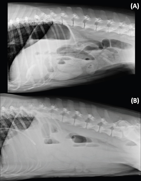
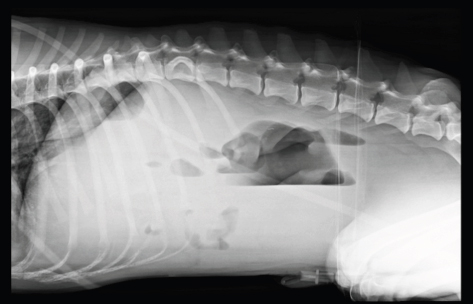
The results of this study shows that the HB view should not be used alone as the sensitivity and specificity is low. The combination of VB view and HB view together did not improve the sensitivity and specificity, probably due to the minimal additional information gained with the HB view. Thus, it is not recommended to incorporate the HB view as a routinely abdominal view to diagnose mechanical ileus according to the ALARA/ALARP principles. Serosal margins visualization, peritoneal effusion, and pneumoperitoneumWe found statistical differences on the evaluation of serosal margins visualization, being VB view superior to HB view. Based on our results, the HB view diagnosed more frequently a decrease in the serosal margins visualization, and an increased presence of peritoneal effusion compared with VB view. Since there are no previous studies in healthy dogs describing the serosal margins visualization using the HB view, we proposed that these findings could be artefactual due to the position of the organs in the most dependent part because of the gravity effect and crowding. Pneumoperitoneum was more often visualized in the HB view compared with the VB views in this study. This was expected, as it is widely described in the literature that HB view has a higher accuracy in detecting small volumes of pneumoperitoneum (Thrall, 2013; Ng et al., 2019). In this study, there was no statistical difference detecting pneumoperitoneum between VB and HB views, probably due to the low number of cases with the presence of pneumoperitoneum. Small intestinal distensionThe authors did not find an explanation for the more frequent visualization of diffuse small intestinal distension in the HB view. One of the hypotheses considered was that gas rises and fluid gravitates, thus generalized gas-distended small intestines in the dorsal aspect of the abdomen is easily identify, compared to fluid filled intestines in the dependent (ventral) aspect of the abdomen. HB view thus allows the reviewers to visualize the diffuse small intestinal distension in the more dorsal aspect of the abdomen. However, the visualization of the small focal distension was lower with the HB view. We consider that it is possibly more difficult to visualize focal distension at the most dependent part of the abdomen with the HB view due to fluid gravitation and a higher degree of crowding of the abdominal structures. Specific assessment of large intestine identification in the different views was not asked to the reviewers; authors hypothesized that the HB view did not supply more information to distinguish large from small intestines compared to VB views, thus colonography (positive or negative) is still needed. Presence of gas-capped fluid linesThe presence of variable gas-capped fluid lines has been reported as a radiographic sign of mechanical gastrointestinal ileus in the HB view (Gibbs and Pearson, 1973; Harlow et al., 1993). This study confirms that 78.8% of the mechanical ileus cases exhibit this sign. However, this is not specific as 30.3% of the without mechanical ileus cases also have this similar radiographic sign (Fig. 2). Thus, the presence of gas-capped fluid lines in the HB view needs to be interpreted carefully and it is not a pathognomonic sign for mechanical ileus. However, it may suggest mechanical ileus and need further investigation. In this study, specific timing between radiographic views was not recorded. The HB view was carried out just after the VB views. The author (C.A.) personally involved in performing the radiographic studies found that it was initially difficult to achieve the HB view mainly due to lack of experience in this technique (collimation, different parameters, use of antiscatter grid, superposition of hind limbs, and rotation/movement of the patient as no sedation was used), but the learning curve was fast (e.g., use of a radiolucent soft pad to increase the height of the patient from the table and enhance comfort in sternal position, and stretching caudally the hind limbs) and the HB view was usually achievable in few minutes. There are no documented data specifying the time (minutes) of recumbency to obtain a better visualization of the fluid-gas levels or redistribution of gas. More conspicuous amounts of gas in the pylorus and duodenum are expected in the LL view after prolonged LL recumbency (Vander Hart and Berry, 2015). Small intestinal dispositionIt is reported that normal small intestinal loops are usually diffusely distributed in the abdominal cavity with a smooth round turn, appearing as continuously curving tubes or as solid circles or rings (Thrall, 2013). In cases of mechanical ileus, the intestinal loops become progressively distended, the segments become crowded into a relatively smaller space, often assuming a stacked appearance (Thrall, 2013). In this study, the use of the HB view did not affect the visualization of small intestinal disposition compared with the VB views, thus a different recumbency seems not to alter the small intestinal disposition. However, organs crowding at the ventral aspect of abdomen make it difficult the evaluation of fluid/soft tissue filled intestines when there is concurrent peritoneal effusion due to border effacement of structures (Fig. 3).
Fig. 2. Left-to-right lateral HB views in sternal recumbency of two different patients. (A) A dog with mechanical ileus due to a gastro-jejunal linear foreign body. Note the presence of different levels of the gas-capped fluid lines in the small intestine. Additionally, there is a curvilinear radiopaque structure in the stomach, which was not identified in the standard VB views but recognized on the HB view. (B) A dog diagnosed with gastrointestinal intoxication secondary to mushroom ingestion in group P. Note the presence of the different level of gas-capped fluid lines in the small intestines’ loops. A potential limitation of this study is that reviewers are not familiar interpreting the HB view in small animals, as its use is not widespread and not all radiographic equipment has this option. The time between viewing sessions could have affected the reviewer’s radiographic evaluation, as they could remember the cases between each sitting. To avoid this limitation, a minimum of 1 week time gap was established, although it might not be long enough. The order of viewing sessions had not been randomized and could be considered a limiting factor. An additional bias of the study is the exclusion of cases with chronic (more than 2 weeks) vomiting, which can be secondary to a partial mechanical obstruction, where maybe the HB view could be beneficial. For future work, a larger number of dogs and a control group of normal dogs may be helpful. In summary, there was no statistical difference between the HB view alone or in combination with VB views to differentiate dogs with or without mechanical gastrointestinal ileus. However, for patients who are not stable enough to be positioned in lateral or VD recumbency, the HB view in sternal recumbency may be valuable as an alternative radiographic view. Conflict of interestThe authors declare that they have no conflict of interest.
Fig. 3. Example of crowding organs at the ventral aspect of the abdomen in a left-to-right lateral HB view. Evaluation of the ventral abdomen is challenging due to presence of fluid/soft tissue-filled intestines and concurrent peritoneal effusion. Authors’ contributionsConceptualization and design: H.G.H. and C.A.; acquisition of data: H.G.H., C.A., R.N.T., and Y.E.G.; formal analysis and interpretation of data: M.F.T. and C.A.; writing-original draft preparation: M.F.T.; writing-review and editing: M.F.T., H.G.H., C.A., R.N.T., and Y.E.G. All authors have read and agreed to the published version of the manuscript. ReferencesButler, J.A., Colles, C.M., Dyson, S.J., Kold, S.E. and Poulos, P.W. 2017. Clinical radiology of the horse, 4th ed. Hoboken, NJ: Wiley Blackwell, pp: 688–689. Ciasca, T.C., David, F.H. and Lamb, C.R. 2013. Does measurement of small intestinal diameter increase diagnostic accuracy of radiography in dogs with suspected intestinal obstruction? Vet. Radiol. Ultrasound 54(3), 207–211. Dennis, R., Kirberger R.M., Barr, F. and Wrigley. R.H. 2010. Handbook of small animal radiology and ultrasound, 2nd ed. Edinburgh, UK: Churchill Livingstone Elsevier, pp: 280–281. Garcia, D.A.A. and Froes, T.R. 2014. Importance of fasting in preparing dogs for abdominal ultrasound examination of specific organs. J. Small Anim. Pract. 55(12), 630–634. Geng, W.Z.M., Fuller, M. and Thoirs, M. 2018. The value of the erect abdominal radiograph for the diagnosis of mechanical bowel obstruction and paralytic ileus in adults presenting with acute abdominal pain. J. Med. Rad. Sci. 65(4), 1–8. Gibbs, C. and Pearson, H. 1973. The radiological diagnosis of gastrointestinal obstruction in the dog. J. Small Anim. Pract. 14(2), 61–82. Harlow, C. L., Stears, R.L.G, Zeligman, B.E. and Archer, P.G. 1993. Diagnosis of bowel obstruction on plain abdominal radiographs: significance of air-fluid levels at different heights in the same loop of bowel. Am. J. Roentgenol. 161(2), 291–295. Kurlander, G. and Helmen, C. 1966. Subpulmonary pneumothorax. Am. J. Roentgenol. 96(4), 1019–1021. Landis, J. R. and Koch, G. G. 1977. The measurement of observer agreement for categorical data. Biometrics 33(1), 159–174. Lynch, K.C., Oliveira, C.R., Matheson, J.S., Mitchell, M.A. and O’brien, R.T. 2012. Detection of pneumothorax and pleural effusion with horizontal beam radiography. Vet. Radiol. Ultrasound 53(1), 38–43. Ng, J., Linn, K.A., Shmon, C.L., Parker, S. and Zwicker, L.A. 2019. The left lateral projection is comparable to horizontal beam radiography for identifying experimental small volume pneumoperitoneum in the canine abdomen. Vet. Radiol. Ultrasound. 61(2), 130–136. Shanaman, M.M., Schwarz, T., Gal, A. and O’Brien, R.T. 2013. Comparison between survey radiography, b-mode ultrasonography, contrast-enhanced ultrasonography and contrast-enhanced multi-detector computed tomography findings in dogs with acute abdominal signs. Vet. Radiol. Ultrasound 54(6), 591–604. Sharma, A., Thompson, M.S., Scrivani, P.V., Dykes, N.L., Yeager, A.E., Freer, S.R. and Erb, H.N. 2011. Comparison of radiography and ultrasonography for diagnosing small-intestinal mechanical obstruction in vomiting dogs. Vet. Radiol. Ultrasound 52(3), 248–255. Thrall, D. 2013. Textbook of Veterinary Diagnostic Radiology, Journal of Chemical Information and Modeling, 6th ed. Maryland heights. Philadelphia, PA: Elsevier Saunders, pp: 770–772. Vander Hart, D. and Berry, C. R. 2015. Initial influence of right versus left lateral recumbency on the radiographic finding of duodenal gas on subsequent survey ventrodorsal projections of the canine abdomen. Vet. Radiol. Ultrasound 56(1), 12–17. | ||
| How to Cite this Article |
| Pubmed Style Tascon MF, Heng HG, Torroja RN, Gerlach YE, Anselmi C. Horizontal and vertical beam radiographs in vomiting dogs to diagnose mechanical gastrointestinal ileus: a diagnostic imaging comparative study. Open Vet. J.. 2022; 12(2): 281-289. doi:10.5455/OVJ.2022.v12.i2.17 Web Style Tascon MF, Heng HG, Torroja RN, Gerlach YE, Anselmi C. Horizontal and vertical beam radiographs in vomiting dogs to diagnose mechanical gastrointestinal ileus: a diagnostic imaging comparative study. https://www.openveterinaryjournal.com/?mno=40103 [Access: January 25, 2026]. doi:10.5455/OVJ.2022.v12.i2.17 AMA (American Medical Association) Style Tascon MF, Heng HG, Torroja RN, Gerlach YE, Anselmi C. Horizontal and vertical beam radiographs in vomiting dogs to diagnose mechanical gastrointestinal ileus: a diagnostic imaging comparative study. Open Vet. J.. 2022; 12(2): 281-289. doi:10.5455/OVJ.2022.v12.i2.17 Vancouver/ICMJE Style Tascon MF, Heng HG, Torroja RN, Gerlach YE, Anselmi C. Horizontal and vertical beam radiographs in vomiting dogs to diagnose mechanical gastrointestinal ileus: a diagnostic imaging comparative study. Open Vet. J.. (2022), [cited January 25, 2026]; 12(2): 281-289. doi:10.5455/OVJ.2022.v12.i2.17 Harvard Style Tascon, M. F., Heng, . H. G., Torroja, . R. N., Gerlach, . Y. E. & Anselmi, . C. (2022) Horizontal and vertical beam radiographs in vomiting dogs to diagnose mechanical gastrointestinal ileus: a diagnostic imaging comparative study. Open Vet. J., 12 (2), 281-289. doi:10.5455/OVJ.2022.v12.i2.17 Turabian Style Tascon, Maria Frau, Hock Gan Heng, Rosa Novellas Torroja, Yvonne Espada Gerlach, and Carlo Anselmi. 2022. Horizontal and vertical beam radiographs in vomiting dogs to diagnose mechanical gastrointestinal ileus: a diagnostic imaging comparative study. Open Veterinary Journal, 12 (2), 281-289. doi:10.5455/OVJ.2022.v12.i2.17 Chicago Style Tascon, Maria Frau, Hock Gan Heng, Rosa Novellas Torroja, Yvonne Espada Gerlach, and Carlo Anselmi. "Horizontal and vertical beam radiographs in vomiting dogs to diagnose mechanical gastrointestinal ileus: a diagnostic imaging comparative study." Open Veterinary Journal 12 (2022), 281-289. doi:10.5455/OVJ.2022.v12.i2.17 MLA (The Modern Language Association) Style Tascon, Maria Frau, Hock Gan Heng, Rosa Novellas Torroja, Yvonne Espada Gerlach, and Carlo Anselmi. "Horizontal and vertical beam radiographs in vomiting dogs to diagnose mechanical gastrointestinal ileus: a diagnostic imaging comparative study." Open Veterinary Journal 12.2 (2022), 281-289. Print. doi:10.5455/OVJ.2022.v12.i2.17 APA (American Psychological Association) Style Tascon, M. F., Heng, . H. G., Torroja, . R. N., Gerlach, . Y. E. & Anselmi, . C. (2022) Horizontal and vertical beam radiographs in vomiting dogs to diagnose mechanical gastrointestinal ileus: a diagnostic imaging comparative study. Open Veterinary Journal, 12 (2), 281-289. doi:10.5455/OVJ.2022.v12.i2.17 |


