


| Case Report | ||
Open Vet J. 2023; 13(7): 948-954 Open Veterinary Journal, (2023), Vol. 13(7): 948-954 Case Report Intra-articular replacement of the caudal cruciate ligament using a UHMWPE ligament under arthroscopic guidance in a dog: A case reportFrançois Fauqueux1*, Bastien Goin2,3,4, Mathilde Agbalé5, Antonin Jean Johan Crumière4, Philippe Buttin6, Eric Viguier2 and Thibaut Cachon21Clinique Vétérinaire Animed, Gap, France 2Université de Lyon, VetAgro Sup, Interactions Cellules Environnement (ICE), Marcy l’Etoile, France 3Univ Lyon, Univ Gustave Eiffel, Univ Claude Bernard Lyon 1, Lyon, France 4Novetech Surgery, Monaco, Monaco 5National Veterinary School of Alfort, Maisons-Alfort, France 6Itinerant Surgeon, Villaz, France *Corresponding Author: François Fauqueux. Clinique Vétérinaire Animed, Gap, France. Email: fauqueux [at] animed.fr Submitted: 16/03/2023 Accepted: 19/06/2023 Published: 31/07/2023 © 2023 Open Veterinary Journal
AbstractBackground: As isolated ruptures of the caudal cruciate ligament (CdCL) are rare in dogs, there is no consensus on the indications and the gold-standard surgical technique for treatment. Case Description: A 2-year-old Shepherd dog with an isolated rupture of the CdCL was treated with a new surgical technique for synthetic reconstruction. Three bone tunnels were drilled in the femur and the tibia under arthroscopic guidance to make sure the anatomical insertions of the physiological ligament were respected. An ultra-high molecular weight polyethylene (UHMWPE) implant was fixed with interference screws to reconstruct the CdCL. A synovial inflammation remained present on radiographs for 6 months after the surgery, together with a mild lameness. However, the dog fully recovered clinically and recovered a normal level of activity after 6 months. Liverpool osteoarthritis in dogs questionnaire results at 6 months and 1 year postoperatively were excellent. Conclusion: The use of a UHMWPE implant fixed with interference screws to reconstruct the CdCL allowed a return to full function of the knee without complications, despite a persistent synovial inflammation and mild lameness for a 6-month period after the surgery. The success of this isolated surgical technique could lead to improvements in the surgical management of CdCL rupture, if these initial results are confirmed by a prospective study with a larger number of patients. Keywords: Arthroscopy, Caudal cruciate ligament, Dog, Synthetic ligament reconstruction, UHMWPE. IntroductionRupture of the caudal cruciate ligament (CdCL) is an uncommon cause of lameness in dogs and is characterized by a caudal drawer motion, a thickening of the stifle, and a painful mobilization of the joint, especially in hyperextension (Johnson and Olmstead, 1987; Johnson and Johnson, 1993). Historically, conservative treatment with non-steroidal anti-inflammatory drugs (NSAIDs) and activity restriction was recommended for isolated and moderate lesions in small dogs and for chronic lesions associated with osteoarthritis in old dogs from large breeds (Harari et al., 1987; Arnoczky, 1988). Since this condition is rather rare, the literature only reports few surgical techniques that specifically treat isolated CdCL ruptures. The need for CdCL repair is also questioned as dogs can heal without surgical management. Some authors thus recommended a one-month observation period before attempting surgery, especially in small dogs, geriatric dogs, and animals with significant medical problems (Vasseur, 1993). The surgical approaches to treat the rupture of one or both cruciate ligaments are either extra-articular or intra-articular (Yoshiya et al., 1986; Johnson and Olmstead, 1987; Arnoczky, 1988; Lopez et al., 2003; Cook et al., 2017; Mao et al., 2019; Smith et al., 2019; Soreide et al., 2019; Biskup and Conzemius, 2020). Most of these techniques were developed for the surgical treatment of cranial cruciate ligament ruptures. We hypothesized that they are theoretically transposable to the treatment of CdCL ruptures by adapting the location of the fixations of intra- and extra-articular techniques to the physiological insertions of the CdCL. Extra-articular techniques are feasible in dogs weighing less than 15 kg and consist in stabilizing the joint by relying on different sutures or transpositions of extra-articular structures (Johnson and Olmstead, 1987, Arnoczky, 1988). Intra-articular techniques aim to replace the damaged ligament with either an autograft from a transposition of the patellar ligament or fascia lata, or with an allograft (Johnson and Olmstead, 1987, Arnoczky, 1988, Lopez et al., 2003, Biskup and Conzemius, 2020), a synthetic material (Julinder and Ödman, personal communication) (Smith et al., 2019; Soreide et al., 2019), or a combination of both (Yoshiya et al., 1986, Cook et al., 2017, Mao et al., 2019). In the event of avulsion at the level of the ligament attachment, a reduction using a screw may be considered (Knecht et al., 1976; Livet and Cachon, 2017). The reference examination in the context of suspected ligament rupture is arthroscopy as it allows a complete and precise evaluation of intra-articular lesions (Bleedorn et al., 2011; Little et al., 2014). Intra-articular techniques are frequently performed under arthroscopy, which makes it possible to repair eventual cartilage and meniscal lesions or remove bone fragments, and position the substitution ligament more precisely. Arthroscopy also reduces the invasiveness of the surgical procedure and, therefore, the risk of comorbidity. There is no consensus regarding the indications for surgical management of isolated ruptures of the CdCL and no recommended reference techniques. In light of the satisfactory results obtained during ex-vivo biomechanical tests for the reconstruction of the cranial cruciate ligament (Blanc et al., 2019; Goin et al., 2019, 2021, 2022; Rafael et al., 2020) and its clinical utilization in tendon reconstructions (Buttin et al., 2020), we posited that the use of an ultra-high molecular weight polyethylene (UHMWPE) implant should be efficient for the synthetic reconstruction of the CdCL. This case report describes the arthroscopically-assisted reconstruction of the CdCL in a dog with a UHMWPE implant and its outcome over a 12-month period. Case DetailsA 2-year-old female mixed-breed Shepherd weighing 15 kg, who suffered a trauma during play 3 months before, was presented with severe chronic lameness of the right hindlimb nonresponsive to NSAIDs. The orthopedic examination revealed a positive drawer test. The stress radiographs confirmed the suspicion of CdCL rupture with a displacement of the tibia relative to the femur (Fig. 1). A Liverpool osteoarthritis in dogs (LOAD) questionnaire was completed with the owner of the animal during the preoperative consultation, with a score of 22/52 indicating severe mobility impairment (Walton et al., 2013). A replacement of the CdCL by a synthetic UHMWPE ligament was decided using an arthroscopic approach. The dog was anesthetized with tiletamine (0.5 mg/kg)/zolazepam (0.5 mg/kg) + medetomidine (0.01 mg/kg) + morphine hydrochloride (0.5 mg/kg) by induction. An isoflurane relay (between 0.8 and 1%) in half-closed circuit was carried out at a flow rate of 1 l/minute. A constant rate infusion (2 ml/kg/hour) of a mixture of 250 ml of physiological serum + 30 mg of morphine + 202.75 mg of lidocaine + 75 mg of ketamine was administered. A single injection of cephalexin at 30 mg/kg was given at the start of the surgical preparation. The dog was placed in dorsal recumbency and prepared for aseptic surgery on the right hindlimb using chlorhexidine. A joint distractor supported by two 2.5-mm pins was placed on the stifle. The arthroscopic port was placed on the lateral side through a 5-mm lateral parapatellar incision performed with an 11-mm blade. Arthroscopic evaluation of the joint revealed inflammation of the medial meniscus without structural damage (Fig. 2A) and confirmed the complete rupture of the CdCL in its distal part at the tibial insertion (Fig. 2B). The lateral meniscus and the cranial cruciate ligament were intact. The lateral parapatellar incision was made 3 cm wider to allow the complete resection of the damaged CdCL with an 11-mm blade under arthroscopic control. The bone tunnels for femoral and tibial fixation of the synthetic ligament were drilled under arthroscopic control. A 2-mm guide wire was inserted at the femoral footprint of the origin of the CdCL in a caudo-medial direction to exit on the medial side of the femoral condyle under arthroscopic guidance. The femoral tunnel was drilled over the guide wire using a 3.6-mm cannulated drill bit (Fig. 3A). The tibial tunnel was created with the help of a tibial drilling guide. A stab incision was made at the caudal aspect of the joint to insert the tip of the tibial drilling guide, which was positioned at the footprint of the proximal tibial insertion of the CdCL under arthroscopic control, and oriented in a distal and cranio-lateral direction. A stab incision was made on the proximal aspect of the craniolateral aspect of the tibia to position the sleeve of the drilling guide. The appropriate position of the tip of the tibial drilling guide was assessed arthroscopically before drilling. The same technique as that used for the femoral tunnel was used to drill the tibial tunnel (Fig. 3B). The UHMWPE implant (Novalig 4000, Novetech Surgery, Monaco) was passed through the tibial and femoral tunnels using a passing tube and a wire loop (Fig. 4A). A 4.5 × 20-mm interference screw was placed from inside-out (Fig. 4B) (Rafael et al., 2020). The UHMWPE implant was then passed through a 3-mm-wide sliding tunnel, drilled perpendicularly through the tibial crest using a medial approach and located near the distal exit of the tibial tunnel. This setup increased the precision of the tensioning of the prosthesis using the created counter-support and facilitated the placement of the interference screw. Once satisfactory tension was obtained, the implant was locked with a second 4.5 × 20-mm interference screw placed from outside-in (Fig. 4C) (Rafael et al., 2020). Once the implant was securely locked, no residual drawer sign was observed and a normal range of motion was reported.
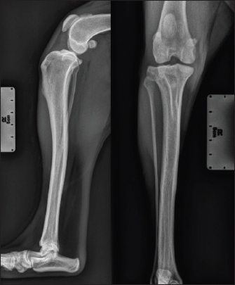
Fig. 1. Radiographic views of the dog's right pelvic limb from side (left) and front (right) at the time of diagnosis.
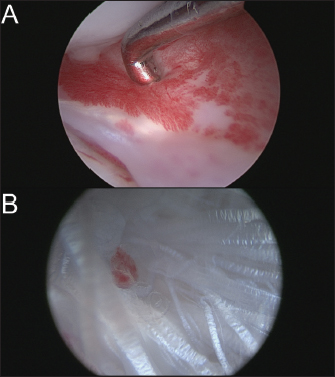
Fig. 2. (A) Arthroscopic view of inflammation of medial meniscus without structural damage. (B) Arthroscopic view of ruptured CdCL. Postoperative radiographs showed a satisfactory position of the tunnels and a good implantation of the interference screws along the drilling axes (Fig. 5). The patient was placed on full rest for eight weeks, with short lead walks only. Pain was managed with NSAIDs (Meloxicam (0.05 mg/kg)) for 6 days, and a painkiller (fentanyl patch (12 μg/hour)) for 72 hours. On day 1 post-surgery, the dog was walking on her operated leg with moderate lameness. During each postoperative consultation, two x-rays were performed (face and profile), as well as an orthopedic examination. A LOAD questionnaire was also completed. After 2 weeks, moderate lameness was observed, and radiographs showed thickening of the patellar tendon and moderate synovial inflammation; the LOAD score was 21/52. After 1 month, moderate lameness was persistent, radiographs still showed thickening of the patellar tendon and moderate synovial inflammation; the LOAD score was 13/52. After 2 months, moderate lameness was still observed, radiographs had not evolved, and the LOAD score was 7/52. After 3 months, mild lameness was observed, a mild posterior drawer was reported at orthopedic examination and moderate synovial inflammation was observed on radiographs (Fig. 6); the LOAD score was 7/52. After 6 months, very mild lameness was observed, a mild posterior drawer was reported at orthopedic examination and a resolution of the synovial inflammation was seen on radiographs (Fig. 7); the LOAD score was 5/52. After a year, a persistent yet stable mild posterior drawer was reported at the orthopedic examination but was not associated with any lameness, and the LOAD score was down to 2/52.
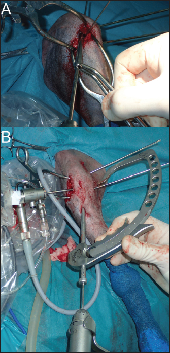
Fig. 3. (A) Femoral tunnel drilling. (B) Insertion of the 2-mm guide wire from lateral tibial metaphysis to CdCL tibia insertion under arthroscopic guidance.
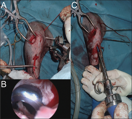
Fig. 4. (A) Placement of UHMWPE implant. (B) Arthroscopic view of femoral interference screw insertion. (C) Prosthesis tensioning and insertion of tibial interference screw. DiscussionThis case report describes a complete rupture of the CdCL treated with a UHMWPE synthetic ligament placed under arthroscopic guidance. In view of the clinical and radiographic outcomes over 12 months postoperatively, we conclude that this technique of intra-articular reconstruction of the CdCL yields satisfactory results. There is no real consensus in the literature regarding the surgical treatment of isolated CdCL rupture. The choice of this synthetic reconstruction technique was based on the satisfactory biomechanical results published on the interference screw fixation system of this UHMWPE implant (Rafael et al., 2020), combined with the encouraging clinical results of this device used in tendon reconstructions (Buttin et al., 2020).
Fig. 5. Immediate postoperative radiographic views of the dog's right pelvic limb from side (left) and front (right).
Fig. 6. 3-month postoperative radiographic views of dog's right pelvic limb from side (left) and front (right).
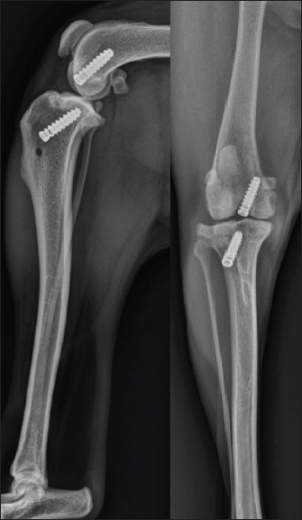
Fig. 7. 6-month postoperative radiographic views of dog's right pelvic limb from side (left) and front (right). In human medicine, the surgical indications for posterior cruciate ligament reconstruction include symptomatic severe posterior knee instability, posterior cruciate ligament avulsion with large bony fragments, or the presence of multiple ligament injuries (Veltri et al., 1995; Chen, 2007). As in dogs, there is no consensus about the surgical approach to adopt in humans. The techniques described in the literature vary according to the approach, the type of sutures, the nature of the substitution ligament, the use of arthroscopy, the orientation of the bone tunnels, and the type of screws when applicable (Sasaki et al., 2007; Gui et al., 2009; Zhang et al., 2013; Gwinner et al., 2014; Sabat et al., 2016). In dogs, synthetic reconstruction seems indicated for isolated ligament lesions in large individuals (>15–20 kg) whose growth is complete and for whom conservative treatment does not allow the resolution of clinical signs (Goin et al., 2019). Synthetic reconstruction of the ligament was performed arthroscopically as it is the most sensitive and specific technique for the evaluation of intra-articular lesions, especially meniscal lesions (Pozzi et al., 2008; Bleedorn et al., 2011; Little et al., 2014). Arthroscopy-assisted surgery also helped to reduce potential damage to the surgical site and to identify the tibial and femoral footprints of the CdCL, allowing the synthetic implant to fit the physiological localization of the ligament (Bedi and Altchek, 2009). Furthermore, arthroscopy-assisted surgery is aimed at minimizing perioperative trauma for the patient and at optimizing the chances of early recovery (Hoelzler et al., 2004). This goal was achieved with a return to weight bearing on the day following the surgery. However, using an intra-articular synthetic implant may also present risks. Barnhart and colleagues reconstructed ruptured cranial cruciate ligaments in 50 dogs using a braided synthetic material (multi-filamentous UHMWPE terathalate core contained within a braided porous non-expanded polytetrafluoroethylene sheath) (Barnhart et al., 2016). Among these 50 patients, 16 presented major complications, and nine presented minor ones (Barnhart et al., 2016). The most common complication was septic arthritis (Barnhart et al., 2016). Although no major complication occurred in this clinical case, a synovial inflammation remained present 6 months after the surgery together with mild lameness. Since the UHMWPE implant is also braided, one can hypothesize that this feature increases the risk of developing a major complication. The absence of an analysis of joint fluid makes it impossible to formally rule out septic arthritis, although no other clinical sign was observed. Finally, the implant tensioning technique used was probably the cause of excessive tension in the case presented in this study. However, the dog fully recovered clinically and recovered a normal level of activity after 6 months. LOAD results at 6 months and 1 year postoperatively were excellent. The limitations of this study include the uniqueness of the case and the absence of a magnetic resonance imaging examination, which would have provided a better postoperative assessment of soft tissues and suspected synovitis (D'Anjou et al., 2008). Similarly, carrying out a control arthroscopic examination would have helped to assess the precise positioning of the implant, perform a direct evaluation of the intra-articular structures, take a sample of the synovial membrane for histological analysis, and ensure that there was no impingement with the cranial cruciate ligament. A cytological analysis of synovial fluid puncture and the search for free synthetic particles in the joint would have completed the exploration of potential complications (Little et al., 2014). In light of the scarcity of clinical cases presenting this condition and the limited number of surgical techniques published, we believe that this technique should be considered as a possible treatment not only for isolated rupture of the CdCL but also for multiple ligament-injured stifles in dogs (Buttin et al., 2022). AcknowledgmentsThe authors thank Mrs. Cooke-Martageix for English copyediting of this article Conflicts of interestThe authors have no conflicts of interest to declare. FundingThis study was supported by the company Novetech Surgery, which provided human resources in the persons of Bastien Goin and Antonin Crumière for data processing and writing of this scientific article. Data availabilityAll the data are presented in figures directly in this article. Authors contributionsFauqueux F., DVM, performed the surgical management and follow-up controls, and wrote the article. Goin B., PhD student, and Agbalé M., DVM, wrote the article. Crumière A. J. J., PhD, Buttin P., DVM, Viguier E., DVM, Ph.D., ECVS, and Cachon T., DVM, Ph.D., ECVS, revised and improved the manuscript. ReferencesArnoczky, S.P. 1988. BSAVA Education committee commissioned article: the cruciate ligaments: the enigma of the canine stifle. J. Small Anim. Pract. 29, 71–90. Barnhart, M.D., Maritato, K., Schankereli, K., Wotton, H. and Naber, S. 2016. Evaluation of an intra-articular synthetic ligament for treatment of cranial cruciate ligament disease in dogs: a six-month prospective clinical trial. Vet. Comp. Orthop. Traumatol. 29, 491–498. Bedi, A., and Altchek, D.W. 2009. The "Footprint" anterior cruciate ligament technique: an anatomic approach to anterior cruciate ligament reconstruction. J. Arthrosc. Relat. Surg. 25, 1128–1138. Biskup, J.J. and Conzemius, M.G. 2020. Long-term arthroscopic assessment of intra-articular allografts for treatment of spontaneous cranial cruciate ligament rupture in the dog. Vet. Surg. 49, 764–771. Blanc, Q., Goin, B., Rafael, P., Moissonnier, P., Carozzo, C., Buttin, P., Cachon, T. and Viguier, E. 2019. Effect of the number of interference screws for the fixation of an intra-articular cranial cruciate ligament prosthesis in dogs: biomechanical study. Comput. Methods Biomech. Biomed. Eng. 22, S102–S104. Bleedorn, J.A., Greuel, E.N., Manley, P.A., Schaefer, S.L., Markel, M.D., Holzman, G. and Muir, P. 2011. Synovitis in dogs with stable stifle joints and incipient cranial cruciate ligament rupture: a cross-sectional study. Vet. Surg. 40, 531–543. Buttin, P., Goin, B., Cachon, T. and Viguier, E. 2020. Repair of tendon disruption using a novel synthetic fiber implant in dogs and cats: the surgical procedure and three case reports. Vet. Med. Int. 2020, 4146790. Buttin, P., Vallefuoco, R. and Le Pommellet, H. 2022. Stabilization of multiple ligament-injured stiffle using an ultra-high molecular weight polyethylen (UHMWPE) ligament in one dog and three cats. In 21st Congress of the European Society of Veterinary Orthopaedics and Traumatology (ESVOT), Nice, France. Chen, CH. 2007. Surgical treatment of posterior cruciate ligament injury. Chang. Gung. Med. J. 30, 480–492. Cook, J.L., Smith, P., Stannard, J.P., Pfeiffer, F., Kuroki, K., Bozynski, C.C., and Cook, C. 2017. A canine arthroscopic anterior cruciate ligament reconstruction model for study of synthetic augmentation of tendon allografts. J. Knee Surg. 30, 704–711. D'Anjou, MA., Moreau, M., Troncy, É., Martel-Pelletier, J., Abram, F., Raynauld, JP. and Pelletier, JP. 2008. Osteophytosis, subchondral bone sclerosis, joint effusion and soft tissue thickening in canine experimental stifle osteoarthritis: comparison between 1.5 T magnetic resonance imaging and computed radiography. Vet. Surg. 37, 166–177. Goin, B., Buttin, P., Lafon, Y., Massenzio, M., Viguier, E. and Cachon, T. 2022. Biomechanical cyclic loading test of a synthetic ligament fixation system used for intra-articular stabilization of deficient canine stifles. Open Vet. J. 12, 341–350. Goin, B., Morvan, V., Buttin, P., Lafon, Y., Massenzio, M., Viguier, E. and Cachon, T. 2021. Biomechanical comparison of two femoral fixation methods for synthetic cranial cruciate ligament reconstruction in canine cadavers. ABSTRACTS 46ème congrès société biomécanique. Comput. Methods Biomech. Biomed. Eng. 24, 127–129. Goin, B., Rafael, P., Blanc, Q., Cachon, T., Buttin, P., Carozzo, C., Chabrand, P. and Viguier, E. 2019. Biomechanical analysis of a ligament fixation system for CCL reconstruction in a canine cadaver model. Comput. Methods Biomech. Biomed. Eng. 22, S109–S111. Gui, J., Wang, L., Jiang, Y., Wang, Q., Yu, Z. and Gu, Q. 2009. Single-tunnel suture fixation of posterior cruciate ligament avulsion fracture. Arthroscopy 25, 78–85. Gwinner, C., Kopf, S., Hoburg, A., Haas, N.P. and Jung, T.M. 2014. Arthroscopic treatment of acute tibial avulsion fracture of the posterior cruciate ligament using the tightRope fixation device. Arthrosc. Tech. 3, e377–e382. Harari, J., Johnson, L., Stein, L.E., Kneller, S.K., and Pijanowski, G. 1987. Evaluation of experimental transection and partial excision of the caudal cruciate ligament in Dogs. Vet. Surg. 16, 151–154. Hoelzler, M.G., Millis, D.L., Francis, D.A. and Weigel, J.P. 2004. Results of arthroscopic versus open arthrotomy for surgical management of cranial cruciate ligament deficiency in dogs. Vet. Surg. 33, 146–153. Johnson, A.L. and Olmstead, M.L. 1987. Caudal cruciate ligament rupture a retrospective analysis of 14 Dogs. Vet. Surg. 16, 202–206. Johnson, J.M. and Johnson, A.L. 1993. Cranial cruciate ligament rupture: pathogenesis, diagnosis, and postoperative rehabilitation. Vet. Clin. North Am. Small Anim. Pract. 23, 717–733. Knecht, C.D., Crise, R. and Cechner, P.E. 1976. Repair of avulsion of the caudal cruciate ligament in a dog using a bone screw. J. Am. Anim. Hosp. Assoc. 12, 784–787. Little, J.P., Bleedorn, J.A., Sutherland, B.J., Sullivan, R., Kalscheur, V.L., Ramaker, M.A., Schaefer, S.L., Hao, Z. and Muir, P. 2014. Arthroscopic assessment of stifle synovitis in dogs with cranial cruciate ligament rupture. PLoS One. 9, e97329. Livet, V. and Cachon, T. 2017. Avulsion fracture of the femoral attachment of the caudal cruciate ligament treated by arthroscopy in a bernese mountain puppy. Vet. Rec. Case Rep. 4, e000348. Lopez, M.J., Markel, M.D., Kalscheur, V., Lu, Y. and Manley, P.A. 2003. Hamstring graft technique for stabilization of canine cranial cruciate ligament deficient stifles. Vet. Surg. 32, 390–401. Mao, G., Qin, Z., Li, Z., Li, X., Qiu, Y. and Bian, W. 2019. A tricalcium phosphate/polyether ether ketone anchor bionic fixation device for anterior cruciate ligament reconstruction: Safety and efficacy in a beagle model. J. Biomed. Materials Res. Part B: Appl. Biomaterials. 107, 554–563. Pozzi, A., Hildreth, B.E. and Rajala-Schultz, P.J. 2008. Comparison of arthroscopy and arthrotomy for diagnosis of medial meniscal pathology: an Ex Vivo study. Vet. Surg. 37, 749–755. Rafael, P., Goin, B., Buttin, P., Cachon, T. and Viguier, E. 2020. Comparison of two methods of fixation with interference screw for cranial cruciate ligament reconstruction in canine cadaver model. Computer Methods Biomech. Biomed. Engin. 23, S247–S249. Sabat, D., Jain, A. and Kumar, V. 2016. Displaced posterior cruciate ligament avulsion fractures: a retrospective comparative study between open posterior approach and arthroscopic single-tunnel suture fixation. Arthroscopy 32, 44–53. Sasaki, S.U., da Mota e Albuquerque, R.F., Amatuzzi, M.M. and Pereira, C.A.M. 2007. Open screw fixation versus arthroscopic suture fixation of tibial posterior cruciate ligament avulsion injuries: a mechanical comparison. Arthroscopy 23, 1226–1230. Smith, P.A., Bozynski, C.C., Kuroki, K., Henrich, S.M., Wijdicks, C.A. and Cook, J.L. 2019. Intra-articular biocompatibility of multistranded, long-chain polyethylene suture tape in a Canine ACL model. J. Knee Surg. 32, 525–531. Soreide, E., Denbeigh, J.M., Lewallen, E.A., Thaler, R., Xu, W., Berglund, L., Yao, J.J., Martinez, A., Nordsletten, L., van Wijnen, A.J. and Kakar, S. 2019. In vivo assessment of high-molecular-weight polyethylene core suture tape for intra-articular ligament reconstruction. Bone Joint J. 101-B, 1238–1247. Vasseur, P.B. 1993. Stifle Joint. In Textbook of small animal surgery. Eds., Slatter, D. Philadelphia, PA: Saunders, pp: 1817–1865. Veltri, D.M., Deng, X.H., Torzilli, P.A., Warren, R.F. and Maynard, M.J. 1995. The role of the cruciate and posterolateral ligaments in stability of the Knee: a biomechanical study. Am. J. Sports Med. 23, 436–443. Walton, M.B., Cowderoy, E., Lascelles, D. and Innes, J.F. 2013. Evaluation of construct and criterion validity for the ‘liverpool osteoarthritis in dogs’ (LOAD) clinical metrology instrument and comparison to two other instruments. PLoS One 8, e58125. Yoshiya, S., Andrish, J.T., Manley, M.T. and Kurosaka, M. 1986. Augmentation of anterior cruciate ligament reconstruction in dogs with prostheses of different stiffnesses. J. Orthop. Res. 4, 475–485. Zhang, X., Cai, G., Xu, J. and Wang, K. 2013. A minimally invasive postero-medial approach with suture anchors for isolated tibial avulsion fracture of the posterior cruciate ligament. Knee. 20, 96–99. | ||

| How to Cite this Article |
| Pubmed Style Fauqueux F, Goin B, Agbalé M, Crumière AJJ, Buttin P, Viguier E, Cachon T. Intra-articular replacement of the caudal cruciate ligament using an UHMWPE ligament under arthroscopic guidance in a dog: A case report. Open Vet J. 2023; 13(7): 948-954. doi:10.5455/OVJ.2023.v13.i7.15 Web Style Fauqueux F, Goin B, Agbalé M, Crumière AJJ, Buttin P, Viguier E, Cachon T. Intra-articular replacement of the caudal cruciate ligament using an UHMWPE ligament under arthroscopic guidance in a dog: A case report. https://www.openveterinaryjournal.com/?mno=146563 [Access: July 17, 2025]. doi:10.5455/OVJ.2023.v13.i7.15 AMA (American Medical Association) Style Fauqueux F, Goin B, Agbalé M, Crumière AJJ, Buttin P, Viguier E, Cachon T. Intra-articular replacement of the caudal cruciate ligament using an UHMWPE ligament under arthroscopic guidance in a dog: A case report. Open Vet J. 2023; 13(7): 948-954. doi:10.5455/OVJ.2023.v13.i7.15 Vancouver/ICMJE Style Fauqueux F, Goin B, Agbalé M, Crumière AJJ, Buttin P, Viguier E, Cachon T. Intra-articular replacement of the caudal cruciate ligament using an UHMWPE ligament under arthroscopic guidance in a dog: A case report. Open Vet J. (2023), [cited July 17, 2025]; 13(7): 948-954. doi:10.5455/OVJ.2023.v13.i7.15 Harvard Style Fauqueux, F., Goin, . B., Agbalé, . M., Crumière, . A. J. J., Buttin, . P., Viguier, . E. & Cachon, . T. (2023) Intra-articular replacement of the caudal cruciate ligament using an UHMWPE ligament under arthroscopic guidance in a dog: A case report. Open Vet J, 13 (7), 948-954. doi:10.5455/OVJ.2023.v13.i7.15 Turabian Style Fauqueux, Francois, Bastien Goin, Mathilde Agbalé, Antonin Jean Johan Crumière, Philippe Buttin, Eric Viguier, and Thibaut Cachon. 2023. Intra-articular replacement of the caudal cruciate ligament using an UHMWPE ligament under arthroscopic guidance in a dog: A case report. Open Veterinary Journal, 13 (7), 948-954. doi:10.5455/OVJ.2023.v13.i7.15 Chicago Style Fauqueux, Francois, Bastien Goin, Mathilde Agbalé, Antonin Jean Johan Crumière, Philippe Buttin, Eric Viguier, and Thibaut Cachon. "Intra-articular replacement of the caudal cruciate ligament using an UHMWPE ligament under arthroscopic guidance in a dog: A case report." Open Veterinary Journal 13 (2023), 948-954. doi:10.5455/OVJ.2023.v13.i7.15 MLA (The Modern Language Association) Style Fauqueux, Francois, Bastien Goin, Mathilde Agbalé, Antonin Jean Johan Crumière, Philippe Buttin, Eric Viguier, and Thibaut Cachon. "Intra-articular replacement of the caudal cruciate ligament using an UHMWPE ligament under arthroscopic guidance in a dog: A case report." Open Veterinary Journal 13.7 (2023), 948-954. Print. doi:10.5455/OVJ.2023.v13.i7.15 APA (American Psychological Association) Style Fauqueux, F., Goin, . B., Agbalé, . M., Crumière, . A. J. J., Buttin, . P., Viguier, . E. & Cachon, . T. (2023) Intra-articular replacement of the caudal cruciate ligament using an UHMWPE ligament under arthroscopic guidance in a dog: A case report. Open Veterinary Journal, 13 (7), 948-954. doi:10.5455/OVJ.2023.v13.i7.15 |

