


| Research Article | ||
Open Vet. J.. 2025; 15(7): 3269-3276 Open Veterinary Journal, (2025), Vol. 15(7): 3269-3276 Research Article Ultrasound imaging identifies the antemortem and postmortem features: PMI in euthanized dogs with an implication of cause of death may mask the imaging findingsMohammed Eleid1, Mahmoud Elalfy1*, Ahmed M.A. Meligy1, Alaa Fehaid2, Mostafa A. Elmadawy3, Zakriya A. Al Mohamad1, Khalid M. Alkhodair4 and Ayman Elnahas11Clinical Science Department, College of Veterinary, King Faisal University, Ahsa, Saudi Arabia 2Forensic Medicine and Toxicology Department, Faculty of Veterinary Medicine, Mansoura University, Mansoura, Egypt 3Forensic Medicine and Toxicology Department, Faculty of Veterinary Medicine, Kafr Elsheikh University, Kafr Elsheik, Egypt 4Anatomy Department, College of Veterinary, King Faisal University, Ahsa, Saudi Arabia *Corresponding Author: Mahmoud Elalfy. Clinical Science Department, College of Veterinary, King Faisal University, Ahsa, Saudi Arabia. Email: malhefnawy [at] kfu.edu.sa Submitted: 08/06/2025 Revised: 15/06/2025 Accepted: 17/06/2025 Published: 31/07/2025 © 2025 Open Veterinary Journal
ABSTRACTBackground: The use of ultrasound imaging in veterinary forensic medicine is a growing field that offers a noninvasive and efficient method for assessing postmortem changes in dogs. Aim: This study aimed to explore the usefulness’ of ultrasound image modality to identify the PMI and distinguish between antemortem and postmortem features in euthanized dogs. Methods: This study investigated the utility of ultrasonography in estimating the postmortem time interval (PMI), which is a critical factor in determining the time since death in forensic cases involving canines. Traditional PMI estimation methods rely heavily on external signs, such as rigor mortis, livor mortis, and body temperature, which can be influenced by environmental factors. Results: Ultrasound provides a more consistent and objective tool by allowing internal visualization of changes in soft tissue and organs over time. Ultrasound was examined on canine cadavers at specific intervals postmortem to monitor internal decomposition processes. Key ultrasonographic markers, such as the presence of intraorgan gas, fluid accumulation, and changes in organ echogenicity, were analyzed and correlated with the elapsed time since death. These internal indicators progress in a predictable sequence, providing a reliable framework for PMI estimation. The study demonstrated that Ultrasound has proven effective in detecting minor internal alterations during PMI studies, especially in the liver, owing to its substantial blood volume and anatomical location in the lower abdomen that contribute to improved accuracy in forensic assessments. Conclusion: The findings support the integration of ultrasonography into standard forensic protocols for veterinary cases. The ability to differentiate antemortem from postmortem changes and estimate PMI noninvasively makes it a valuable tool for veterinary pathologists, legal authorities, and animal welfare investigators. Rapid, repeatable, and ultrasound imaging has significant promise in advancing the field of veterinary forensic science. Keywords: Euthanized dogs, Ultrasound imaging, Antemortem, Postmortem, PMI. IntroductionRadiology and imaging have historically worked together in synergy in “post-mortem assessment,” alongside the core field of Legal Medicine, which is fundamentally known as Forensic Pathology (Ferrara et al., 2017). Ultrasound imaging has become a cornerstone diagnostic tool in veterinary medicine (Grabherr et al., 2017), offering a noninvasive, real-time method for visualizing internal structures in dogs. It plays a critical role in the diagnosis of several conditions, including abdominal, cardiac, and musculoskeletal disorders (Papazoglou et al., 2004; Haers et al., 2009). Ultrasound enables veterinarians to detect abnormalities such as tumors, organ enlargement, and fluid accumulation without the need for surgical exploration, thereby minimizing stress and risk for canine patients (Szatmári et al., 2003). The versatility and rapid diagnostic capabilities of this system make it indispensable in both emergency and routine veterinary care. Beyond diagnostics in living animals, ultrasound has emerged as an important tool in forensic veterinary medicine, particularly in estimating the postmortem time interval (PMI). The PMI refers to the time since death, and accurate determination of the PMI is crucial for legal investigations involving animal abuse, neglect, or insurance claims (Brooks, 2016). In such cases, traditional methods such as rigor mortis, livor mortis, and body cooling provide only rough estimates. The integration of ultrasound into postmortem examinations allows for more precise evaluations based on organ decomposition and other internal changes. In postmortem settings, ultrasound can detect early decomposition changes in soft tissues, such as gas formation, tissue liquefaction, and structural breakdown, which correlate with the time since death. For instance, gas bubbles in the liver or intestines, which are visible on ultrasound, are indicative of bacterial activity and can suggest a specific PMI range. Thus, postmortem ultrasonography complements external assessments and strengthens the accuracy of forensic timelines for canine deaths. Furthermore, postmortem ultrasonography is advantageous because it is a nondestructive technique that preserves the integrity of the body, which is important when further examinations, necropsy, or legal autopsies are necessary. It also allows for repeated evaluations over time, enabling a dynamic study of decomposition in different environmental conditions (Grabherr et al., 2016). This adaptability enhances its utility across a variety of cases, from clinical necropsy to criminal investigations involving dogs. Advanced ultrasonography offers significant potential for distinguishing between antemortem and postmortem changes in dogs by providing high-resolution imaging of soft tissues and internal organs. Antemortem injuries typically trigger physiological responses such as hemorrhage, edema, and inflammatory infiltrates, which are visible on ultrasound as heterogeneous echogenic areas, fluid collections, and changes in tissue vascularity using Doppler modes. In contrast, postmortem changes lack these responses and instead present with predictable structural alterations, including loss of vascular signals, tissue homogeneity, and gas formation within organs due to bacterial activity. Forensic ultrasonography can thus differentiate traumatic injuries sustained during life from those inflicted after death, a distinction critical in abuse or cruelty investigations (Grabherr et al., 2016). Moreover, advanced ultrasound techniques, including elastography and contrast-enhanced ultrasound, are increasingly being explored to assess tissue stiffness and perfusion status, respectively—parameters that further help in differentiating between vital and non-vital tissue reactions. These modalities, when applied in a time-sequenced manner postmortem, can also be used to track the degradation of organ architecture, thereby assisting in estimating PMI with greater accuracy. Such innovations elevate ultrasound from a basic imaging tool to a forensic diagnostic platform capable of supporting both legal and scientific determination in veterinary pathology. Materials and MethodsSeven male dogs were used in the training of students in the Clinical Science Department, Veterinary Teaching Hospital, King Faisal University, for student training and at the end of the semester. We performed ultrasound imaging before and after the induction of euthanasia. The current study was carried out on male dogs undergoing euthanization or after failure of veterinarian treatment. We used xylazine 2% (Pang, 2024) for the induction of sedation at doses of 0.4 and 40 mg/kg for the induction of death as the method of euthanization. Furthermore, we performed ultrasound imaging at sedation as antemortem, and at this stage, the dogs showed signs of viability with normal heartbeat and respiration, and then we gave them an overdose of anesthesia and ensured induction of death by complete stop of the heart and complete loss of respiration. Furthermore, we performed imaging of dog cadavers at 0. 2, and 4 hours after the moment of death in Clearfield. Cadavers of various dog breeds, weights, and ages were used to assess postmortem changes through ultrasound imaging. All animals had died of natural causes or were euthanized for humane reasons unrelated to the study, with owner consent and ethical approval from the institutional animal care committee (ETHICS3410). The cadavers were stored under controlled conditions (room temperature of 25°C–30°C) to standardize decomposition rates. Before imaging, each subject was identified, and external examinations were conducted to record baseline physical findings, such as rigor mortis, livor mortis, and body temperature. Ultrasound was examined using a portable veterinary ultrasound machine equipped with a microconvex and a linear transducer operating at frequencies between 5 and 10 MHz. Imaging was conducted at regular intervals postmortem (e.g., 0, 2, 4 hours) to monitor internal changes over time. Standardized anatomical regions, including the liver, heart, kidneys, intestines, and bladder, were selected for scanning. To ensure consistency, the same operator performed all ultrasound scans, and the coupling gel was generously applied to optimize image quality without applying undue pressure to the tissues. Ultrasound images were evaluated for specific postmortem signs such as gas formation, fluid accumulation, changes in tissue echogenicity, and loss of organ definition. All findings were documented, photographed, and correlated with the estimated PMI. Additional environmental data, such as ambient temperature and humidity, were recorded to assess their influence on decomposition. The results were statistically analyzed to determine patterns of ultrasonographic changes relative to time since death. Ethical approvalEthical approval was obtained from the Institutional Animal Care and Use Committee (ETHICS3410). ResultsUltrasound imaging has emerged as a valuable noninvasive tool for postmortem identification in canine cadavers, particularly in forensic veterinary medicine. While traditionally used in live animals, ultrasonography can be adapted for use after death to assess anatomical structures and pathological conditions that may aid in identifying unknown remains. Internal features such as organ morphology, pathological lesions, implants (such as microchips or surgical materials), and musculoskeletal variations can often be visualized postmortem. These elements may provide unique identifiers when matched against medical or veterinary records. Additionally, ultrasound can help confirm the presence of species-specific anatomy and breed-related traits, assisting in narrowing down the identity of an unknown dog, especially when external features are degraded or decomposed. Moreover, postmortem ultrasonography supports forensic examination by detecting injuries, disease processes, or signs of surgical intervention that may be referenced in prior veterinary histories. For example, identifying the site of splenectomy, healed fractures, or internal neoplasms can help link cadavers to documented veterinary cases. Although not a replacement for microchip scanning or DNA analysis, ultrasound offers a practical, real-time method to gather soft tissue-based clues that can support identification in conjunction with other forensic techniques. The ability to preserve the body intact for potential legal review or secondary analysis further reinforces its role in humane and professional postmortem examinations in dogs. In the illustrated Figure 1, organ boundaries are clearly distinguishable before death; however, following euthanasia, tissue echogenicity diminishes as blood pools in the lower regions, resulting in reduced tissue vitality. This pattern was even observed in the early postmortem period (group 0), when PMI was reported as less than 24 hours. Between-group comparisons of mean echogenicity values showed that there was only a significant difference in the liver echogenicity among groups 0, 2, and 4 hours postmortem. Ultrasound could distinguish the antemortem and postmortem ultrastructure (Table 1) Notably, in one dog, a foreign body was detected in the gastrointestinal tract during the antemortem stage, which obscured the ultrasound imaging. During sedation, the dog expelled a large piece of gauze, and this foreign body was no longer visible in the postmortem imaging. This highlights a limitation of using ultrasound as a tool for PMI estimation, even though it can still be valuable for identifying surgical complications or failure of veterinarian treatment in dogs (Figs. 2 and 3). Necropsy of all dogs showed normal internal organs in all cadavers of dogs without regarding the previous veterinarian or surgical operation performed. DiscussionUltrasound imaging has gained increasing relevance in legal medicine due to its noninvasive nature and ability to detect internal physiological and pathological changes (Möbius et al. 2021). In the context of forensic investigations involving dogs, ultrasound can be used not only during life but also after death to assess internal organs and soft tissues. This technique allows forensic veterinarians to evaluate trauma, disease, or unnatural death without the immediate need for invasive dissection. As a result, ultrasound is a valuable first step in forensic assessments, especially when external signs are minimal or ambiguous. One of the most significant contributions of ultrasound in forensic cases is its role in estimating the PMI. PMI is critical in cases of suspected abuse, poisoning, or neglect because it helps determine the time of death. Using ultrasound, forensic investigators can observe postmortem changes, such as gas formation in the liver and intestines, fluid redistribution, and tissue degradation, which correlate with the decomposition timeline. These internal findings can be used in combination with external signs (such as rigor mortis or body cooling) to improve the accuracy of PMI estimation. Ultrasound also aids in distinguishing between antemortem and postmortem injuries and is an essential aspect in legal cases. Antemortem injuries typically exhibit signs of physiological response, such as hemorrhage, inflammation, or edema, which are often visible via ultrasound as increased echogenicity or fluid accumulation. In contrast, postmortem injuries usually lack these features because of the absence of blood circulation and cellular response, appearing more uniform or “clean” on ultrasound (Grabherr et al., 2017). This differentiation helps determine whether trauma occurred before or after death, thereby influencing forensic interpretations and legal conclusions. Additionally, ultrasound offers the advantage of preserving the cadaver’s integrity, through further analysis of images, which is essential in sensitive cases requiring reevaluation or expert testimony. Unlike invasive necropsy, which may alter or destroy tissue evidence, ultrasonography allows repeated observations and can be stored as digital images for future reference. This approach is particularly beneficial in multidisciplinary investigations involving pathologists, legal authorities, and animal welfare organizations (Hoyer et al., 2016) Ultrasound plays a significant role in identifying the cause of death in dogs, particularly in cases involving internal trauma, fluid accumulation, or organ pathology. By providing real-time imaging of soft tissues and internal organs, ultrasonography can detect abnormalities, such as internal hemorrhaging, ruptured organs, or tumors that may not be visible externally. For example, in cases of blunt force trauma, ultrasound can reveal free fluid in the abdominal cavity (suggestive of internal bleeding), whereas in poisoning cases, characteristic changes in liver or kidney echotexture can support a toxicological cause (Chandravanshi and Pal 2018). These findings help forensic veterinarians establish a more definitive cause of death before necropsy.
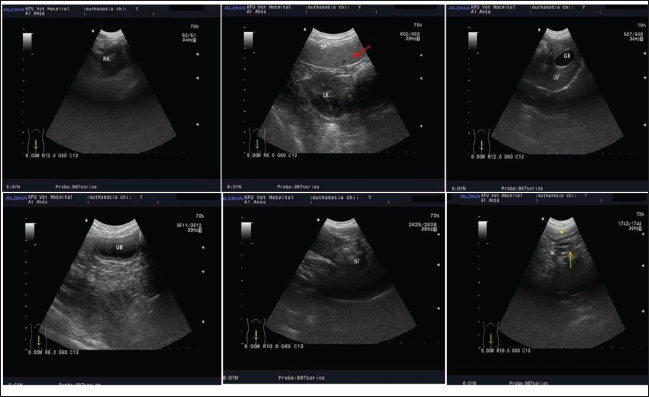
Fig. 1. Identification of antemortem features of internal organs in dogs under light anesthesia with wide normal small and large intestines and the presence of cleared splenic blood vessels that could be easily identified by ultrasound imaging. Table 1. Differences in antemortem and postmortem features identified by ultrasound imaging modality.
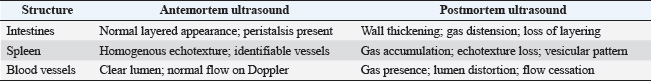
Ultrasound is also crucial for estimating the PMI in veterinary imaging at a university specialist hospital by monitoring decomposition-related changes in the body over time (Bryce et al., 2021). Postmortem ultrasonography can detect gas accumulation in tissues, fluid shifts, and structural breakdown of organs, such as the liver and intestines—changes that occur in predictable patterns depending on environmental conditions and time since death. These internal alterations, which are often missed by external examination alone, serve as valuable indicators of forensic timelines. When used alongside other PMI estimation techniques, such as rigor mortis and body cooling, ultrasound enhances the precision of time-of-death assessments in veterinary forensic investigations. Moreover, the noninvasive nature of ultrasonography makes it especially valuable in forensic contexts, where preserving the body’s condition is often critical. Unlike traditional necropsy for estimation of PMI in dogs (Erlandsson et al., 2007). which can alter tissue evidence, ultrasound can be performed repeatedly without damaging the cadaver, allowing investigators to document findings and reevaluate them later if needed. This is particularly important in legal cases involving suspected abuse or malpractice where objective, reproducible evidence is essential (Brooks et al., 2016). As such, ultrasound is not only a diagnostic tool but also a forensic asset for identifying both the cause and timing of death in dogs (Kazantsev et al., 2024).
Fig. 2. Antemortem imaging of dogs under light anesthesia shows foreign body and masks the imaging for identification the boundary and demarcation of internal organs.
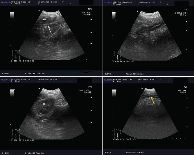
Fig. 3. The internal organs show reduction in echogenicity with unclear organ architecture at 2 and 4 hours after induction of euthanization at different times. Notably, the irregularity in the mucosa of gall bladder (whit arrow), irregularity of mucosa of small and large intestine (yellow arrow), changes in liver echogenicity, and unclear of vascularity in splenic paryenchyma were noticed at 2 and 4 hours after induction of death. Ultrasound is a promising yet underused imaging technique in legal medicine. Research on its feasibility has shown positive results, particularly for minimally invasive procedures. Ultrasound has shown significant improvements in the acquisition of high-quality biopsy samples, leading to more accurate diagnostics. In addition, ultrasound-based evaluation of epiphyses for age estimation has provided valuable insights. However, further investigation into the potential of ultrasound in forensic applications is required (Möbius et al., 2021). Postmortem decomposition is influenced by variables such as ambient temperature, humidity, and body size, which can significantly affect the rate of internal changes observed via ultrasound. Furthermore, ultrasonography is a highly operator-dependent technique, and its effectiveness relies on the skill and interpretation ability of the examiner, which may introduce variability (Grabherr, 2016). As decomposition progresses, gas formation and tissue liquefaction often reduce image clarity, making it difficult to interpret late-stage postmortem findings accurately. Additionally, ultrasound lacks the molecular and cellular resolution offered by histopathology or biochemical assays, limiting its ability to detect early or subtle postmortem alterations (Michaud et al., 2023). Finally, there is a shortage of longitudinal veterinary studies that validate and calibrate ultrasound findings against exact PMIs, reducing its current reliability as a standalone forensic tool (Brooks et al., 2016). Tissue rigidity after death is a fundamental indicator used in forensic medicine to estimate the time of death. However, qualitative approaches for assessing body stiffness can be limited in terms of accuracy and reliability. For liver echogenicity, only quantitative shear-wave elastography can be used to estimate the time of death (Mosadegh et al., 2022). Ultrasound imaging plays a crucial role in both clinical and forensic veterinary medicine, allowing for the detailed evaluation of abdominal organs in dogs. Antemortem ultrasonography provides vital diagnostic information about the gastrointestinal tract, spleen, and blood vessels. The intestine has a characteristic layered wall structure with visible peristalsis and normal gas patterns. The spleen appears as a homogenous, hyperechoic organ with identifiable splenic veins at the hilus. Major abdominal vessels, such as the aorta and portal vein, are routinely visualized with the aid of Doppler ultrasound to assess blood flow and detect abnormalities such as thrombosis or vascular anomalies (Spaulding, 1997). In contrast, postmortem ultrasound imaging revealed significant changes due to the onset of decomposition. As early as, gas accumulation becomes evident in the liver, intestines, portal vein, and caudal vena cava, with the aorta showing gas presence at later stages. These changes alter the appearance of abdominal structures—intestinal walls lose their normal layered pattern, become distended and echogenic, and the spleen undergoes echotexture changes, developing a vesicular appearance with internal gas pockets. These findings can interfere with interpretation if not recognized as postmortem changes and are especially relevant in forensic investigations to estimate time since death (Charlier et al., 2013). A comparison of antemortem and postmortem findings revealed significant differences in the structure and echogenicity of the intestines and spleen, as well as in the appearance of blood vessels. In live animals, the presence of perfused vessels and motile bowel loops is a key diagnostic feature, whereas postmortem changes reflect vascular gas formation and tissue decay. Understanding these differences enhances diagnostic accuracy in clinical practice and provides forensic veterinarians with a framework to evaluate postmortem intervals and cause of death (Watson et al., 2017). Notably, the irregularity in the mucosa of the gallbladder is a hallmark of early autolytic changes. The gallbladder is particularly prone to postmortem alterations due to its high content of digestive enzymes and bile, which can lead to enzymatic digestion of the mucosal lining. Histological studies have shown that mucosal desquamation and epithelial breakdown can occur within 1–4 hours after death, particularly in warm environments (Klimkowski et al., 2022). Similarly, the irregularity of the mucosa in the small and large intestines (yellow arrow) reflects the early stages of autolysis. The gastrointestinal tract is one of the first organ systems to undergo decomposition because of its high bacterial load. The intestinal mucosa is rapidly disrupted as bacteria proliferate and tissue hypoxia progresses. Mucosal sloughing and submucosal edema can be observed as early as 2–4 hours postmortem (Pérez et al., 2020). In addition, changes in liver echogenicity observed via ultrasonography indicate progressive hepatic cell lysis and the accumulation of intrahepatic gas or fluid due to autolysis and early putrefaction. Due to its large volume and rich blood supply, the liver undergoes predictable postmortem changes that can be detected within hours after death. The echogenicity may initially decrease due to fluid infiltration and increased cellular breakdown, but it later becomes heterogeneous due to gas formation (Wu et al., 2021). Moreover, unclear vascularity in the splenic parenchyma may reflect early autolytic disruption of the splenic tissue architecture. The spleen typically softens and loses its definition as proteolytic enzymes break down cellular and connective tissues. Moreover, loss of vascular clarity can be associated with hemolysis and blood settling (livor mortis), which distorts normal imaging features (Roccarina et al., 2024). In conclusion, ultrasound imaging significantly enhances clinical and forensic veterinary practice. The proposed method offers real-time insights into a dog’s internal condition during life and provides critical data for estimating PMI after death during life and provides critical data for estimating PMI after death Munro and Munro (2013), Yitbarek and Dagnaw (2022). By enabling non-invasive, detailed visualization of internal changes, ultrasound supports more accurate diagnoses, forensic evaluations, and legal proceedings. As technology continues to advance, the role of ultrasonography in veterinary science, in both life and postmortem, is poised to grow even further. Moreover, ultrasound is an essential tool in forensic veterinary medicine, providing valuable support for estimating PMI and distinguishing between antemortem and postmortem findings. Its non-invasive, real-time imaging capability provides critical information without compromising the physical state of the body, making it highly effective in both clinical and legal veterinary investigations. The integration of ultrasound is likely to become a standard component of postmortem examinations in dogs as the field of veterinary forensics expands. AcknowledgmentsThis work was supported by the Deanship of Scientific Research, Vice Presidency for Graduate Studies and Scientific Research, King Faisal University, Saudi Arabia (Project No. GRANT- kfu-251849). Conflict of interestAll authors declare that they have no conflicts of interest regarding any aspect of this publication. FundingDeanship of Scientific Research, Vice Presidency for Graduate Studies and Scientific Research, King Faisal University, Saudi Arabia (Project No. GRANT- kfu-251849). Authors’ contributionsAll authors contributed to this study. Data availabilityData will be available upon reasonable request. ReferencesBrooks, J.W. 2016. Postmortem changes in animal carcasses and estimation of the postmortem interval. Vet. Pathol. 53(5), 929–940. Bryce, A.J., Dandrieux, J.R., Tyrrell, D. and Milne, M.E. 2021. The evolving use of post-mortem veterinary imaging in a university specialist hospital. Foren. Imag. 26, 200475. Chandravanshi, L.P. and Pal, M., 2018. Assessment and diagnosis of poisoning with characteristic features in living or dead. J. Foren. Sci. Crim. Invest. 10, 1–12. Charlier, P., Chaillot, P.F., Watier, L., Ménétrier, M., Carlier, R., Cavard, S., Hervé, C., Grandmaison, G.L.D.L. and Huynh-Charlier, I. 2013. Is post-mortem ultrasonography a useful tool for forensic purposes?. Med. Sci. Law. 53(4), 227–234. Erlandsson, M. and Munro, R. 2007. Estimation of the post-mortem interval in beagle dogs. Sci. Jus. 47(4), 150–154. Ferrara, S.D., Cecchetto, G., Cecchi, R., Favretto, D., Grabherr, S., Ishikawa, T., Kondo, T., Montisci, M., Pfeiffer, H., Bonati, M.R. and Shokry, D. 2017. Back to the future-part 2. Post-mortem assessment and evolutionary role of the bio-medicolegal sciences. Inter. J. Leg. Med. 131, 1085–1101. Grabherr, S., Baumann, P., Minoiu, C., Fahrni, S. and Mangin, P. 2016. Post-mortem imaging in forensic investigations: current utility, limitations, and ongoing developments. Res. Rep. Foren. Med. Sci. 6, 25–37. Grabherr, S., Egger, C., Vilarino, R., Campana, L., Jotterand, M. and Dedouit, F. 2017. Modern post-mortem imaging: an update on recent developments. Foren. Sci. Res. 2(2), 52–64. Haers, H. and Saunders, J.H. 2009. Review of clinical characteristics and applications of contrast-enhanced ultrasonography in dogs. J. Amer. Vet. Med. Ass. 234(4), 460–470. Hoyer, R., Means, R., Robertson, J., Rappaport, D., Schmier, C., Jones, T., Stolz, L.A., Kaplan, S.J., Adamas-Rappaport, W.J. and Amini, R. 2016. Ultrasound-guided procedures in medical education: a fresh look at cadavers. Inter. Emerg. Med. 11, 431–436. Kazantsev, R. and Yatsenko, I. 2024. Forensic veterinary diagnosis of systemic hemostatic disorders and sudden death in cats and dogs: thanatogenetic aspect of critical conditions. Reg. Mech. Biosyst. 15(4), 808–820. Klimkowski, S.P., Fung, A., Menias, C.O. and Elsayes, K.M. 2022. Gallbladder imaging interpretation pearls and pitfalls: ultrasound, computed tomography, and magnetic resonance imaging. Radiol. Clin. 60(5), 809–824. Michaud, K., Jacobsen, C., Basso, C., Banner, J., Blokker, B.M., de Boer, H.H., Dedouit, F., O’Donnell, C., Giordano, C., Magnin, V. and Grabherr, S. 2023. Application of postmortem imaging modalities in cases of sudden death due to cardiovascular diseases–current achievements and limitations from a pathology perspective: endorsed by the Association for European Cardiovascular Pathology and by the International Society of Forensic Radiology and Imaging. Virchows Archiv. 482(2), 385–406. Möbius, D., Fitzek, A., Hammer, N., Heinemann, A., Ron, A., Schädler, J., Zwirner, J. and Ondruschka, B. 2021. Ultrasound in legal medicine—a missed opportunity or simply too late? A narrative review of ultrasonic applications in forensic contexts. Inter. J. Legal Med. 135(6), 2363–2383. Mosadegh, M., Khazaei, M., Abdollahpour, Z.D., Alahyari, S., Moharamzad, Y., Emamhadi, M., Aram, S., Abolbaghaei, M. and Sanei Taheri, M. 2022. Ultrasound shear-wave elastography applicability in estimation of post-mortem time. Ultrasound 30(2), 134–140. Munro, R. and Munro, H.M.C. 2013. Some challenges in forensic veterinary pathology: a review. J. Com. Pathol. 149(1), 57–73. Pang, D.S. 2024. Anesthetic and analgesic adjunctive drugs. In Veterinary anesthesia and analgesia the sixth edition of Lumb and Jones. Eds., Grimm, K.A., Lamont, L.A., Tranquilli, W.J., Greene, S.A. and Robertson, S.A. Hoboken, NJ, pp: 420–447. Papazoglou, L.G., Patsikas, M.N., Papadopoulou, P., Savas, I., Petanides, T. and Rallis, T. 2004. Intestinal obstruction due to sand in a dog. Vet. Rec. 155(25), 809. Pérez, M.M., García, E.B. and Bonilla, J.M. 2020. Bowel ultrasound: examination techniques and normal and pathologic patterns. Radiología (English Edition) 62(6), 517--527. Roccarina, D., Deganello, A., Buscemi, P., Cidoni, D. and Meloni, M.F. 2024. Diagnostic insights into splenic pathologies: the role of multiparametric ultrasound. Abdom. Radiol. 50, 1–12. Spaulding, K.A. 1997. A review of sonographic identification of abdominal blood vessels and juxtavascular organs. Vet. Radiol. Ultrasound 38(1), 4–23. Szatmári, V., Harkaanyi, Z. and Vöaröas, K. 2003. A review of nonconventional ultrasound techniques and contrast-enhanced ultrasonography of noncardiac canine disorders. Vet. Radiol. Ultrasound 44(4), 380–391. Watson, E. and Heng, H.G. 2017. Forensic radiology and imaging for veterinary radiologists. Vet. Radiol. Ultrasound 58(3), 245–258. Wu, M., Sharma, P.G. and Grajo, J.R. 2021. The echogenic liver: steatosis and beyond. Ultrasound Quar. 37(4), 308–314. Yitbarek, D. and Dagnaw, G.G. 2022. Application of advanced imaging modalities in veterinary medicine: a review. Vet. Med. Res. Rep. 117–130. | ||
| How to Cite this Article |
| Pubmed Style Eleid M, Elalfy M, Meligy AM, Fehaid A, Elmadawy MA, Mohamad ZAA, Alkhodair KM, Elnahas A. Ultrasound imaging identifies the antemortem and postmortem features: PMI in euthanized dogs with an implication of cause of death may mask the imaging findings. Open Vet. J.. 2025; 15(7): 3269-3276. doi:10.5455/OVJ.2025.v15.i7.37 Web Style Eleid M, Elalfy M, Meligy AM, Fehaid A, Elmadawy MA, Mohamad ZAA, Alkhodair KM, Elnahas A. Ultrasound imaging identifies the antemortem and postmortem features: PMI in euthanized dogs with an implication of cause of death may mask the imaging findings. https://www.openveterinaryjournal.com/?mno=263369 [Access: January 25, 2026]. doi:10.5455/OVJ.2025.v15.i7.37 AMA (American Medical Association) Style Eleid M, Elalfy M, Meligy AM, Fehaid A, Elmadawy MA, Mohamad ZAA, Alkhodair KM, Elnahas A. Ultrasound imaging identifies the antemortem and postmortem features: PMI in euthanized dogs with an implication of cause of death may mask the imaging findings. Open Vet. J.. 2025; 15(7): 3269-3276. doi:10.5455/OVJ.2025.v15.i7.37 Vancouver/ICMJE Style Eleid M, Elalfy M, Meligy AM, Fehaid A, Elmadawy MA, Mohamad ZAA, Alkhodair KM, Elnahas A. Ultrasound imaging identifies the antemortem and postmortem features: PMI in euthanized dogs with an implication of cause of death may mask the imaging findings. Open Vet. J.. (2025), [cited January 25, 2026]; 15(7): 3269-3276. doi:10.5455/OVJ.2025.v15.i7.37 Harvard Style Eleid, M., Elalfy, . M., Meligy, . A. M., Fehaid, . A., Elmadawy, . M. A., Mohamad, . Z. A. A., Alkhodair, . K. M. & Elnahas, . A. (2025) Ultrasound imaging identifies the antemortem and postmortem features: PMI in euthanized dogs with an implication of cause of death may mask the imaging findings. Open Vet. J., 15 (7), 3269-3276. doi:10.5455/OVJ.2025.v15.i7.37 Turabian Style Eleid, Mohammed, Mahmoud Elalfy, Ahmed M.a. Meligy, Alaa Fehaid, Mostafa A. Elmadawy, Zakriya A. Al Mohamad, Khalid M. Alkhodair, and Ayman Elnahas. 2025. Ultrasound imaging identifies the antemortem and postmortem features: PMI in euthanized dogs with an implication of cause of death may mask the imaging findings. Open Veterinary Journal, 15 (7), 3269-3276. doi:10.5455/OVJ.2025.v15.i7.37 Chicago Style Eleid, Mohammed, Mahmoud Elalfy, Ahmed M.a. Meligy, Alaa Fehaid, Mostafa A. Elmadawy, Zakriya A. Al Mohamad, Khalid M. Alkhodair, and Ayman Elnahas. "Ultrasound imaging identifies the antemortem and postmortem features: PMI in euthanized dogs with an implication of cause of death may mask the imaging findings." Open Veterinary Journal 15 (2025), 3269-3276. doi:10.5455/OVJ.2025.v15.i7.37 MLA (The Modern Language Association) Style Eleid, Mohammed, Mahmoud Elalfy, Ahmed M.a. Meligy, Alaa Fehaid, Mostafa A. Elmadawy, Zakriya A. Al Mohamad, Khalid M. Alkhodair, and Ayman Elnahas. "Ultrasound imaging identifies the antemortem and postmortem features: PMI in euthanized dogs with an implication of cause of death may mask the imaging findings." Open Veterinary Journal 15.7 (2025), 3269-3276. Print. doi:10.5455/OVJ.2025.v15.i7.37 APA (American Psychological Association) Style Eleid, M., Elalfy, . M., Meligy, . A. M., Fehaid, . A., Elmadawy, . M. A., Mohamad, . Z. A. A., Alkhodair, . K. M. & Elnahas, . A. (2025) Ultrasound imaging identifies the antemortem and postmortem features: PMI in euthanized dogs with an implication of cause of death may mask the imaging findings. Open Veterinary Journal, 15 (7), 3269-3276. doi:10.5455/OVJ.2025.v15.i7.37 |

