


| Case Report | ||
Open Vet. J.. 2025; 15(9): 4759-4762 Open Veterinary Journal, (2025), Vol. 15(9): 4759-4762 Case Report Ultrasonographic findings of an ovarian bursal abscess in an intact female dogClara Pagá-Casanova* and Vicente Cervera-CastellanosDiagnostic Imaging Department, AniCura Valencia Sur Veterinary Hospital, Valencia, Spain *Corresponding Author: Clara Pagá-Casanova. Diagnostic Imaging Department, Hospital Veterinario AniCura Valencia Sur Avenida Picassent, Valencia, Spain. Email: clarapagacasanova [at] gmail.com Submitted: 24/06/2025 Revised: 14/08/2025 Accepted: 29/08/2025 Published: 30/09/2025 © 2025 Open Veterinary Journal
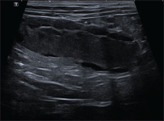
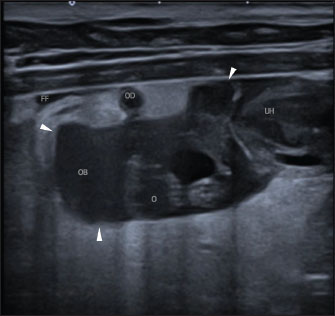
AbstractBackground: Ovarian bursal abscess (OBA) is a rarely described, potentially life-threatening condition with a nonspecific clinical presentation, inconclusive first-line diagnostic tests, and no well-defined ultrasonographic features. Case Description: A 3.5-year-old intact female miniature Schnauzer was presented with acute lethargy, vomiting, and diarrhea. The physical examination, blood analyses, and abdominal radiographs were nonspecific. Abdominal ultrasound revealed an irregular hypoechoic fluid-filled lesion surrounding the right ovary, focal peritoneal hyperechogenicity, scant free peritoneal fluid, a thin, tubular structure with no Doppler color signal partially encircling the ovary, and bilaterally distended uterine horns with irregular cystic walls. Free fluid obtained by ultrasound-guided aspiration was consistent with septic exudate. A right OBA was presumptively diagnosed. Surgery, histopathology, and culture confirmed an acute suppurative right-sided peri-oophoritis, salpingitis, pyometra, chronic endometritis, endometrial cystic hyperplasia, and septic peritonitis, with Escherichia coli isolation. The patient’s recovery was uneventful. Conclusion: Although rare, OBA should be considered a differential diagnosis in intact female dogs due to its potential evolution to septic peritonitis if left untreated. To the best of the authors’ knowledge, this is the first report describing specific ultrasonographic features of an OBA in an intact female dog, which, together with ultrasound-guided abdominocentesis, allowed an early diagnosis. The ultrasonographic findings described here may serve as a reference for similar future cases. Keywords: Escherichia coli, Intact female dog, Ovarian bursal abscess, Oviduct, Septic peritonitis. IntroductionAlthough genital pathology is frequent in female dogs, ovarian bursal abscess (OBA) has only been described in four case reports. In these cases, clinical presentation, blood analyses, and abdominal radiographs were non-specific and often not indicative of severity. To the best of the author’s knowledge, no characteristic ultrasonographic findings have been described (Van Israël et al., 2002; Park et al., 2013; Kumar et al., 2017; Eurell and Peacock, 2019). Case DetailsA 3.5-year-old intact female Miniature Schnauzer was presented with an acute onset of lethargy along with one episode of vomiting and diarrhea. The only previous clinical history was recurrent gastrointestinal signs, which were treated symptomatically without investigation. Her last estrus had ended 7 days before the presentation. On physical examination, the patient exhibited panting, pain upon cranial abdomen palpation, and increased rectal temperature (39.9°C). No vaginal discharge was noted. Bloodwork (complete blood count, biochemistry, electrolytes, and coagulation times) revealed the presence of band neutrophils (without abnormalities in total white cell count or differential count), mild anemia (hematocrit 34%, reference range 37%-62%), and a slight increase in the citrate partial thromboplastin time (126 seconds, reference range 72-102 seconds). On abdominal radiographs, a peritoneal left caudolateral, longitudinal structure, compatible with a dilated left uterine horn and a mild loss of adjacent serosal detail, was found (Fig. 1). No lesions were observed in the right cranial abdomen. A Canon Aplio i600 ultrasound machine with convex (9-10.8 MHz) and linear (12-18 MHz) probes was used. The conscious patient was positioned in both the lateral and dorsal recumbencies. Moderate distension of both uterine horns (up to 17 mm in diameter) with immobile hyperechoic intraluminal content and irregular walls with multiple mural ovoid anechoic lesions were noted (Fig. 2). Cranial to the ovarian end of the right uterine horn and surrounding the right ovary, a 25-mm, well-defined, irregular lesion filled with mildly echogenic fluid was observed, which was compatible with a fluid-filled ovarian bursa. The adjacent peritoneum was moderately hyperechoic with a mild amount of free echogenic fluid (Fig. 3). A scant amount of free anechoic fluid was also present next to the uterine horns. A slightly tortuous tubular structure (up to 4 mm in diameter) with no color Doppler signal was identified running longitudinally adjacent to the ovary up to the uterine horn’s ovarian end, which was interpreted as the oviduct (Figs. 3 and 4). Moreover, both ovaries showed several rounded structures that were compatible with the corpora lutea. Vascularization near the lesion preserved the color Doppler signal.
Fig. 1. Ventrodorsal (left) and left laterolateral (right) abdominal radiographs. A peritoneal left caudolateral, longitudinal structure, compatible with a dilated left uterine horn (arrowheads) and a mild loss of adjacent serosal detail (asterisk) are noted. No evident lesions are observed in the region of the right ovary.
Fig. 2. Abdominal ultrasonographic longitudinal examination in the left lateral recumbency using the B-mode with a microconvex probe. Cranial is to the left of the image. The right uterine horn is moderately distended with immobile hyperechoic intraluminal content and irregular walls with multiple mural ovoid anechoic lesions. Analysis of the peritoneal fluid next to the ovarian bursa, obtained via ultrasound-guided abdominocentesis, was consistent with a septic exudate, showing abundant toxic neutrophils, intracellular bacilli, total protein of 7.4 g/dl, decreased glucose, and increased lactate compared to peripheral blood (36 mg/dl vs. 105 mg/dl and 8.17 mmol/l vs. 1.59 mmol/l, respectively). A presumptive diagnosis of OBA with focal right-sided septic peritonitis, salpingitis, pyometra, mucometra, or hemometra, and cystic endometrial hyperplasia was made. An exploratory laparotomy was performed, followed by ovariohysterectomy. Surgery confirmed a septic focus in the right ovarian bursa. Culture and sensitivity testing of the bursal fluid isolated Escherichia coli, which was sensitive to multiple antibiotics. Histopathological examination of the reproductive tract revealed acute suppurative right-sided peri-oophoritis, salpingitis, and pyometra, bilateral cystic endometrial hyperplasia, and chronic endometritis. The dog had an uneventful recovery, with no observed adverse events during the 15-day post-surgery period. DiscussionThe ovarian bursa surrounds the ovary and contains a variable amount of fat in intact female dogs. It has a small medial opening that directly communicates with the peritoneal cavity. The oviduct (also known as the uterine tube or fallopian tube) connects the ovarian bursa to the uterine horn, with a papilla in the uterotubal junction, which remains open during the estrous period for the passage of sperm and blastocysts (Park et al., 2013; Rubio et al., 2014; König and Liebich, 2020).
Fig. 3. Abdominal ultrasonographic longitudinal examination in left lateral recumbency using a lineal high-frequency transducer in B-mode. Cranial is to the left of the image. The ovarian end of the right uterine horn (“UH”); the right ovary with nodular lesions (“O”), surrounded by a fluid-filled, well-defined lesion consistent with the right ovarian bursa (“OB”; the arrowheads mark the limits of the ovarian bursa). Surrounding this lesion, there is increased echogenicity of the peritoneal fat, as well as a mild volume of free fluid (“FF”). The thin, tortuous tubular structure, which is transversally imaged in this figure, is compatible with the oviduct (“OD”). Several routes of ovarian bursal infection have been proposed in the literature: ascending (via the uterine horn and oviduct), hematogenous, lymphatic, and transmural (Van Israël et al., 2002; Park et al., 2013; Rubio et al., 2014; Eurell and Peacock, 2019). An ascending route of infection was most likely in our case. This was based on the isolation of E. coli, the bacterium most commonly associated with canine pyometra, and the presence of ipsilateral pyometra with cystic endometrial hyperplasia and chronic endometritis, which are frequently linked to this disease (Van Israël et al., 2002; Park et al., 2013; Rubio et al., 2014; Eurell and Peacock, 2019). In two previously reported cases, the ascending route was also considered the most likely route, given that the pyometra was unilateral and ipsilateral to the OBA. In one of these cases, E. coli was also isolated from the ovarian bursa (Van Israël et al., 2002; Eurell and Peacock, 2019). Although the ovarian bursa is not ultrasonographically distinguished per se, a small volume of fluid can be observed within the ovarian bursa after ovulation, likely due to intra-follicular fluid accumulating after follicular opening (Lévy and Fontbonne 2007; Mogheiseh et al., 2017; Nogueira et al., 2021; Rault and Hecht, 2025). Normal oviducts are generally not visible with diagnostic imaging techniques, likely due to their small size (Russo et al., 2021). To the best of the author’s knowledge, this is the first report of ultrasonographic identification of a non-neoplastic oviduct in a female dog.
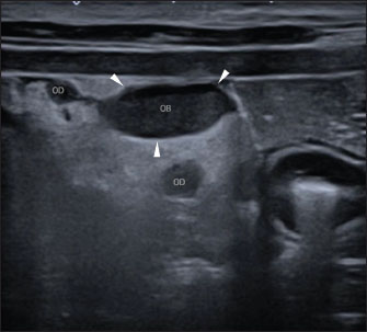
Fig. 4. Abdominal ultrasonographic transversal examination in left lateral recumbency using a lineal high-frequency probe in B-mode. Lateral is to the left of the image. Note the right ovarian bursa (“OB”; the arrowheads mark the limits of the ovarian bursa), and parts of the thin, tortuous, tubular structure, compatible with the oviduct (“OD”; the highest one in the image is near the ovarian bursa, and the lowest one is next to the uterine horn’s ovarian end). To the best of the authors’ knowledge, OBA is rare in dogs, having been described in only four case reports, in which the clinical presentation was nonspecific and diagnosis was not achieved through first-line diagnostic tools (bloodwork and radiographs) alone (Van Israël et al., 2002; Park et al., 2013; Kumar et al., 2017; Eurell and Peacock, 2019). Moreover, the ultrasound studies were limited and showed nonspecific findings, such as an abdominal fluid-filled lesion of undetermined origin (Van Israël et al., 2002; Eurell and Peacock, 2019), uterine abnormalities (Park et al., 2013; Kumar et al., 2017), and signs of peritonitis and/or free peritoneal fluid (Van Israël et al., 2002; Park et al., 2013). An accurate presumptive diagnosis was not made before surgery in any of these cases, which was delayed in at least two cases due to the absence of initial indicators of severity or surgical need (Van Israël et al., 2002; Park et al., 2013; Kumar et al., 2017; Eurell and Peacock, 2019). In our case, the signalment, history, physical examination, and initial tests neither allowed a diagnosis nor were indicative of severity, with more likely differential diagnoses including acute pancreatitis, gastrointestinal disease, pyometra, peritonitis, or referred pain. However, our case is unique because abdominal ultrasound and ultrasound-guided abdominocentesis clearly indicated an infectious process involving the ovarian bursa, resulting in septic peritonitis. These findings allowed for an early and accurate diagnosis, leading to surgery with a better prognosis and preventing patient deterioration if conservative symptomatic treatment was initially pursued. ConclusionIn conclusion, this is the first report of specific ultrasonographic findings of an OBA in an intact female dog. Although infrequent, OBA can be life-threatening if left undiagnosed; thus, it should be considered as a differential diagnosis in intact female dogs with acute abdomen or other non-specific signs. In our case, abdominal ultrasound and ultrasound-guided abdominocentesis were critical for an early and accurate diagnosis, allowing prompt surgical management with a better prognosis. The ultrasonographic findings described here could be useful for diagnosing similar cases in the future, and the identification of the oviduct could be useful in confirming the genital origin of lesions. AcknowledgmentsNone. Conflict of interestThe authors declare no conflict of interest. FundingThis study received no specific grant. Authors' contributionsClara Pagá Casanova: The main author of the manuscript and literature review. Vicente Cervera Castellanos: reviewed the process and manuscript. All authors have read and approved the published version of the manuscript. Data availabilityAll data supporting this study’s findings are available within the manuscript. ReferencesEurell, T.E. and Peacock, R.E. 2019. Persistent vaginal hemorrhage in a dog with an ovarian bursal abscess. Aust. Vet. Pract. 49(2), 40-3. König, H.E. and Liebich, H.G. 2020. Female genital organs (organa genitalia feminina). In Veterinary anatomy of domestic animals: textbook and color atlas. 7th ed. Stuttgart, Germany: Thieme, 449-470. Kumar, A., Tyagi, S.P. and Kumar, A. 2017. Surgical management of unilateral ovario-bursal abscess in a dog. Intas Polivet 18(I), 162-163. Lévy, X. and Fontbonne, A. 2007. Determining the optimal mating time in bitches: particularities. Rev. Bras. Reprod. Anim. 31(1), 128-134. Mogheiseh, A., Nikahval, B., Ahmadi, N., Yazdanpanah, R., Sadat, Z. and Nazifi, S. 2017. Bilateral ovarian pedicle ligation as an alternative to ovariectomy and ovarian response to eCG treatment. Comp. Clin. Pathol. 26(1), 197-202. Nogueira Aires, L.P., Pavan, L., Gasser, B., Silva, P., Maronezi, M.C., Del Aguila Da Silva, P., Vinícius Silveira, M., Correia Santos, V.J., Padilha-Nakaghi, L.C., Pozzobon, R. and Rossi Feliciano, M.A. 2021. Ultrasonographic aspects of the uterus and ovaries of bitches during the estrous cycle - paper review. Rev. Bras. Reprod. Anim. 45(1), 3-11. Park, E., Park, J., Jeong, S., Choi, H., Lee, Y., Song, K., Park, S., Yoon, K., Chung, T., Shin, S. and Cho, J. 2013. Peritonitis secondary to pyometra & ovarian bursal abscess in a dog. J. Vet. Clin. 30(5), 387-389. Rault, D. and Hecht, S. 2015. Female reproductive tract. 3rd ed. In Atlas of small animal ultrasonography. Eds., Penninck, D. and d’Anjou, M.A. Hoboken, USA: John Wiley & Sons, pp 481-510. Rubio, A., Boyen, F., Tas, O., Kitshoff, A., Polis, I., Van Goethem, B. and De Rooster, H. 2014. Bacterial colonization of the ovarian bursa in dogs with clinically suspected pyometra and in controls. Theriogenology 82(7), 966-971. Russo, M., England, G.C.W., Catone, G. and Marino, G. 2021. Imaging of canine neoplastic reproductive disorders. Animals 11(5), 1213. Van Israël, N., Kirby, B.M. and Munro, E.A.C. 2002. Septic peritonitis secondary to unilateral pyometra and ovarian bursal abscessation in a dog. J. Small Anim. Pract. 43 (10), 452-455. | ||

| How to Cite this Article |
| Pubmed Style Pagá-casanova C, Cervera-castellanos V. Ultrasonographic findings of an ovarian bursal abscess in an intact female dog. Open Vet. J.. 2025; 15(9): 4759-4762. doi:10.5455/OVJ.2025.v15.i9.82 Web Style Pagá-casanova C, Cervera-castellanos V. Ultrasonographic findings of an ovarian bursal abscess in an intact female dog. https://www.openveterinaryjournal.com/?mno=266657 [Access: June 22, 2026]. doi:10.5455/OVJ.2025.v15.i9.82 AMA (American Medical Association) Style Pagá-casanova C, Cervera-castellanos V. Ultrasonographic findings of an ovarian bursal abscess in an intact female dog. Open Vet. J.. 2025; 15(9): 4759-4762. doi:10.5455/OVJ.2025.v15.i9.82 Vancouver/ICMJE Style Pagá-casanova C, Cervera-castellanos V. Ultrasonographic findings of an ovarian bursal abscess in an intact female dog. Open Vet. J.. (2025), [cited June 22, 2026]; 15(9): 4759-4762. doi:10.5455/OVJ.2025.v15.i9.82 Harvard Style Pagá-casanova, C. & Cervera-castellanos, . V. (2025) Ultrasonographic findings of an ovarian bursal abscess in an intact female dog. Open Vet. J., 15 (9), 4759-4762. doi:10.5455/OVJ.2025.v15.i9.82 Turabian Style Pagá-casanova, Clara, and Vicente Cervera-castellanos. 2025. Ultrasonographic findings of an ovarian bursal abscess in an intact female dog. Open Veterinary Journal, 15 (9), 4759-4762. doi:10.5455/OVJ.2025.v15.i9.82 Chicago Style Pagá-casanova, Clara, and Vicente Cervera-castellanos. "Ultrasonographic findings of an ovarian bursal abscess in an intact female dog." Open Veterinary Journal 15 (2025), 4759-4762. doi:10.5455/OVJ.2025.v15.i9.82 MLA (The Modern Language Association) Style Pagá-casanova, Clara, and Vicente Cervera-castellanos. "Ultrasonographic findings of an ovarian bursal abscess in an intact female dog." Open Veterinary Journal 15.9 (2025), 4759-4762. Print. doi:10.5455/OVJ.2025.v15.i9.82 APA (American Psychological Association) Style Pagá-casanova, C. & Cervera-castellanos, . V. (2025) Ultrasonographic findings of an ovarian bursal abscess in an intact female dog. Open Veterinary Journal, 15 (9), 4759-4762. doi:10.5455/OVJ.2025.v15.i9.82 |

